

doi: 10.1097/GOX.0000000000000035.
eCollection 2013 Dec.
The clinical importance of the fat compartments in midfacial aging
Affiliations
- PMID: 25289286
- PMCID: PMC4174112
- DOI: 10.1097/GOX.0000000000000035
Item in Clipboard
The clinical importance of the fat compartments in midfacial aging
Plast Reconstr Surg Glob Open.
.
Abstract
The recent identification of the facial fat compartments has greatly affected our understanding of midfacial aging. This article chronicles the discovery of these fat compartments including the shift of attention from a purely gravitational to a volumetric approach to facial aging and the series of methodologies attempted to ultimately define the anatomy of these compartments. The revived interest in volumetric facial rejuvenation including compartment-guided augmentation techniques is discussed. Lastly, the article discusses interesting distributional patterns noted in these fat compartments likely related to the different mechanical and biologic environments of the deep and superficial facial fat pads.
Figures
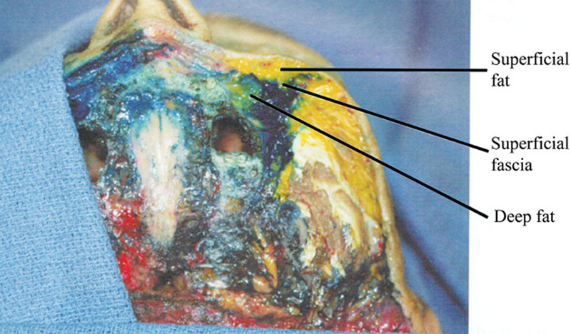
Midfacial fat is broadly divided into superficial and deep layers relative to the superficial fascia or superficial musculoaponeurotic system (SMAS). Reprinted with permission from QMP. Adaptations are themselves works protected by copyright. So in order to publish this adaptation, authorization must be obtained both from the owner of the copyright in the original work and from the owner of copyright in the translation or adaptation.
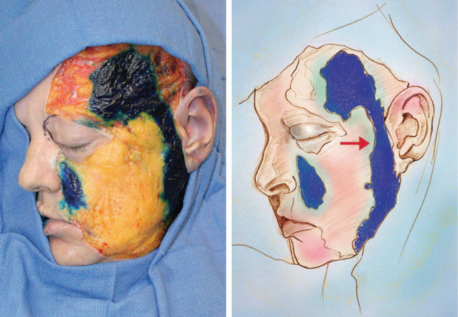
Rohrich and Pessa injected methylene blue dye into cadaveric specimens, allowing dye diffusion to dictate the natural septal boundaries of the facial fat compartments. The nasolabial fat (blue) and lateral temporal cheek fat (arrow) are partitioned in this manner. Reprinted with permission from Plast Reconstr Surg. 2007;119:2219–2227. Adaptations are themselves works protected by copyright. So in order to publish this adaptation, authorization must be obtained both from the owner of the copyright in the original work and from the owner of copyright in the translation or adaptation.
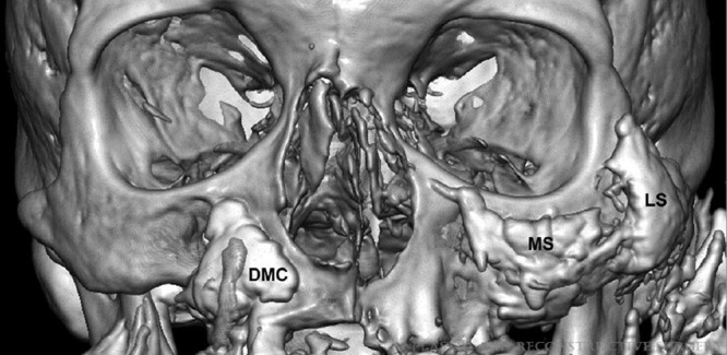
Gierloff’s group injected iodinated contrast medium into cadaveric specimens followed by CT scanning. The homogenous distribution of the medium within the facial fat compartments allowed these compartments to be isolated and volumetrically measured on 3D CT images. The medial part of the deep medial cheek fat (DMC) and the medial (MS) and lateral (LS) sub–orbicularis oculi fat pads are portrayed in this CT image. Reprinted with permission from Plast Reconstr Surg. 2012;129:263–273. Adaptations are themselves works protected by copyright. So in order to publish this adaptation, authorization must be obtained both from the owner of the copyright in the original work and from the owner of copyright in the translation or adaptation.
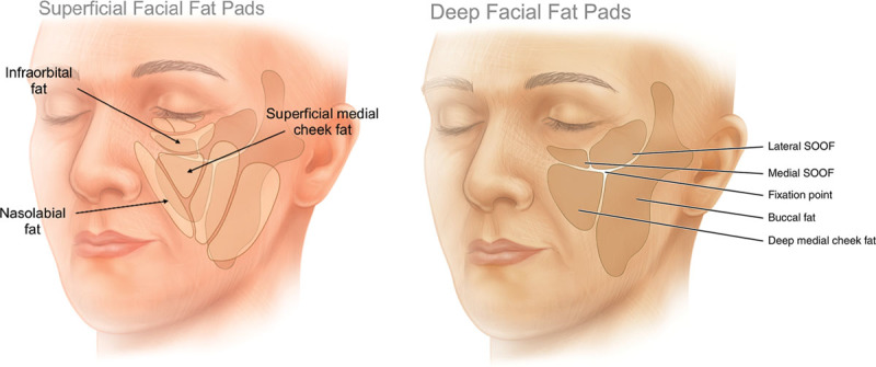
Schematic depiction of the superficial (light beige) and deep (dark tan) facial fat compartments. A, The superficial midfacial fat compartments include the infraorbital fat, superficial medial cheek fat, and nasolabial fat. B, The deep midfacial fat compartments include the medial and lateral SOOF, deep medial cheek fat which can be further divided into medial and lateral parts, and the medial portion of the buccal fat pad. Reprinted with permission from QMP. Adaptations are themselves works protected by copyright. So in order to publish this adaptation, authorization must be obtained both from the owner of the copyright in the original work and from the owner of copyright in the translation or adaptation.
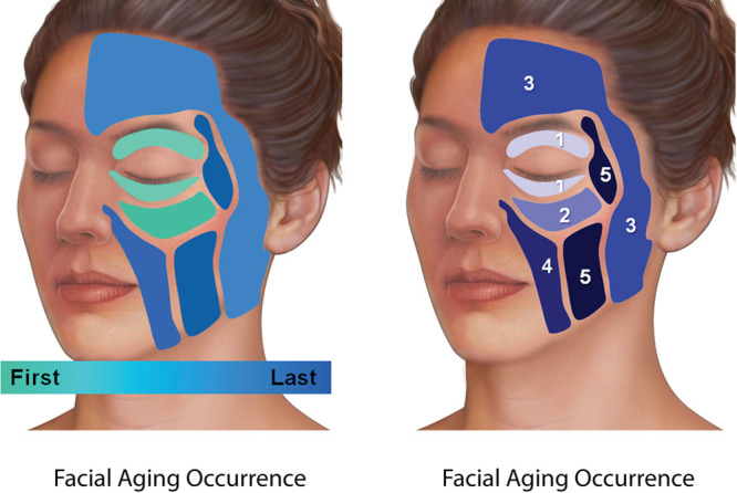
Sequence of fat compartment deflation commonly observed in facial aging. Reprinted with permission from Dr. Rohrich’s lecture from Plastic Surgery—The Meeting at San Diego on October 12, 2013. (CO205) The “lift and fill” face/necklift—Obtaining natural results in facelifts using facial fat augmentation.
References
-
- Gonzalez-Ulloa M, Flores ES. Senility of the face—basic study to understand its causes and effects. Plast Reconstr Surg. 1965;36:239–246. - PubMed
-
- Wulc AE, Sharma P, Czyz CN. The anatomic basis of midfacial aging. In: Hartstein ME, Wulc AE, Holck DEE, editors. Midfacial Rejuvenation. New York: Springer Science+Business Media; 2012. pp. 15–28.
-
- Pessa JE. An algorithm of facial aging: verification of Lambros’s theory by three-dimensional stereolithography, with reference to the pathogenesis of midfacial aging, scleral show, and the lateral suborbital trough deformity. Plast Reconstr Surg. 2000;106:479–488. discussion 489–490. - PubMed
-
- Shaw RB, Jr, Katzel EB, Koltz PF, et al. Aging of the facial skeleton: aesthetic implications and rejuvenation strategies. Plast Reconstr Surg. 2011;127:374–383. - PubMed
LinkOut - more resources
Full Text Sources
Other Literature Sources