. 2014 May 7;2(4):e132.
doi: 10.1097/GOX.0000000000000083. eCollection 2014 Apr.

Overexpressed HIF-2α in Endothelial Cells Promotes Vascularization and Improves Random Pattern Skin Flap Survival

Affiliations

Overexpressed HIF-2α in Endothelial Cells Promotes Vascularization and Improves Random Pattern Skin Flap Survival

Atsushi Morimoto et al. Plast Reconstr Surg Glob Open. .

Abstract

Background: The local skin flap procedure is very useful for reconstruction. However, flap necrosis caused by circulatory failure can occur at its distal portion. Hypoxia-inducible factors (HIFs) in endothelial cells (ECs) help to maintain ECs and promote vascularization, and HIF-2α is abundantly expressed in ECs. However, the mechanisms of action of HIF-2α in ECs are not yet fully understood. The aim of this study was to evaluate the in vivo effects of overexpression of HIF-2α in ECs on skin flap survival.

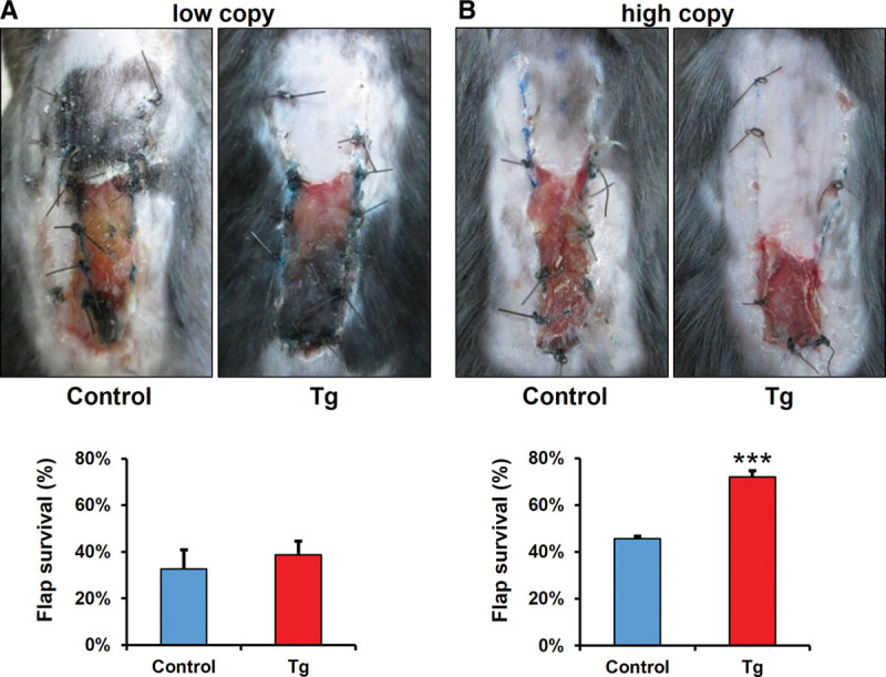
Methods: A random pattern skin flap (1.0 × 3.0 cm) was elevated on the dorsum of transgenic mice (Tg mice) with EC-specific HIF-2α conditional overexpression and wild-type littermate control mice (n = 6). Flap survival was evaluated on postoperative day 7. Tissue samples from the skin flaps were harvested and analyzed using Western blotting, quantitative reverse transcriptase-polymerase chain reaction, and immunohistochemistry.

Results: The HIF-2α mRNA and protein levels were significantly increased in the Tg mice when compared with control mice. Tg mice had significantly increased skin flap survival areas (72.0% ± 2.7%) when compared with wild-type mice (45.7% ± 1.1%). Moreover, histological examination revealed an increase in the subcutaneous blood vessel counts in the Tg mice.

Conclusions: Specific overexpression of HIF-2α in ECs promoted vascularization and enhanced skin flap survival in vivo in a mouse model.


Figures

Fig. 1.
Fig. 1.
A, Schematic representation of the LSL-HIF-2α transgenic mouse gene. Set3 (for2α) and td Tomato (solid triangles) indicate the sites of PCR primers used for mouse genotyping. B, Quantitative RT-PCR-based genotyping for td Tomato. Y axis shows relative mRNA level. PCR was performed with primers td Tomato. C, Quantitative RT-PCR-based genotyping for HIF-2α. Y axis shows relative copy number of HIF-2α to Arnt gene. PCR was performed with primers Set3 (for2α). Transgene copy number in 2 transgenic mice lines was compared. D, HIF-1α and HIF-2α mRNA levels in the lung. High-copy HIF-2α transgenic lines maintained under normoxic (21% O2) conditions. Data in bar graphs are presented as means ± SEM (n = 6). **P < 0.01. E and F, Western blot analysis of HIF-1α and HIF-2α protein in the lung. High-copy HIF-2α transgenic lines maintained under hypoxic (10% O2, 90% N2) conditions. β-Actin was used as a loading control. Values are means ± SEM (n = 6). *P < 0.05. RT-PCR indicates reverse transcriptase-polymerase chain reaction; Tg, endothelial cell–specific HIF-2α conditional overexpression transgenic mice.
Fig. 2.
Fig. 2.
Comparison of flap viability between the control mice and the Tg mice. A, Low-copy HIF-2α transgenic lines. B, High-copy HIF-2α transgenic lines. Flap survival was evaluated on postoperative day 7. Data in bar graphs are presented as percentages of flap survival ± SEM (n = 6). ***P < 0.001.
Fig. 3.
Fig. 3.
Expression of HIF-2α protein in a mouse skin flap model. A and B, Western blot analyses were performed on tissue lysates from dorsal skin taken preoperatively and from the proximal parts of the skin flaps on postoperative day 7. High-copy HIF-2α transgenic lines and wild-type littermate control mice were subjected to analysis. β-Actin was used as a loading control. Values are means ± SEM (n = 6). *P < 0.05, **P < 0.01, ***P < 0.001.
Fig. 4.
Fig. 4.
HIFs, VEGF, and PHD mRNA levels in the mouse skin flap model. Transcript levels of angiogenesis-related factors were assessed by quantitative RT-PCR analysis. A, Total RNA was isolated from dorsal skin taken before operation. Values are means ± SEM (n = 6). B and C, Total RNA was isolated from the proximal parts of the skin flaps on postoperative day 1 and 7. Data in bar graphs are presented as means ± SEM (n = 6). *P < 0.05, **P < 0.01, ***P < 0.001. RT-PCR indicates reverse transcriptase-polymerase chain reaction.
Fig. 5.
Fig. 5.
Effect of specific HIF-2α overexpression in ECs on postoperative angiogenesis and vasculature in mouse subcutaneous plexus. Section of proximal portions of skin flaps preoperatively (A) and on postoperative day 7 (B) were stained with hematoxylin and eosin (HE) and rhodamine-lectin. C, The number of lectin-positive (red) vessels was counted per high-power field. Scale bar indicates 200 μm. ***P < 0.001.

References

    1. McGregor IA, Morgan G. Axial and random pattern flaps. Br J Plast Surg. 1973;26:202–213. - PubMed
    1. Fujihara Y, Koyama H, Nishiyama N, et al. Gene transfer of bFGF to recipient bed improves survival of ischemic skin flap. Br J Plast Surg. 2005;58:511–517. - PubMed
    1. Zacchigna S, Papa G, Antonini A, et al. Improved survival of ischemic cutaneous and musculocutaneous flaps after vascular endothelial growth factor gene transfer using adeno-associated virus vectors. Am J Pathol. 2005;167:981–991. - PMC - PubMed
    1. Toutain CE, Brouchet L, Raymond-Letron I, et al. Prevention of skin flap necrosis by estradiol involves reperfusion of a protected vascular network. Circ Res. 2009;104:245–254. - PubMed
    1. Shalom A, Friedman T, Westreich M. The effect of postoperative aspirin on random pattern flaps in rats. Am Surg. 2007;73:1126–1128. - PubMed

LinkOut - more resources