. 2014 Oct 7;9(10):e109486.
doi: 10.1371/journal.pone.0109486. eCollection 2014.

Cell turnover and detritus production in marine sponges from tropical and temperate benthic ecosystems

Affiliations

Cell turnover and detritus production in marine sponges from tropical and temperate benthic ecosystems

Brittany E Alexander et al. PLoS One. .

Abstract

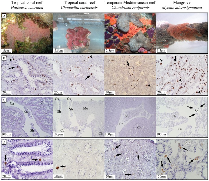
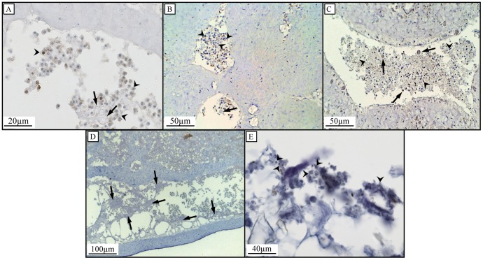
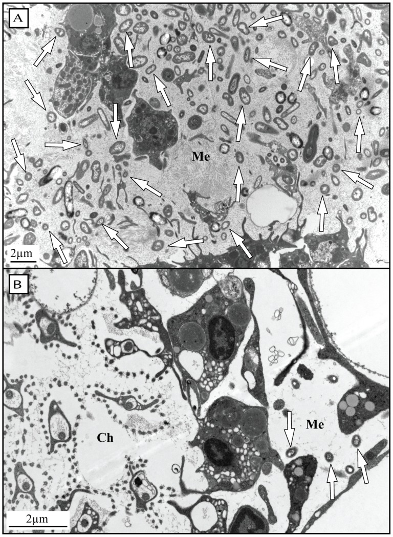
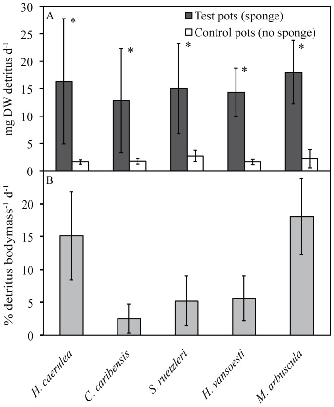
This study describes in vivo cell turnover (the balance between cell proliferation and cell loss) in eight marine sponge species from tropical coral reef, mangrove and temperate Mediterranean reef ecosystems. Cell proliferation was determined through the incorporation of 5-bromo-2'-deoxyuridine (BrdU) and measuring the percentage of BrdU-positive cells after 6 h of continuous labeling (10 h for Chondrosia reniformis). Apoptosis was identified using an antibody against active caspase-3. Cell loss through shedding was studied quantitatively by collecting and weighing sponge-expelled detritus and qualitatively by light microscopy of sponge tissue and detritus. All species investigated displayed substantial cell proliferation, predominantly in the choanoderm, but also in the mesohyl. The majority of coral reef species (five) showed between 16.1±15.9% and 19.0±2.0% choanocyte proliferation (mean±SD) after 6 h and the Mediterranean species, C. reniformis, showed 16.6±3.2% after 10 h BrdU-labeling. Monanchora arbuscula showed lower choanocyte proliferation (8.1±3.7%), whereas the mangrove species Mycale microsigmatosa showed relatively higher levels of choanocyte proliferation (70.5±6.6%). Choanocyte proliferation in Haliclona vansoesti was variable (2.8-73.1%). Apoptosis was negligible and not the primary mechanism of cell loss involved in cell turnover. All species investigated produced significant amounts of detritus (2.5-18% detritus bodyweight(-1)·d(-1)) and cell shedding was observed in seven out of eight species. The amount of shed cells observed in histological sections may be related to differences in residence time of detritus within canals. Detritus production could not be directly linked to cell shedding due to the degraded nature of expelled cellular debris. We have demonstrated that under steady-state conditions, cell turnover through cell proliferation and cell shedding are common processes to maintain tissue homeostasis in a variety of sponge species from different ecosystems. Cell turnover is hypothesized to be the main underlying mechanism producing sponge-derived detritus, a major trophic resource transferred through sponges in benthic ecosystems, such as coral reefs.


Conflict of interest statement

Competing Interests: BEA, RO, and JMdG are affiliated with Porifarma B.V. Porifarma B.V. received funding from the European Union Seventh Framework Programme (FP7/2007–2013) under grant agreement no KBBE-2010-266033 to undertake the research leading to these results. BEA is employed by Porifarma B.V. under grant agreement no KBBE-2010-266033 from the European Union Seventh Framework Programme (FP7/2007–2013). There are no patents, products in development or marketed products to declare. This does not alter the authors' adherence to all the PLOS ONE policies on sharing data and materials.

Figures

Figure 1
Figure 1. Cell proliferation in eight marine sponge species after continuous BrdU-labeling.
Mean percentages (±SD) of BrdU-positive cells in the choanoderm (open bars) and mesohyl (solid bars) are shown. Significant differences between cell proliferation in the choanoderm versus mesohyl are indicated for each species (* p<0.001; 95%-CI are given in Table 2). All species were labeled with BrdU for 6 h except for C. reniformis which was labeled for 10 h.
Figure 2
Figure 2. Cell proliferation and cell loss in a selection of four sponge species.
Sponge species from three benthic ecosystems; tropical coral reef, temperate Mediterranean reef, and mangroves. (A) In situ (H. caerulea, C. reniformis and M. microsigmatosa) and ex situ (C. caribensis) photographs of test species. (B) BrdU-positive choanocytes (arrows) and mesohyl cells (arrowheads) of sponges BrdU-labeled for 6 h (10 h for C. reniformis) in vivo as a measure for proliferation. Areas of non-specific BrdU-labeling are occasionally seen in the cytoplasm of cells or extracellularly. (C) High amounts of cell shedding (Sh) in the lumen of excurrent canals (Ca) in specimens of H. caerulea, C. caribensis and C. reniformis sampled in situ. Choanocyte chambers (Ch), oscula (Os), the mesohyl (Me) and pinacoderm (Pi) are shown. Minor amounts of cell shedding (arrows) in the tropical mangrove sponge M. microsigmatosa sampled in situ. (D) Active caspase-3 activity of in vivo tissue was found in cells located in the mesohyl (arrows) resembling spherulous cells and, occasionally, archeocytes.
Figure 3
Figure 3. Histological investigations of cell shedding and identification of cellular debris in sponge-derived detritus.
Arrowheads indicate shed choanocytes and arrows indicate shed spherulous cells in H. caerulea (A), C. caribensis (B) and C. reniformis (C). Shed cells in H. caerulea are present as mucal sheets (arrows) of cellular debris when close to the outflow openings (D). Light microscopy of detritus samples shows the presence of degraded cellular material, indicated by arrowheads (E).
Figure 4
Figure 4. TEM images indicating microbial abundances in two sponge species.
Arrows indicate selected bacterial symbionts. (A) Many associated microorganisms are present in the mesohyl (Me) of the high microbial abundance (HMA) species C. caribensis. (B) Choanoderm (Ch) and mesohyl (Me) of the low microbial abundance (LMA) species M. microsigmatosa. Only a few associated microorganisms are present.
Figure 5
Figure 5. Daily detritus production of five tropical coral reef sponge species.
(A) Daily detritus production (mg) and controls (mean±SD). Significant differences were found between detritus collected from pots containing sponge and control pots without sponge (* p<0.001). (B) Percentage bodyweight produced in detritus per day in five tropical coral reef species.

References

    1. Richter C, Wunsch M, Rasheed M, Kötter I, Badran M (2001) Endoscopic exploration of Red Sea coral reefs reveals dense populations of cavity-dwelling sponges. Nature 413: 726–730. - PubMed
    1. De Goeij JM, van den Berg H, van Oostveen MM, Epping EHG, Van Duyl FC (2008) Major bulk dissolved organic carbon (DOC) removal by encrusting coral reef cavity sponges. Mar Ecol Prog Ser 357: 139–151.
    1. De Goeij JM, van Oevelen D, Vermeij MJA, Osinga R, Middelburg JJ, et al. (2013) Surviving in a marine desert: The sponge loop retains resources within coral reefs. Science 342: 108–110. - PubMed
    1. Yahel G, Sharp J, Marie D, Hase C, Genin A (2003) In situ feeding and element removal in the symbiont-bearing sponge Theonella swinhoei: Bulk DOC is the major source for carbon. Limnol Oceanogr 48: 141–149.
    1. Mueller B, de Goeij JM, Vermeij MJA, Mulders Y, van der Ent E, et al. (2014) Natural diet of coral-excavating sponges consists mainly of dissolved organic carbon (DOC). PLoS One 9 (2): e90152. - PMC - PubMed

Publication types