. 2014 Oct 7;9(10):e109524.
doi: 10.1371/journal.pone.0109524. eCollection 2014.

Angiopoietin-like 4 is over-expressed in rheumatoid arthritis patients: association with pathological bone resorption

Affiliations

Angiopoietin-like 4 is over-expressed in rheumatoid arthritis patients: association with pathological bone resorption

Catherine Swales et al. PLoS One. .

Abstract

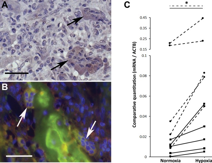
Introduction: Osteoclasts are responsible for the bone loss associated with rheumatoid arthritis (RA). The secreted adipokine angiopoietin-like 4 (ANGPTL4) specifically increases osteoclast-mediated bone resorption. We have investigated expression of ANGPTL4 and its regulatory transcription factor, hypoxia-inducible factor-1 alpha (HIF-1α), in osteoclasts and other cells within rheumatoid synovium. We have also examined whether circulating levels of ANGPTL4 differ in RA patients compared with that in normal controls or patients with osteoarthritis (OA).

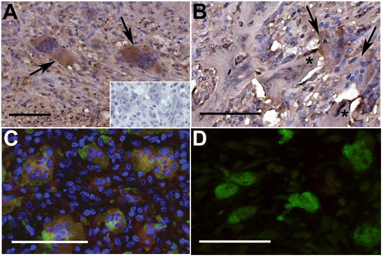
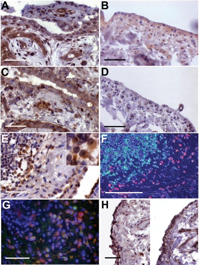
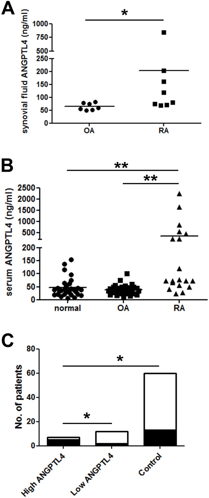
Results: Immunohistochemical analysis revealed that bone-apposing osteoclasts within the rheumatoid synovium express both ANGPTL4 and HIF-1α. ANGPTL4 was also strongly expressed in synovial lining cells, endothelial cells, stromal cells, CD68+ macrophages and plasma cells within RA synovium. Little ANGPTL4 was evident in normal synovial tissue. This reflected the over-expression of HIF-1α in rheumatoid versus normal synovial tissue. The concentration of ANGPTL4 was higher in both the serum and the synovial fluid of RA patients than in patients with OA or normal controls. High serum ANGPTL4 associated with elevated levels of the serum marker of bone resorption, receptor activator for nuclear factor κB ligand (RANKL).

Conclusions: Over-expression of ANGPTL4 in multiple cell types within the rheumatoid synovium potentially provides a local pool of ANGPTL4 to stimulate osteoclast-mediated bone resorption in RA. Additionally, correlation of high serum ANGPTL4 with circulating RANKL suggests that ANGPTL4 may represent a novel marker for bone destruction in RA.


Conflict of interest statement

Competing Interests: The authors have declared that no competing interests exist.

Figures

Figure 1
Figure 1. ANGPTL4 is expressed by osteoclasts in RA.
(A) ANGPTL4 is strongly expressed by osteoclasts (arrows) in the RA synovium. Inset: Representative isotype control image. (B) Resorbing osteoclasts adjacent to bone (starred) express ANGPTL4. (C) ANGPTL4 (red) expression co-localises with cathepsin K-positive (green) osteoclasts. Representative images from n = 3 cases. (D) Cathepsin K-positive (green) osteoclasts in the OA synovium do not express detectable ANGPTL4 (red). All scale bars represent 100 µM.
Figure 2
Figure 2. HIF-1α is expressed by osteoclasts in RA.
(A) HIF-1α expression by osteoclasts in the RA synovium (arrows). Representative image from n = 3 cases. (B) HIF-1α (green) and ANGPTL4 (red) expression co-localises in 2 bone-apposing osteoclasts (arrows). All scale bars represent 100 µM. (C) ANGPTL4 (solid lines) and SLC2A1 (Glut-1; dashed lines) mRNA expression in monocyte-derived osteoclasts from RA patients following 24 h exposure to normoxia or hypoxia (2% O2). Hypoxic fold-change in mRNA expression; *, p<0.05.
Figure 3
Figure 3. ANGPTL4 and HIF-1α are expressed in RA synovial tissue.
(A) The RA synovium is strongly positive for ANGPTL4, as are adjacent blood vessels and surrounding stromal cells; (B) normal synovium shows weak, heterogeneous expression of ANGPTL4. (C) RA synovium is strongly positive for HIF-1α, as are adjacent blood vessels; (D) normal synovium generally does not express HIF-1α. (E) Synovial lining cells and stromal cells adjacent to lymphoid aggregates express ANGPTL4. Inset: ANGPTL4-positive plasma cells. (F) ANGPTL4 (red) is not expressed by B cells (CD20, green). (G) ANGPTL4 (red) is expressed by CD68-positive (green) macrophages adjacent to a lymphoid aggregate. (H) The OA synovium expresses elevated levels of both ANGPTL4 (left panel) and HIF-1α (right panel) compared with the normal synovium. Scale bars (A–F, H) represent 100 µM, scale bar (G) represents 50 µM.
Figure 4
Figure 4. Serum and synovial fluid ANGPTL4 concentrations are elevated in RA.
(A) Synovial fluid from RA patients contains more ANGPTL4 (203.3±264.8 ng/ml, range 68.7–847.3 ng/ml) than that from patients with non-inflammatory OA (64.4±13.6 ng/ml, range 50.4–82.2 ng/ml). *, p<0.05. (B) Serum from RA patients contains more ANGPTL4 (363.4±138.7 ng/ml, range 24.0–2235.0) than that from OA patients (39.0±3.4 ng/ml, range 11.9–100.5 ng/ml) or normal controls (45.8±6.7 ng/ml, range 6.5–154.7 ng/ml). **, p<0.01. (C) Serum from ‘high ANGPTL4’ RA patients is more likely to have detectable RANKL (black shading; RANKL-positive) than either serum from ‘low ANGPTL4’ RA patients or controls. White shading; RANKL-negative. *, p<0.05.

References

    1. Kaarela K (1985) Prognostic factors and diagnostic criteria in early rheumatoid arthritis. Scand J Rheumatol 57: 1–54. - PubMed
    1. Gough AK, Lilley J, Eyre S, Holder RL, Emery P (1994) Generalised bone loss in patients with early rheumatoid arthritis. Lancet 344: 23–27. - PubMed
    1. Bromley M, Woolley DE (1984) Chondroclasts and osteoclasts at subchondral sites of erosion in the rheumatoid joint. Arthritis Rheum 27: 968–975. - PubMed
    1. Gough A, Sambrook P, Devlin J, Huissoon A, Njeh C, et al. (1998) Osteoclastic activation is the principal mechanism leading to secondary osteoporosis in rheumatoid arthritis. J Rheumatol 25: 1282–1289. - PubMed
    1. Tanaka S (2013) Regulation of bone destruction in rheumatoid arthritis through RANKL-RANK pathways. World J Orthop 4: 1–6. - PMC - PubMed

Publication types

MeSH terms