. 2014 Oct 7;9(10):e109944.
doi: 10.1371/journal.pone.0109944. eCollection 2014.

Human adenovirus-specific γ/δ and CD8+ T cells generated by T-cell receptor transfection to treat adenovirus infection after allogeneic stem cell transplantation

Affiliations

Human adenovirus-specific γ/δ and CD8+ T cells generated by T-cell receptor transfection to treat adenovirus infection after allogeneic stem cell transplantation

Jan Dörrie et al. PLoS One. .

Abstract

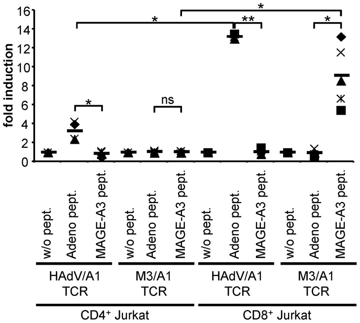
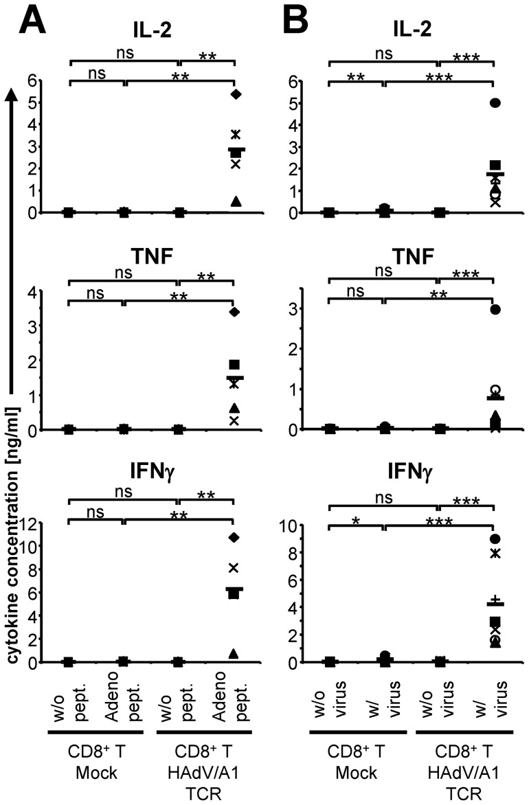
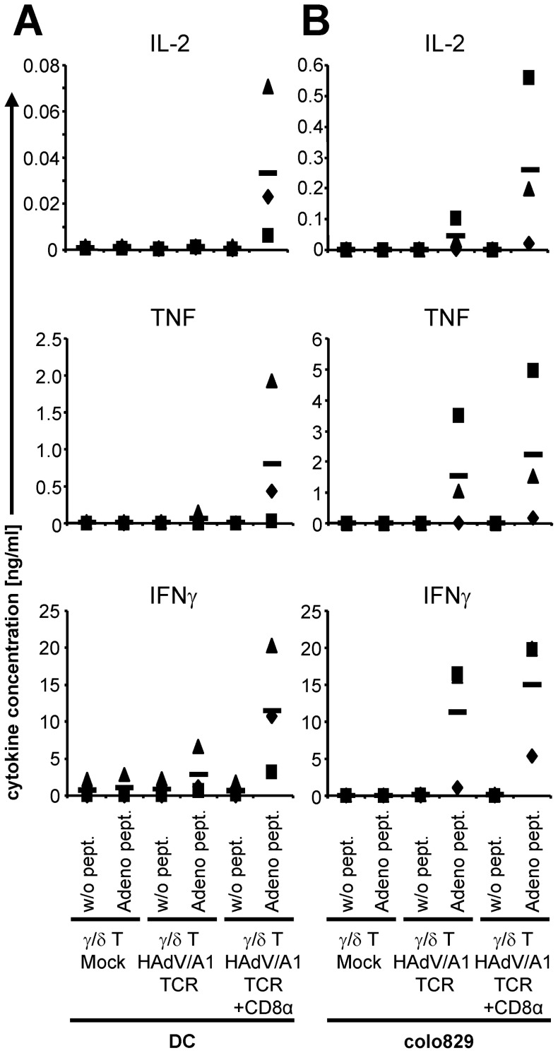
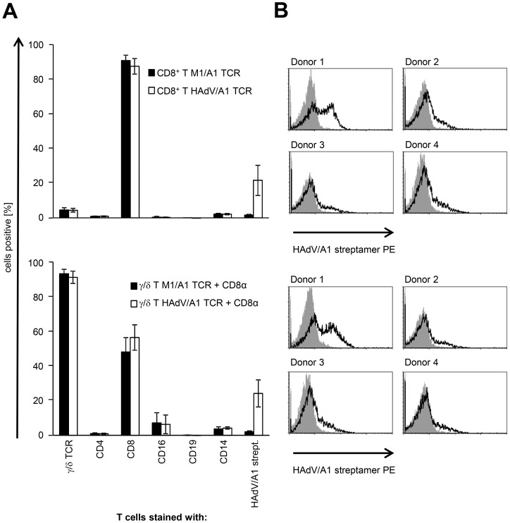
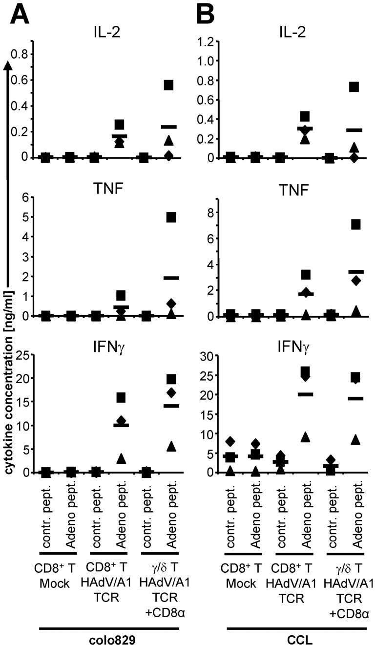
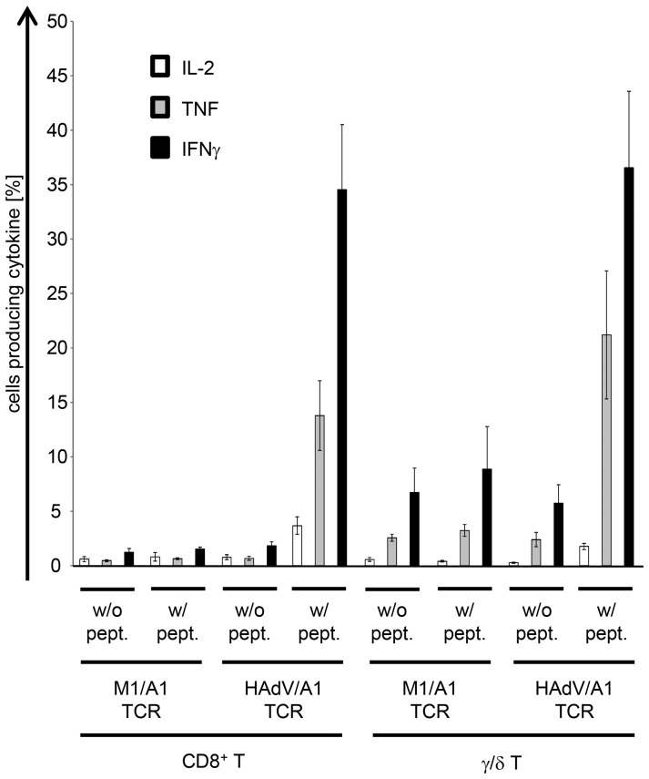
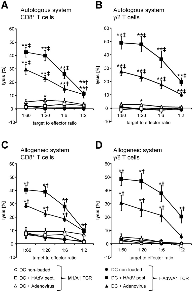
Human adenovirus infection is life threatening after allogeneic haematopoietic stem cell transplantation (HSCT). Immunotherapy with donor-derived adenovirus-specific T cells is promising; however, 20% of all donors lack adenovirus-specific T cells. To overcome this, we transfected α/β T cells with mRNA encoding a T-cell receptor (TCR) specific for the HLA-A*0101-restricted peptide LTDLGQNLLY from the adenovirus hexon protein. Furthermore, since allo-reactive endogenous TCR of donor T lymphocytes would induce graft-versus-host disease (GvHD) in a mismatched patient, we transferred the TCR into γ/δ T cells, which are not allo-reactive. TCR-transfected γ/δ T cells secreted low quantities of cytokines after antigen-specific stimulation, which were increased dramatically after co-transfection of CD8α-encoding mRNA. In direct comparison with TCR-transfected α/β T cells, TCR-CD8α-co-transfected γ/δ T cells produced more tumor necrosis factor (TNF), and lysed peptide-loaded target cells as efficiently. Most importantly, TCR-transfected α/β T cells and TCR-CD8α-co-transfected γ/δ T cells efficiently lysed adenovirus-infected target cells. We show here, for the first time, that not only α/β T cells but also γ/δ T cells can be equipped with an adenovirus specificity by TCR-RNA electroporation. Thus, our strategy offers a new means for the immunotherapy of adenovirus infection after allogeneic HSCT.


Conflict of interest statement

Competing Interests: The authors have declared that no competing interests exist.

Figures

Figure 1
Figure 1. The newly cloned HAdV/HLA-A1-specific TCR is functional in Jurkat T cells.
Parental CD4+ and transgenic CD8+ Jurkat T cells were co-electroporated with RNA encoding the HAdV/A1-specific TCR or MAGE-A3/A1 (M3/A1)-specific TCR and an NFAT-inducible luciferase reporter plasmid. These Jurkat T cells were stimulated with DC either non-loaded (w/o pept.) or loaded with the adenovirus peptide (Adeno pept.) or the MAGE-A3 peptide (MAGE-A3 pept.) (as indicated). The luciferase activity was measured, and the specific activation of the Jurkat T cells was calculated as fold induction by dividing the luciferase activity induced by peptide-loaded DC by that of similarly electroporated Jurkat T cells stimulated with non-loaded DC. Data of 4 (CD4+ Jurkat T cells) and 5 (CD8+ Jurkat T cells) individual experiments are shown. Bars indicate mean values. P-values were calculated by the Mann-Whitney U test. ns  =  not significant; ** p≤0.01; * p≤0.05. Raw data are summarized in Table S1 in File S1.
Figure 2
Figure 2. Antigen-specific cytokine production by HAdV/A1-TCR-transfected CD8+ T cells in response to peptide-loaded and adenovirus-infected targets.
CD8+ T cells were either mock electroporated or electroporated with HAdV/A1-TCR-RNA and were stimulated with DC, which were either left unloaded (A: w/o pept., B: w/o virus), or were loaded with the adenovirus peptide (Adeno pept.) (A), or were infected with adenovirus (w/virus) (B). Cytokine concentrations (IL-2, TNF, and IFNγ) in the supernatant after over-night co-incubation are depicted. Data of 5 (A) and 7 (B) individual experiments are shown. Bars indicate mean values. P-values were calculated by the Mann-Whitney U test. ns  =  not significant, *** p≤0.001; ** p≤0.01; * p≤0.05. Raw data are summarized in Table S2A and B in File S1.
Figure 3
Figure 3. Antigen-specific cytokine production by HAdV/A1-TCR-transfected γ/δ T cells in response to peptide-loaded target cells.
γ/δ T cells were either mock electroporated, electroporated with HAdV/A1-TCR-RNA alone, or electroporated with HAdV/A1-TCR-RNA and CD8α-RNA. These cells were stimulated with DC (A) or colo829 cells (B), which were either left unloaded (w/o pept.), or were loaded with the adenovirus peptide (Adeno pept.). Cytokine concentrations (IL-2, TNF, and IFNγ) in the supernatant after overnight co-incubation are depicted. Data of 3 individual experiments are shown. Bars indicate mean values. Raw data are summarized in Table S3A and B in in File S1.
Figure 4
Figure 4. Surface marker and TCR expression on HAdV/A1-TCR-transfected CD8+ and γ/δ T cells.
CD8+ T cells (A and B; upper panels) and γ/δ T cells (A and B; lower panels) were either electroporated with M1/A1-TCR RNA (+CD8α RNA in case of γ/δ T cells) or with HAdV/A1-TCR-RNA (+CD8α RNA in case of γ/δ T cells) and were cryoconserved 4 h after electroporation. Surface marker and TCR expression on thawed T cells was determined by staining with anti-CD4, anti-CD8, anti-CD16, anti-CD19, anti-CD14, and anti-pan γ/δ TCR antibodies, and the expression of the HAdV/A1-specific TCR was determined by staining with HAdV/A1 streptamer. Expression levels were analyzed by flow-cytometry and shown as % positive cells; isotype control stainings were subtracted (A; average values of 4 experiments ± SEM) or as histograms (B; grey histogram: T cells electroporated with M1/A1-TCR RNA, black line: T cells electroporated with HAdV/A1-TCR-RNA). Raw data are summarized in Table S4A and B in File S1.
Figure 5
Figure 5. Direct comparison of cytokine production by HAdV/A1-TCR-transfected CD8+ T cells and HAdV/A1-TCR-transfected γ/δ T cells.
CD8+ T cells and γ/δ T cells of the same donors were either mock electroporated, electroporated with HAdV/A1-TCR-RNA alone, or electroporated with HAdV/A1-TCR-RNA and CD8α-RNA. These cells were stimulated with colo829 cells (A) or CCL cells (B), which were either loaded with a control peptide (contr. pept.) or the adenovirus peptide (Adeno pept.). Cytokine concentrations (IL-2, TNF, and IFNγ) in the supernatant after overnight co-incubation are depicted. Data of 3 individual experiments are shown. Bars indicate mean values. Raw data are summarized in Table S5A and B in File S1.
Figure 6
Figure 6. Antigen-specific cytokine production by HAdV/A1-TCR-transfected CD8+ and γ/δ T cells in response to peptide-loaded target cells.
CD8+ T cells and γ/δ T cells were either electroporated with M1/A1-TCR RNA (+CD8α RNA in case of γ/δ T cells) or with HAdV/A1-TCR-RNA (+CD8α RNA in case of γ/δ T cells) and were cryoconserved 4 h after electroporation. After thawing, these cells were stimulated with CCL cells, which were either left unloaded (w/o pept.), or were loaded with the adenovirus peptide (w/pept.). Intracellular cytokine stainings for IL-2 (white bars), TNF (grey bars), and IFNγ (black bars) were performed and analyzed by flow-cytometry. The percentages of cytokine containing cells are depicted. Average values of 4 (CD8+ T cells) and 3 (γ/δ T cells) individual experiments ± SEM are shown. Raw data are summarized in Table S6 in File S1.
Figure 7
Figure 7. Cytolytic capacity of HAdV/A1-TCR-transfected CD8+ T cells and γ/δ T cells against adenovirus-infected target cells.
CD8+ T cells (A and C) and γ/δ T cells (B and D) of the same donors were either electroporated with mRNA encoding the MAGE-1/A1-specific TCR (open symbols) or electroporated with mRNA encoding the HAdV/A1-specific TCR (closed symbols) either in combination with CD8α mRNA (γ/δ T cells) or not (CD8+ T cells). These cells were used as effector cells in standard 4–6 h cytotoxicity assays. Autologous (A and B) and allogenic (C and D) DC either untreated (non-loaded), loaded with the adenovirus peptide (Adeno pept.), or infected with adenovirus (adenovirus) were used as target cells and the percentage of lysed cells was calculated. The target to effector cell ratios were 1∶60, 1∶20, 1∶6, and 1∶2. Average values of 6 (A and B) or 4 (C and D) individual experiments (each performed in triplicates) +/− SEM are shown. P-values were calculated by the Mann-Whitney U test. For statistics, conditions were compared to the non-loaded DC condition (* and **) and to T cells transfected with the M1/A1 control TCR ( and ). (** or p≤0.01; *or p≤0.05). Raw data are summarized in Table S7A to D in File S1.

References

    1. Eiz-Vesper B, Maecker-Kolhoff B, Blasczyk R (2012) Adoptive T-cell immunotherapy from third-party donors: characterization of donors and set up of a T-cell donor registry. Front Immunol 3: 410 10.3389/fimmu.2012.00410 [doi] - PMC - PubMed
    1. Matthes-Martin S, Feuchtinger T, Shaw PJ, Engelhard D, Hirsch HH, et al. (2012) European guidelines for diagnosis and treatment of adenovirus infection in leukemia and stem cell transplantation: summary of ECIL-4 (2011). Transpl Infect Dis 14: 555–563 10.1111/tid.12022 [doi] - PubMed
    1. Symeonidis N, Jakubowski A, Pierre-Louis S, Jaffe D, Pamer E, et al. (2007) Invasive adenoviral infections in T-cell-depleted allogeneic hematopoietic stem cell transplantation: high mortality in the era of cidofovir. Transpl Infect Dis 9: 108–113 TID184 [pii];10.1111/j.1399-3062.2006.00184.x [doi] - PubMed
    1. Feuchtinger T, Matthes-Martin S, Richard C, Lion T, Fuhrer M, et al. (2006) Safe adoptive transfer of virus-specific T-cell immunity for the treatment of systemic adenovirus infection after allogeneic stem cell transplantation. Br J Haematol 134: 64–76 BJH6108 [pii];10.1111/j.1365-2141.2006.06108.x [doi] - PubMed
    1. Leen AM, Christin A, Myers GD, Liu H, Cruz CR, et al. (2009) Cytotoxic T lymphocyte therapy with donor T cells prevents and treats adenovirus and Epstein-Barr virus infections after haploidentical and matched unrelated stem cell transplantation. Blood 114: 4283–4292 blood-2009-07-232454 [pii];10.1182/blood-2009-07-232454 [doi] - PMC - PubMed

Publication types

MeSH terms