

Critical cerebral perfusion pressure at high intracranial pressure measured by induced cerebrovascular and intracranial pressure reactivity
- PMID: 25289933
- PMCID: PMC6395276
- DOI: 10.1097/CCM.0000000000000655
Critical cerebral perfusion pressure at high intracranial pressure measured by induced cerebrovascular and intracranial pressure reactivity
Abstract
Objectives: The lower limit of cerebral blood flow autoregulation is the critical cerebral perfusion pressure at which cerebral blood flow begins to fall. It is important that cerebral perfusion pressure be maintained above this level to ensure adequate cerebral blood flow, especially in patients with high intracranial pressure. However, the critical cerebral perfusion pressure of 50 mm Hg, obtained by decreasing mean arterial pressure, differs from the value of 30 mm Hg, obtained by increasing intracranial pressure, which we previously showed was due to microvascular shunt flow maintenance of a falsely high cerebral blood flow. The present study shows that the critical cerebral perfusion pressure, measured by increasing intracranial pressure to decrease cerebral perfusion pressure, is inaccurate but accurately determined by dopamine-induced dynamic intracranial pressure reactivity and cerebrovascular reactivity.
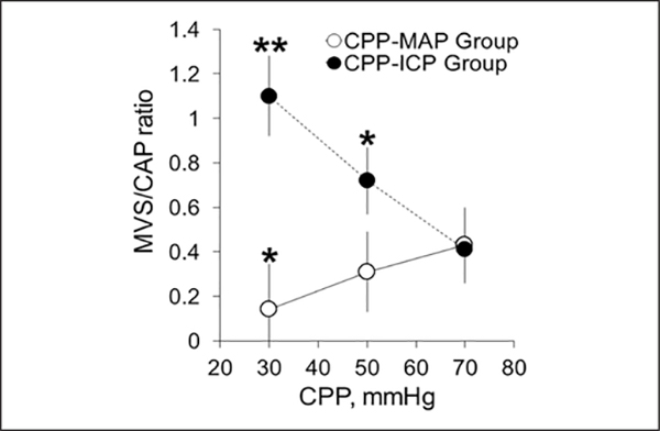
Design: Cerebral perfusion pressure was decreased either by increasing intracranial pressure or decreasing mean arterial pressure and the critical cerebral perfusion pressure by both methods compared. Cortical Doppler flux, intracranial pressure, and mean arterial pressure were monitored throughout the study. At each cerebral perfusion pressure, we measured microvascular RBC flow velocity, blood-brain barrier integrity (transcapillary dye extravasation), and tissue oxygenation (reduced nicotinamide adenine dinucleotide) in the cerebral cortex of rats using in vivo two-photon laser scanning microscopy.
Setting: University laboratory.
Subjects: Male Sprague-Dawley rats.
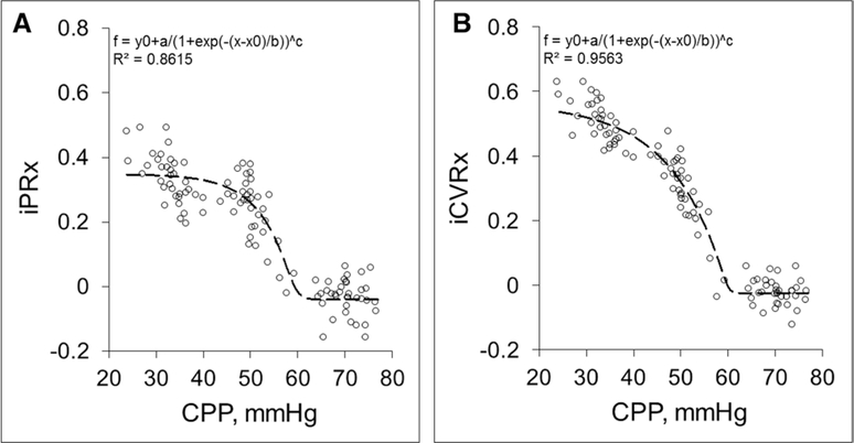
Interventions: At each cerebral perfusion pressure, dopamine-induced arterial pressure transients (~10 mm Hg, ~45 s duration) were used to measure induced intracranial pressure reactivity (Δ intracranial pressure/Δ mean arterial pressure) and induced cerebrovascular reactivity (Δ cerebral blood flow/Δ mean arterial pressure).
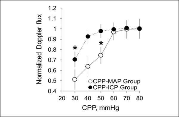
Measurements and main results: At a normal cerebral perfusion pressure of 70 mm Hg, 10 mm Hg mean arterial pressure pulses had no effect on intracranial pressure or cerebral blood flow (induced intracranial pressure reactivity = -0.03 ± 0.07 and induced cerebrovascular reactivity = -0.02 ± 0.09), reflecting intact autoregulation. Decreasing cerebral perfusion pressure to 50 mm Hg by increasing intracranial pressure increased induced intracranial pressure reactivity and induced cerebrovascular reactivity to 0.24 ± 0.09 and 0.31 ± 0.13, respectively, reflecting impaired autoregulation (p < 0.05). By static cerebral blood flow, the first significant decrease in cerebral blood flow occurred at a cerebral perfusion pressure of 30 mm Hg (0.71 ± 0.08, p < 0.05).
Conclusions: Critical cerebral perfusion pressure of 50 mm Hg was accurately determined by induced intracranial pressure reactivity and induced cerebrovascular reactivity, whereas the static method failed.
Figures
Similar articles
-
Induced Dynamic Intracranial Pressure and Cerebrovascular Reactivity Assessment of Cerebrovascular Autoregulation After Traumatic Brain Injury with High Intracranial Pressure in Rats.Acta Neurochir Suppl. 2018;126:309-312. doi: 10.1007/978-3-319-65798-1_60. Acta Neurochir Suppl. 2018. PMID: 29492580 Free PMC article.
-
Dynamic Cerebrovascular and Intracranial Pressure Reactivity Assessment of Impaired Cerebrovascular Autoregulation in Intracranial Hypertension.Acta Neurochir Suppl. 2016;122:255-60. doi: 10.1007/978-3-319-22533-3_51. Acta Neurochir Suppl. 2016. PMID: 27165917 Free PMC article.
-
Effect of cerebral perfusion pressure on cerebral cortical microvascular shunting at high intracranial pressure in rats.Stroke. 2013 Jan;44(1):177-81. doi: 10.1161/STROKEAHA.112.668293. Epub 2012 Nov 29. Stroke. 2013. PMID: 23204051 Free PMC article.
-
Monitoring of cerebral blood flow autoregulation: physiologic basis, measurement, and clinical implications.Br J Anaesth. 2024 Jun;132(6):1260-1273. doi: 10.1016/j.bja.2024.01.043. Epub 2024 Mar 12. Br J Anaesth. 2024. PMID: 38471987 Review.
-
Monitoring of cerebral autoregulation.Neurocrit Care. 2014 Dec;21 Suppl 2:S95-102. doi: 10.1007/s12028-014-0046-0. Neurocrit Care. 2014. PMID: 25208679 Review.
Cited by
-
Brain Tissue Oxygen and Cerebrovascular Reactivity in Traumatic Brain Injury: A Collaborative European NeuroTrauma Effectiveness Research in Traumatic Brain Injury Exploratory Analysis of Insult Burden.J Neurotrauma. 2020 Sep 1;37(17):1854-1863. doi: 10.1089/neu.2020.7024. Epub 2020 May 4. J Neurotrauma. 2020. PMID: 32253987 Free PMC article.
-
Closed cranial window rodent model for investigating hemodynamic response to elevated intracranial pressure.Animal Model Exp Med. 2021 Nov 19;4(4):391-397. doi: 10.1002/ame2.12187. eCollection 2021 Dec. Animal Model Exp Med. 2021. PMID: 34977490 Free PMC article.
-
Cerebral haemodynamics during experimental intracranial hypertension.J Cereb Blood Flow Metab. 2017 Feb;37(2):694-705. doi: 10.1177/0271678X16639060. Epub 2016 Jul 21. J Cereb Blood Flow Metab. 2017. PMID: 26994043 Free PMC article.
-
High Intracranial Pressure Induced Injury in the Healthy Rat Brain.Crit Care Med. 2016 Aug;44(8):e633-8. doi: 10.1097/CCM.0000000000001625. Crit Care Med. 2016. PMID: 26974548 Free PMC article.
-
Induced Dynamic Intracranial Pressure and Cerebrovascular Reactivity Assessment of Cerebrovascular Autoregulation After Traumatic Brain Injury with High Intracranial Pressure in Rats.Acta Neurochir Suppl. 2018;126:309-312. doi: 10.1007/978-3-319-65798-1_60. Acta Neurochir Suppl. 2018. PMID: 29492580 Free PMC article.
References
-
- Harper AM: The inter-relationship between aPco-2 and blood pressure in the regulation of blood flow through the cerebral cortex. Acta Neurol Scand Suppl 1965; 14:94–103 - PubMed
-
- Rapela CE, Green HD: Autoregulation of canine cerebral blood flow. Circ Res 1964; 15(Suppl):205–212 - PubMed
-
- Panerai RB: Assessment of cerebral pressure autoregulation in humans—A review of measurement methods. Physiol Meas 1998; 19:305–338 - PubMed
-
- Miller JD, Stanek A, Langfitt TW: Concepts of cerebral perfusion pressure and vascular compression during intracranial hypertension. Prog Brain Res 1972; 35:411–432 - PubMed
-
- Grubb RL Jr, Raichle ME, Phelps ME, et al.: Effects of increased intracranial pressure on cerebral blood volume, blood flow, and oxygen utilization in monkeys. J Neurosurg 1975; 43:385–398 - PubMed
Publication types
MeSH terms
Substances
Grants and funding
LinkOut - more resources
Full Text Sources
Other Literature Sources
