. 2014;25(17):1973-85.
doi: 10.1080/09205063.2014.965997. Epub 2014 Oct 7.

Biodegradable lysine-derived polyurethane scaffolds promote healing in a porcine full-thickness excisional wound model

Affiliations

Biodegradable lysine-derived polyurethane scaffolds promote healing in a porcine full-thickness excisional wound model

Elizabeth J Adolph et al. J Biomater Sci Polym Ed. 2014.

Abstract

Lysine-derived polyurethane scaffolds (LTI-PUR) support cutaneous wound healing in loose-skinned small animal models. Due to the physiological and anatomical similarities of human and pig skin, we investigated the capacity of LTI-PUR scaffolds to support wound healing in a porcine excisional wound model. Modifications to scaffold design included the addition of carboxymethylcellulose (CMC) as a porogen to increase interconnectivity and an additional plasma treatment (Plasma) to decrease surface hydrophobicity. All LTI-PUR scaffold and formulations supported cellular infiltration and were biodegradable. At 15 days, CMC and plasma scaffolds simulated increased macrophages more so than LTI PUR or no treatment. This response was consistent with macrophage-mediated oxidative degradation of the lysine component of the scaffolds. Cell proliferation was similar in control and scaffold-treated wounds at 8 and 15 days. Neither apoptosis nor blood vessel area density showed significant differences in the presence of any of the scaffold variations compared with untreated wounds, providing further evidence that these synthetic biomaterials had no adverse effects on those pivotal wound healing processes. During the critical phase of granulation tissue formation in full thickness porcine excisional wounds, LTI-PUR scaffolds supported tissue infiltration, while undergoing biodegradation. Modifications to scaffold fabrication modify the reparative process. This study emphasizes the biocompatibility and favorable cellular responses of PUR scaffolding formulations in a clinically relevant animal model.

Keywords: LTI polyurethane; PUR; lysine triisocyanate; pig model; scaffold; wound repair.


Figures

Figure 1
Figure 1
SEM images showing pore structure of PUR scaffolds. (A) LTI scaffold. (B) CMC scaffold. LTI and CMC scaffolds exhibit similar pore sizes (300 – 500 µm). CMC scaffolds have more openings in pore walls than LTI scaffolds, resulting in higher permeability. Scale bar = 750 µm.
Figure 2
Figure 2
Contact angle of LTI films before and after plasma treatment. Contact decreased from 65° to 45° after plasma treatment, providing evidence that plasma treatment increases surface hydrophilicity. Asterisk indicates significant difference (p < 0.005).
Figure 3
Figure 3
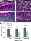
PUR scaffold degradation. (A) Representative images of trichrome staining of PUR scaffolds at day 8 at magnifications of 2X (left) and 20X (right). Scaffold fragments are marked by the letter S. (B) Representative images of trichrome staining of PUR+CMC scaffolds at day 15 at magnifications of 2x (left) and 20x (right). Fewer scaffold fragments are present at day 15 than at day 8. (C) Percentage of wound cross-sectional area occupied by residual polymer measured from images of trichrome staining. Asterisks indicate significant difference (p < 0.05). All scaffold-treated wounds had significantly less scaffold material remaining at day 15 than at day 8, providing evidence that scaffolds were undergoing biodegradation in the period between 8 and and 15 days Intergroup comparison among the 3 different types of scaffolds showed no significant impact on the degree of scaffold degradation.
Figure 4
Figure 4
Analysis of cell proliferation and apoptosis. (A) Ki67 immunostaining was used to analyze the presence of proliferating cells. Data are presented as number of Ki67+ cells per high power field (hpf). Asterisks indicate significant difference (p < 0.05). Dagger indicates significant difference from PUR+CMC and PUR+CMC+P at day 15 (p < 0.02). The number of Ki67+ cells decreased significantly from day 8 to day 15 in all treatment groups. At day 8, there were significant differences among all treatment groups. At day 15, PUR+CMC and PUR+CMC+P groups had significantly more proliferating cells than untreated wounds. (B) TUNEL immunostaining was used to analyze the presence of apoptosing cells. Data are presented as number of TUNEL+ cells per hpf. Asterisks indicate significant difference (p < 0.05). Dagger indicates significant difference from PUR and PUR+CMC at day 8 (p < 0.01). The number of TUNEL+ cells increased significantly from day 8 to day 15 in PUR scaffolds and decreased significantly from day 8 to day 15 in PUR+CMC+P scaffolds. At day 8, wounds treated with Plasma scaffolds had significantly more TUNEL+ cells than wounds treated with PUR and PUR+CMC+P scaffolds. (C) Ratio of proliferation to apoptosis. Dashed line represents a ratio of one. Asterisks indicate significant difference (p < 0.01), and dagger denotes significant difference from PUR and untreated wounds at day 8. By day 15, there were no significant differences in proliferation/apoptosis ratio among treatment groups.
Figure 5
Figure 5
Analysis of the macrophage population using the MAC387immunomarker. MAC387 immunostaining was used to analyze the presence of macrophages. Data are presented as number of MAC387+ cells per hpf. Asterisks (p < 0.02) and dagger (p < 0.005) indicate significant difference. The number of MAC387+ cells increased significantly from day 8 to day 15 in PUR+CMC+P scaffolds. At day 15, wounds treated with PUR+CMC and PUR+CMC+P scaffolds had significantly more MAC397+ cells than untreated wounds.
Figure 6
Figure 6
Blood vessel formation. Factor VIII immunostaining was used to analyze the presence of blood vessels. Data are presented as area% of blood vessels within the wound. Asterisks indicate significant difference (p < 0.05). The blood vessel density increased significantly from day 8 to day 15 in PUR, PUR+CMC+P, and untreated wounds.

References

    1. Clark RAF, Ghosh K, Tonnesen MG. Tissue engineering for cutaneous wounds. Journal of Investigative Dermatology. 2007;127(5):1018–1029. - PubMed
    1. Eccleston GM, Boateng JS, Matthews KH, Stevens HNE. Wound healing dressings and drug delivery systems: A review. Journal of Pharmaceutical Sciences. 2008;97(8):2892–2923. - PubMed
    1. Mostow EN, Haraway GD, Dalsing M, Hodde JP, King D, Grp OVUS. Effectiveness of an extracellular matrix graft (OASIS Wound Matrix) in the treatment of chronic leg ulcers: A randomized clinical trial. Journal of Vascular Surgery. 2005;41(5):837–843. - PubMed
    1. Lim CT, Zhong SP, Zhang YZ. Tissue scaffolds for skin wound healing and dermal reconstruction. Wiley Interdisciplinary Reviews-Nanomedicine and Nanobiotechnology. 2010;2(5):510–525. - PubMed
    1. Raeber GP, Lutolf MP, Hubbell JA. Molecularly engineered PEG hydrogels: a novel model system for proteolytically mediated cell migration. Biophysical journal. 2005;89(2):1374–1388. - PMC - PubMed

Publication types

LinkOut - more resources