Review
. 2014 Oct 7;3(10):e201.
doi: 10.1038/mtna.2014.49.

Nucleic Acid Ligands With Protein-like Side Chains: Modified Aptamers and Their Use as Diagnostic and Therapeutic Agents

Affiliations
Review

Nucleic Acid Ligands With Protein-like Side Chains: Modified Aptamers and Their Use as Diagnostic and Therapeutic Agents

John C Rohloff et al. Mol Ther Nucleic Acids. .

Abstract

Limited chemical diversity of nucleic acid libraries has long been suspected to be a major constraining factor in the overall success of SELEX (Systematic Evolution of Ligands by EXponential enrichment). Despite this constraint, SELEX has enjoyed considerable success over the past quarter of a century as a result of the enormous size of starting libraries and conformational richness of nucleic acids. With judicious introduction of functional groups absent in natural nucleic acids, the "diversity gap" between nucleic acid-based ligands and protein-based ligands can be substantially bridged, to generate a new class of ligands that represent the best of both worlds. We have explored the effect of various functional groups at the 5-position of uracil and found that hydrophobic aromatic side chains have the most profound influence on the success rate of SELEX and allow the identification of ligands with very low dissociation rate constants (named Slow Off-rate Modified Aptamers or SOMAmers). Such modified nucleotides create unique intramolecular motifs and make direct contacts with proteins. Importantly, SOMAmers engage their protein targets with surfaces that have significantly more hydrophobic character compared with conventional aptamers, thereby increasing the range of epitopes that are available for binding. These improvements have enabled us to build a collection of SOMAmers to over 3,000 human proteins encompassing major families such as growth factors, cytokines, enzymes, hormones, and receptors, with additional SOMAmers aimed at pathogen and rodent proteins. Such a large and growing collection of exquisite affinity reagents expands the scope of possible applications in diagnostics and therapeutics.


Figures

Figure 1
Figure 1
Partial listing of modifications at the 5-position of deoxyuridine available for SELEX and post-SELEX optimization. Side chain abbreviations: Bn, benzyl; Pe, 2-phenylethyl; Pp, 3-phenylpropyl; Th, 2-thiophenylmethyl; FBn, 4-fluorobenzyl; Nap, 1-naphthylmethyl; 2Nap, 2-naphthylmethyl; Ne, 1-naphthyl-2-ethyl; 2Ne, 2-naphthyl-2-ethyl; Trp, 3-indole-2-ethyl; Bt, 3-benzothiophenyl-2-ethyl; Bf, 3-benzofuranyl-2-ethyl; Bi, 1-benzimidazol-2-ethyl; Tyr, 4-hydroxyphenyl-2-ethyl; Pyr, 4-pyridinylmethyl; MBn, 3,4-methylenedioxybenzyl; MPe, 3,4-methylenedioxyphenyl-2-ethyl; 3MBn, 3-methoxybenzyl; 4MBn, 4-methoxybenzyl; 3,4MBn, 3,4-dimethoxybenzyl; RTHF, R-tetrahydrofuranylmethyl; STHF, S-tetrahydrofuranylmethyl; Moe, morpholino-2-ethyl; Thr, R-2-hydroxypropyl; iBu, iso-butyl.
Figure 2
Figure 2
Interface area plotted as a function of the total number of hydrogen bonds and charge–charge interactions (polar contacts) for aptamers (gray diamonds) or SOMAmers (blue circles) bound to their targets (from Table 2). The line represents a linear regression fit to points representing the eight conventional aptamers with an R2 = 0.88 and a slope of 0.016. Dashed lines represent the 99% confidence intervals of this trend line for the conventional aptamers (the three SOMAmers fall outside those boundaries).
Figure 3
Figure 3
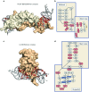
SOMAmers bind their targets with exquisite shape complementarity and utilize hydrophobic modifications at the binding interface. (a and c) Crystal structures of SOMAmers bound to PDGF-BB and IL-6., (b and d) Schematic representations of the corresponding secondary structures, with base pairs annotated according to the Leonitis and Westhof nomenclature. Modified nucleotides are colored brick red; conventional DNA is colored gray; blue squares in d indicate G-quartets.
Figure 4
Figure 4
SOMAmer intra- and intermolecular interactions in the PDGF-BB and IL-6 co-crystal structures. Both the PDGF-BB and the IL-6 SOMAmers exhibit striking shape complementarity at the protein/SOMAmer binding interface. (ac) All eight modified nucleotides of the PDGF-BB SOMAmer cluster in an extensive arrangement of hydrophobic aromatic interactions that primarily contact the aliphatic side chains of the protein residues. Only seven polar interactions are present at the SOMAmer/PDGF-BB interface. (df) The modified nucleotides of the IL-6 SOMAmer form segregated hydrophobic clusters exhibiting face-to-face and edge-to-face aromatic interactions. With only nine polar contacts, the modified nucleotides mainly stack against the methylene portion of charged residues and hydrophobic amino acid side chains. Bn-dU, 5-(N-benzylcarboxamide)-2′-deoxyuridine; Pe-dU, 5-[N-(phenyl-2-ethyl)carboxamide]-2′-deoxyuridine; Th-dU, 5-[N-(2-thiophene-methyl)carboxamide]-2′-deoxyuridine; Nap-dU, 5-(N-(1-naphthylmethyl)carboxamide)-2′-deoxyuridine; dU plus number indicates the uridine ring of specified nucleotide; Bn, Pe, Th, or Nap plus number indicates the modified nucleotide side chain of specified nucleotide; Bn-dU, Pe-dU, Th-dU, or Nap-dU plus number indicates the entire nucleotide. Transparent surface renderings of PDGF-BB and IL-6 are colored gold and wheat; stick renderings of the modified nucleotides are colored brick red.
Figure 5
Figure 5
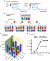
Isolation of SOMAmer pairs for sandwich assays, shown for TSP2 as an example. SOMAmers from a standard SELEX with a first modified nucleotide library (e.g., Bn-dU) were competitive, indicating that they bind to the same epitope. (a) SOMAmer pairs were generated via sandwich SELEX using a SOMAmer–protein complex as the target and a second modified library (e.g., 2Nap-dU) that may favor a distinct second epitope. A pairwise screening assay was developed to distinguish truly noncompetitive SOMAmers from SOMAmers with superior binding properties that simply displaced the first SOMAmer from the target during sandwich SELEX. (b) Such SOMAmer pairs were identified via attaching each individual SOMAmer as capture agent on a different type of LumAvidin beads, which were then pooled for a multiplexed screening assay on the Luminex platform to test each individual SOMAmer as detection agent. In this assay, a fixed target concentration of 10 nmol/l TSP2 was used, and all SOMAmers carried a 5′ biotin for immobilization of the capture agents on streptavidin beads and for labeling of the detection agents with phycoerythrin–streptavidin conjugate. (c) The resulting Luminex signals allowed the identification of the best-performing SOMAmer pairs in this sandwich screening assay. Performance of SOMAmer pair 3339–33 (Bn-dU) for capture and 7574–53 (2Nap-dU) for detection was evaluated via target titration in a Luminex sandwich assay. (d) Background signal in the absence of the protein is generally < 10% of the maximum signal and was subtracted from the total signal. Omission of one of the constituents of the sandwich pair reduced the signal to below background.
Figure 6
Figure 6
Multiplexed SOMAmer affinity assay. (a) SOMAmers labeled with a fluorophore (F), photocleavable linker (L), and biotin (B) are immobilized on streptavidin (SA)-coated beads and incubated with samples containing a complex mixure of proteins (e.g., plasma). (b) Cognate (top and bottom) and noncognate (middle) SOMAmer–target protein complexes form on the beads. (c) The beads are washed removing the unbound proteins and the proteins are tagged with biotin. (d) SOMAmer–protein complexes are released from the beads by photocleavage of the linker with UV light. (e) Incubation in a buffer containing a polyanionic competitor selectively disrupts nonspecific interactions. (f) SOMAmer–protein complexes are recaptured on a second set of streptavidin-coated beads through biotin-tagged proteins followed by additional washing steps that facilitate further removal of nonspecifically bound SOMAmers. (g) SOMAmers are released from the beads in a denaturing buffer. (h) SOMAmers are hybridized to complementary sequences on a microarray chip and quantified by fluorescence. Fluorescence intensity is related to protein amount in the original sample.

References

    1. Tuerk C, Gold L. Systematic evolution of ligands by exponential enrichment: RNA ligands to bacteriophage T4 DNA polymerase. Science. 1990;249:505–510. - PubMed
    1. Ellington AD, Szostak JW. In vitro selection of RNA molecules that bind specific ligands. Nature. 1990;346:818–822. - PubMed
    1. Gold L, Brown D, He Y, Shtatland T, Singer BS, Wu Y. From oligonucleotide shapes to genomic SELEX: novel biological regulatory loops. Proc Natl Acad Sci USA. 1997;94:59–64. - PMC - PubMed
    1. Gold L, Polisky B, Uhlenbeck O, Yarus M. Diversity of oligonucleotide functions. Annu Rev Biochem. 1995;64:763–797. - PubMed
    1. Cerchia L, Giangrande PH, McNamara JO, de Franciscis V. Cell-specific aptamers for targeted therapies. Methods Mol Biol. 2009;535:59–78. - PMC - PubMed