. 2014 Dec;19(12):1691-701.
doi: 10.1007/s10495-014-1038-4.

Glucotoxic and diabetic conditions induce caspase 6-mediated degradation of nuclear lamin A in human islets, rodent islets and INS-1 832/13 cells

Affiliations

Glucotoxic and diabetic conditions induce caspase 6-mediated degradation of nuclear lamin A in human islets, rodent islets and INS-1 832/13 cells

Syeda Khadija et al. Apoptosis. 2014 Dec.

Abstract

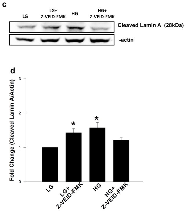
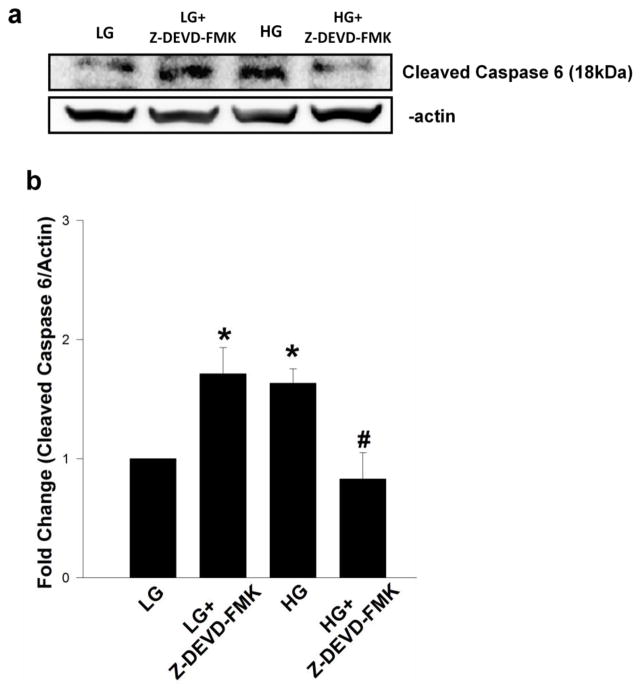
Nuclear lamins form the lamina on the interior surface of the nuclear envelope, and regulate nuclear metabolic events, including DNA replication and organization of chromatin. The current study is aimed at understanding the role of executioner caspase 6 on lamin A integrity in islet β-cells under duress of glucotoxic (20 mM glucose; 24 h) and diabetic conditions. Under glucotoxic conditions, glucose-stimulated insulin secretion and metabolic cell viability were significantly attenuated in INS-1 832/13 cells. Further, exposure of normal human islets, rat islets and INS-1 832/13 cells to glucotoxic conditions leads to caspase 6 activation and lamin A degradation, which is also observed in islets from the Zucker diabetic fatty rat, a model for type 2 diabetes (T2D), and in islets from a human donor with T2D. Z-Val-Glu-Ile-Asp-fluoromethylketone, a specific inhibitor of caspase 6, markedly attenuated high glucose-induced caspase 6 activation and lamin A degradation, confirming that caspase 6 mediates lamin A degradation under high glucose exposure conditions. Moreover, Z-Asp-Glu-Val-Asp-fluoromethylketone, a known caspase 3 inhibitor, significantly inhibited high glucose-induced caspase 6 activation and lamin A degradation, suggesting that activation of caspase 3 might be upstream to caspase 6 activation in the islet β-cell under glucotoxic conditions. Lastly, we report expression of ZMPSTE24, a zinc metallopeptidase involved in the processing of prelamin A to mature lamin A, in INS-1 832/13 cells and human islets; was unaffected by high glucose. We conclude that caspases 3 and 6 could contribute to alterations in the integrity of nuclear lamins leading to metabolic dysregulation and failure of the islet β-cell.


Conflict of interest statement

Conflict of interests

The authors declare no conflict of interests.

Figures

Figure 1
Figure 1. Glucotoxic conditions attenuate GSIS in INS-1 832/13 β-cells
INS-1 832/13 cells were cultured in the presence of low (2.5 mM; LG) or high (20 mM; HG) glucose for 24 hr following which they were stimulated with low (2.5 mM) or high (20 mM) glucose for 45 min. Insulin released into the medium was quantified by ELISA [see Methods for additional details]. The data are expressed as insulin release (ng/ml) and are means ± SEM from three independent experiments. *P < 0.05 vs. LG under 24 hr low glucose treatment; **P < 0.05 vs. HG under 24 hr low glucose treatment.
Figure 2
Figure 2. High glucose treatment induces caspase 6 activation and lamin A cleavage in INS-1 832/13 cells
INS-1 832/13 cells were incubated in the presence of low (2.5 mM; LG) or high (20 mM; HG) glucose for 24 hr. Caspase 6 activation and lamin A cleavage were determined by Western blotting. Protein lysates (40 μg) were resolved by SDS-PAGE and transferred to a nitrocellulose membrane. The membrane was probed for cleaved caspase 6 and cleaved lamin A and immune complexes were identified using ECL detection kit (Panel a). To ensure equal protein loading, the membranes were stripped and probed for β actin. Intensity of protein bands was quantified by densitometry. Data represent mean ± SEM from three independent experiments and expressed as fold change in caspase 6 (Panel b) and lamin A cleavage (Panel c). *P < 0.05 vs. LG.
Figure 3
Figure 3. High glucose treatment results in caspase 6 activation and lamin A cleavage in normal rat islets
Rat islets were incubated in the presence of low (2.5 mM; LG) and high glucose (20 mM; HG) for 24 hr. Approximately 40 μg protein lysate was resolved by SDS-PAGE and the degree of abundance of cleaved caspase 6 and lamin A were determined by Western blot analysis. Immune complexes were identified using ECL detection kit (Panel a). β actin was used as a loading control. Quantitation of caspase 6 activation and lamin A degradation were carried out by densitometry. Data represent mean ± SEM from three independent experiments and are expressed as fold change in caspase 6 (Panel b) and lamin A (Panel c) cleavage. *P < 0.05 vs. LG.
Figure 4
Figure 4. Glucotoxic conditions promote caspase 6 activation and lamin A cleavage in normal human islets treated with high glucose and in diabetic human islets
Normal human islets were incubated in the presence of low (5.8 mM; LG) and high glucose (30 mM; HG) for 24 hr as described in the text (Panel a). Islets obtained from normal and diabetic individuals were lysed using RIPA buffer (Panel b). Lysate proteins (25 μg) were resolved by SDS-PAGE and transferred to a nitrocellulose membrane. The membrane was probed for cleaved caspase 6, and lamin A and immune complexes were identified using ECL detection kit. To check equal protein loading, the membrane was stripped and probed for β actin. Human islet data from normal and diabetic individuals were accrued from a single batch of islet preparation.
Figure 5
Figure 5. Activation of caspase 6 and lamin A degradation are observed in ZDF rat islets
Islets isolated from ZDF and ZLC rats by collagenase digestion method were lysed using RIPA buffer. Lysate proteins (40μg) were resolved by SDS-PAGE and the abundance of cleaved caspase 6 and lamin A was determined by Western blotting. Immune complexes were identified using ECL detection kit. To assess equal protein loading, the membrane was probed for β actin. Representative blots from three ZLC and ZDF islet preparations are shown in Panel a and c. Intensity of protein bands was quantified by densitometry and data represent mean ± SEM from three islet preparations and are expressed as fold change in caspase 6 (Panel b) and lamin A cleavage (Panel d). *P < 0.05 vs. ZLC
Figure 6
Figure 6. High glucose-induced caspase 6 activation and breakdown of lamin A cleavage were attenuated by Z-VEID-FMK, a specific inhibitor of caspase 6, in INS-1 832/13 cells
INS-1 832/13 cells were preincubated with Z-VEID-FMK (3μM) for 1 hr and further treated with low (2.5 mM; LG) or high (20 mM; HG) glucose in the presence and absence of Z-VEID-FMK for 6 hr. Caspase 6 activation and lamin A cleavage were determined by Western blotting. Protein lysates (40 μg) were resolved by SDS-PAGE and transferred to a nitrocellulose membrane. The membrane was probed for cleaved caspase 6 (Panel a) and cleaved lamin A (Panel c), and immune complexes were identified using ECL detection kit. To ensure equal protein loading, the membranes were probed for β actin. Intensity of protein bands was quantified by densitometry. Data represent mean ± SEM from three independent experiments and expressed as fold change in caspase 6 (Panel b) and lamin A cleavage (Panel d). *P < 0.05 vs. LG. NS: Not significant vs. LG
Figure 7
Figure 7. Z-DEVD-FMK, a known inhibitor of Caspase 3, prevented high glucose induced caspase 6 activation and lamin A breakdown in INS-1 832/13 cells
INS-1 832/13 cells were preincubated with Z-DEVD-FMK (3μM) for 1 hr and further treated with low (2.5 mM; LG) or high (20 mM; HG) glucose in the presence and absence of Z-DEVD-FMK for 6 hr. Caspase 6 activation (Panel a) and lamin A cleavage (Panel c) were determined by Western blotting. To check equal protein loading, the membranes were probed for β actin. Intensity of protein bands was quantified by densitometry. Data represent mean ± SEM from three independent experiments and expressed as fold change in caspase 6 (Panel b) and lamin A cleavage (Panel d). *P < 0.05 vs. LG; # P < 0.05 vs. HG.
Figure 8
Figure 8. Immunological evidence to indicate expression of lamin A processing protease (ZMPSTE 24) in INS-1 832/13 cells and normal human islets: Lack of effects of high glucose exposure on the expression of ZMPSTE 24
INS-1 832/13 cells were incubated in the presence of low (2.5 mM; LG) or high (20m M; HG) glucose for 24 hr (as in Figure 2). Protein lysates (40 μg) were resolved by SDS-PAGE and transferred to a nitrocellulose membrane. A representative blot from three studies is shown in Panel a. Intensity of protein bands was quantified by densitometry and data represent mean ± SEM from three independent experiments and are expressed as fold change in ZMPSTE 24 (Panel b). NS: Not significant vs. LG. Normal human islets were incubated in the presence of low (5.8mM; LG) and high glucose (30mM; HG) for 24 hr (as in Figure 4; Panel a). Twenty five μg lysate proteins were separated by SDS-PAGE and transferred to a nitrocellulose membrane. The membranes were probed for ZMPSTE 24 and immune complexes were identified using ECL detection kit. To check equal protein loading, the membranes were probed for β actin. A blot from a single human islet preparation is shown in Panel c.

References

    1. Gruenbaum Y, Wilson KL, Harel A, Goldberg M, Cohen M. Review: nuclear lamins--structural proteins with fundamental functions. J Struct Biol. 2000;129:313–323. - PubMed
    1. Moir RD, Spann TP. The structure and function of nuclear lamins: implications for disease. Cell Mol Life Sci. 2001;58:1748–1757. - PMC - PubMed
    1. Moir RD, Spann TP, Goldman RD. The dynamic properties and possible functions of nuclear lamins. Int Rev Cytol. 1995;162:141–182. - PubMed
    1. Moir RD, Spann TP, Herrmann H, Goldman RD. Disruption of nuclear lamin organization blocks the elongation phase of DNA replication. J Cell Biol. 2000;149:1179–1192. - PMC - PubMed
    1. Shahzidi S, Brech A, Sioud M, Li X, Suo Z, Nesland JM, Peng Q. Lamin A/C cleavage by caspase-6 activation is crucial for apoptotic induction by photodynamic therapy with hexaminolevulinate in human B-cell lymphoma cells. Cancer Lett. 2013;339:25–32. - PubMed

Publication types