Review
. 2014:48:583-611.
doi: 10.1146/annurev-genet-120213-092208. Epub 2014 Oct 6.

Genetic, epigenetic, and environmental contributions to neural tube closure

Affiliations
Review

Genetic, epigenetic, and environmental contributions to neural tube closure

Jonathan J Wilde et al. Annu Rev Genet. 2014.

Abstract

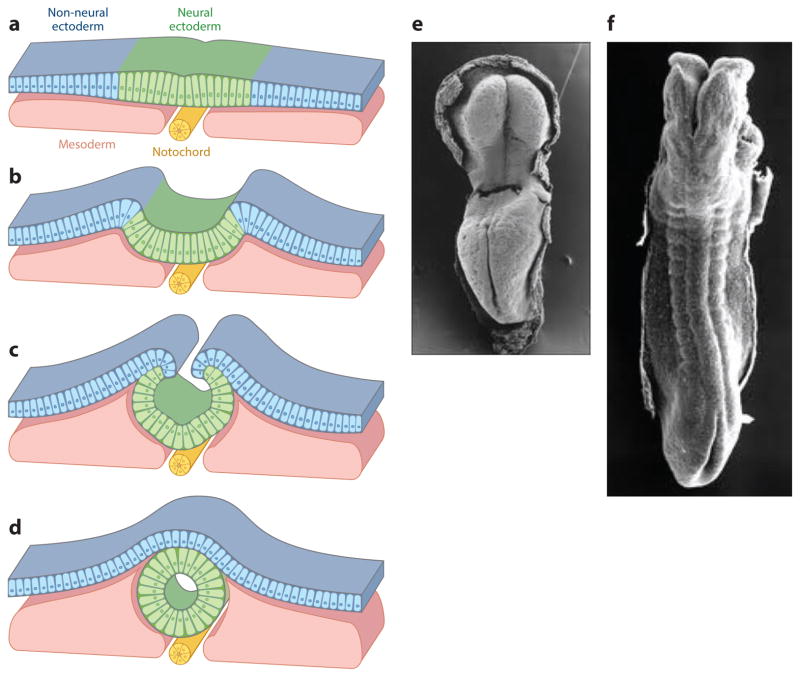
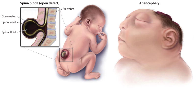
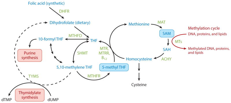
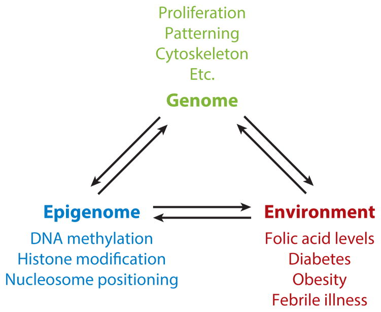
The formation of the embryonic brain and spinal cord begins as the neural plate bends to form the neural folds, which meet and adhere to close the neural tube. The neural ectoderm and surrounding tissues also coordinate proliferation, differentiation, and patterning. This highly orchestrated process is susceptible to disruption, leading to neural tube defects (NTDs), a common birth defect. Here, we highlight genetic and epigenetic contributions to neural tube closure. We describe an online database we created as a resource for researchers, geneticists, and clinicians. Neural tube closure is sensitive to environmental influences, and we discuss disruptive causes, preventative measures, and possible mechanisms. New technologies will move beyond candidate genes in small cohort studies toward unbiased discoveries in sporadic NTD cases. This will uncover the genetic complexity of NTDs and critical gene-gene interactions. Animal models can reveal the causative nature of genetic variants, the genetic interrelationships, and the mechanisms underlying environmental influences.

Keywords: embryonic brain; embryonic spinal cord; gene-environment interactions; neural tube defects, epigenetics.


Figures

Figure 1
Figure 1
Neural tube closure. (ad) Schematic transverse sections that illustrate the (a) flat neural plate stage, (b) hinge-point formation and neural-fold elevation, (c) apposition of the neural folds with the neural ectoderm covered by the non-neural ectoderm (NNE), and the (d) meeting and remodeling of the neural ectoderm and NNE to form a closed neural tube covered by a single layer of NNE. (ef) Carnegie stages 9–11 human embryos (~20–24 days of gestation) just prior to the (e) initiation of neural tube closure and to (f) closure of most of the spinal region. Panels ad adapted from Reference ; panels e and f taken from scanning electron micrographs of early human embryos by Dr. K. Sulik, from embryos collected by Dr. Vekemans and T. Attie-Bitacha, and presented on http://embryology.med.unsw.edu.au/embryology/index.php?title=Embryonic_Development.
Figure 2
Figure 2
Two types of open neural tube defects (NTDs). Spina bifida occurs in the spinal region, whereas failure of cranial neural tube closure is initially called exencephaly but after exposure and degradation of the brain tissue is called anencephaly. Both caudal and cranial NTDs can vary in extent of the opening and the rostral-caudal level. Adapted from Centers for Disease Control and Prevention, National Birth Defects and Developmental Disabilities.
Figure 3
Figure 3
Schematic of folate in one-carbon metabolism. Synthetic and dietary folates are reduced to tetrahydrofolates (THFs), which are converted to the biologically active 5-methyltetrahydrofolate (5-methyl-THF) by SHMT and MTHFR. 5-methyl-THF acts a methyl donor for purine and thymidylate synthesis and generates the major cellular methylation donor S-adenosylmethionine (SAM). Abbreviations: AHCY, S-adenosylhomocysteine hydrolase; DHFR, dihydrofolate reductase; MAT, methionine adenosyltransferase; MTHFD, methylenetetrahydrofolate dehydrogenase; MTHFR, methylenetetrahydrofolate reductase; MTR, methionine synthase; MTRR, methionine synthase reductase; MTs, methyltransferases; SAH, S-adenosylhomocysteine; SAM, S-adenosylmethionine; SHMT, serine-hydroxymethyltransferase.
Figure 4
Figure 4
Interrelationship between the genome, epigenome, and environment, with respect to neural tube closure.

References

    1. Acs N, Bánhidy F, Puhó E, Czeizel AE. Maternal influenza during pregnancy and risk of congenital abnormalities in offspring. Birth Defects Res A Clin Mol Teratol. 2005;73(12):989–96. - PubMed
    1. Aguilar A, Meunier A, Strehl L, Martinovic J, Bonniere M, et al. Analysis of human samples reveals impaired SHH-dependent cerebellar development in Joubert syndrome/Meckel syndrome. Proc Natl Acad Sci USA. 2012;109:16951–56. - PMC - PubMed
    1. Alvarez IS, Schoenwolf GC. Expansion of surface epithelium provides the major extrinsic force for bending of the neural plate. J Exp Zool. 1992;261:340–48. - PubMed
    1. Amir RE, Van den Veyver IB, Wan M, Tran CQ, Francke U, Zoghbi HY. Rett syndrome is caused by mutations in X-linked MECP2, encoding methyl-CpG-binding protein 2. Nat Genet. 1999;23(2):185–88. - PubMed
    1. Aran D, Sabato S, Hellman A. DNA methylation of distal regulatory sites characterizes dysregulation of cancer genes. Genome Biol. 2013;14(3):R21. - PMC - PubMed

Publication types

LinkOut - more resources