. 2014 Oct 7;5(5):e01710-14.
doi: 10.1128/mBio.01710-14.

Pneumolysin activates macrophage lysosomal membrane permeabilization and executes apoptosis by distinct mechanisms without membrane pore formation

Affiliations

Pneumolysin activates macrophage lysosomal membrane permeabilization and executes apoptosis by distinct mechanisms without membrane pore formation

Martin A Bewley et al. mBio. .

Abstract

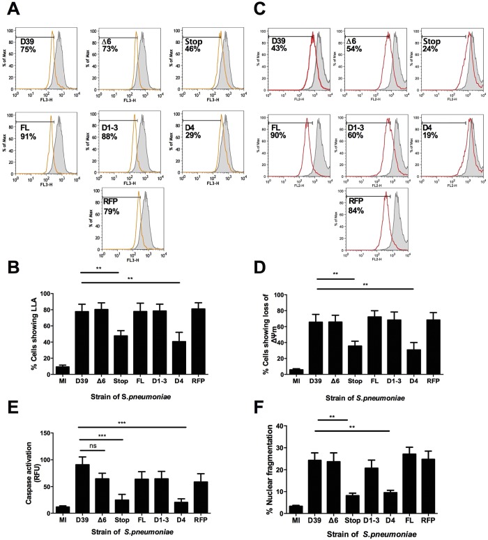
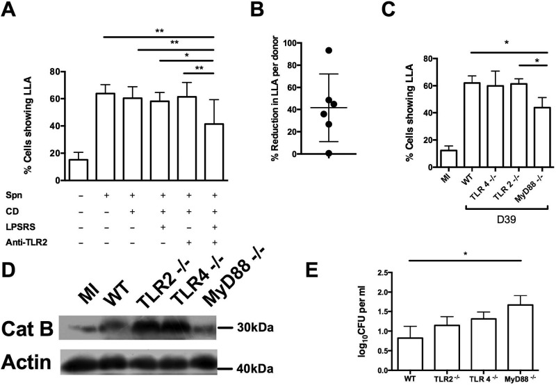
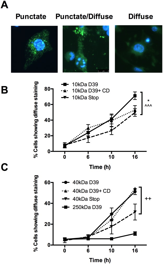
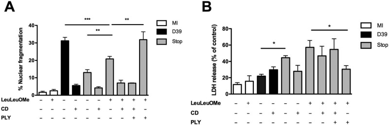
Intracellular killing of Streptococcus pneumoniae is complemented by induction of macrophage apoptosis. Here, we show that the toxin pneumolysin (PLY) contributes both to lysosomal/phagolysosomal membrane permeabilization (LMP), an upstream event programing susceptibility to apoptosis, and to apoptosis execution via a mitochondrial pathway, through distinct mechanisms. PLY is necessary but not sufficient for the maximal induction of LMP and apoptosis. PLY's ability to induce both LMP and apoptosis is independent of its ability to form cytolytic pores and requires only the first three domains of PLY. LMP involves TLR (Toll-like receptor) but not NLRP3/ASC (nucleotide-binding oligomerization domain [Nod]-like receptor family, pyrin domain-containing protein 3/apoptosis-associated speck-like protein containing a caspase recruitment domain) signaling and is part of a PLY-dependent but phagocytosis-independent host response that includes the production of cytokines, including interleukin-1 beta (IL-1β). LMP involves progressive and selective permeability to 40-kDa but not to 250-kDa fluorescein isothiocyanate (FITC)-labeled dextran, as PLY accumulates in the cytoplasm. In contrast, the PLY-dependent execution of apoptosis requires phagocytosis and is part of a host response to intracellular bacteria that also includes NO generation. In cells challenged with PLY-deficient bacteria, reconstitution of LMP using the lysomotrophic detergent LeuLeuOMe favored cell necrosis whereas PLY reconstituted apoptosis. The results suggest that PLY contributes to macrophage activation and cytokine production but also engages LMP. Following bacterial phagocytosis, PLY triggers apoptosis and prevents macrophage necrosis as a component of a broad-based antimicrobial strategy. This illustrates how a key virulence factor can become the focus of a multilayered and coordinated innate response by macrophages, optimizing pathogen clearance and limiting inflammation. Importance: Streptococcus pneumoniae, the commonest cause of bacterial pneumonia, expresses the toxin pneumolysin, which can make holes in cell surfaces, causing tissue damage. Macrophages, resident immune cells essential for responses to bacteria in tissues, activate a program of cell suicide called apoptosis, maximizing bacterial clearance and limiting harmful inflammation. We examined pneumolysin's role in activating this response. We demonstrate that pneumolysin did not directly form holes in cells to trigger apoptosis and show that pneumolysin has two distinct roles which require only part of the molecule. Pneumolysin and other bacterial factors released by bacteria that have not been eaten by macrophages activate macrophages to release inflammatory factors but also make the cell compartment containing ingested bacteria leaky. Once inside the cell, pneumolysin ensures that the bacteria activate macrophage apoptosis, rather than necrosis, enhancing bacterial killing and limiting inflammation. This dual response to pneumolysin is critical for an effective immune response to S. pneumoniae.


Figures

FIG 1
FIG 1
Pneumolysin’s pore-forming ability is not required for macrophage apoptosis. Monocyte-derived macrophages (MDM) were either mock infected (MI) or challenged with wild-type S. pneumoniae (D39), a D39 mutant expressing noncytolytic pneumolysin (Δ6), a pneumolysin-deficient D39 mutant (Stop), or reconstituted mutants expressing full-length pneumolysin (FL), pneumolysin domains 1 to 3 (D1-3), pneumolysin domain 4 only (D4), or red fluorescent protein-tagged pneumolysin (RFPPLY). (A to D) At 16 h postchallenge, cells were analyzed for loss of lysosomal acidification (LLA) (A and B) (n = 10) or for loss of the inner mitochondrial transmembrane potential (Δψm) (C and D) (n = 9). Representative histograms show the mock-infected sample in solid gray and the indicated mutant in white, with the percentage of cells showing loss of the marker indicated (A and C) and summary graphs of all data below (B and D). (E) At 16 h postchallenge, caspase 3 activity was measured (n = 8). (F) At 20 h postchallenge, cells were assessed for nuclear fragmentation (n = 9). In all experiments, ** = P < 0.01, *** = P < 0.001 (one-way ANOVA). All data are expressed as means ± standard errors of the means (SEM). Max, maximum; RFU, relative fluorescence units.
FIG 2
FIG 2
Pneumolysin mediates LLA and apoptosis through distinct pathways. Monocyte-derived macrophages (MDM) were mock infected (MI) or challenged with wild-type S. pneumoniae (D39), pneumolysin-deficient D39 (Stop), exogenous pneumolysin (PLY) alone, or Stop with exogenous pneumolysin (PLY), added at 0.5, 1, or 5 µg/ml (increasing doses are indicated a triangle) (A and C) or 5 µg/ml (all other panels) in the presence (+) or absence (-) of cytochalasin D (CD). At 16 h postchallenge, cells were assessed for loss of lysosomal acidification (LLA) (n = 4) (A), fractionated and the cytosolic fractions blotted for cathepsin B (Cat B), actin, or lysosome-associated membrane protein 1 (LAMP-1) (a representative blot from three independent experiments is shown, with densitometry data below the blot showing fold change in Cat B levels relative to MI cell levels) (n = 3) (B), assessed for loss of inner mitochondrial transmembrane potential (Δψm) (n = 4) (C), or assessed for caspase 3 activity (n = 4) (D), or cells were assessed for nuclear fragmentation at 20 h postchallenge (n = 5) (E). In all experiments, * = P < 0.05, ** = P < 0.01, *** = P < 0.001 (one-way or two-way ANOVA for comparisons within or between CD-negative and CD-positive [-CD and +CD] groups, respectively; data are expressed as means ± SEM). ns, not significant.
FIG 3
FIG 3
Toll-like receptor 2 (TLR2) signaling and TLR4 signaling combine to induce loss of lysosomal acidification. (A and B) Monocyte-derived macrophages (MDM) were challenged in the absence (-) or presence (+) of S. pneumoniae D39 (Spn), cytochalasin D (CD), a TLR4 antagonist (LPSRS), or a TLR2-blocking antibody (Anti-TLR2). At 16 h postchallenge, cells were analyzed for loss of lysosomal acidification (LLA) by flow cytometry (n = 6). * = P < 0.05, ** = P < 0.01 (one-way ANOVA). (B) The percentages of reduction in LLA after D39 challenge for individual donors, comparing untreated cells and those treated with both anti-TLR2 and LPSRS in the absence of CD. (C to E) Bone marrow-derived macrophages (BMDM) from wild-type (WT) or Toll-like receptor 2 (TLR2)-, TLR4-, or myeloid differentiation factor 88 (MyD88)-deficient mice were challenged with strain D39. At 16 h postchallenge, cells were analyzed for loss of lysosomal acidification (LLA) (C) and lysosomal membrane permeabilization, as characterized by cathepsin B (Cat B) translocation to the cytosol (D). (E) At 8 h postchallenge, early bacterial killing was determined by a gentamicin protection assay. In all experiments, n = 4. * = P < 0.05 (one-way ANOVA). Data are expressed as means ± SEM.
FIG 4
FIG 4
Macrophages exposed to S. pneumoniae develop a progressive increase in lysosomal membrane permeabilization. (A to C) Monocyte-derived macrophages (MDM) were preloaded with 5 mg/ml of FITC-dextran molecules of the designated molecular mass and challenged with S. pneumoniae (D39) or pneumolysin-deficient (Stop) S. pneumoniae or with D39 in the presence of cytochalasin D (D39+CD). At the designated time postchallenge, cells were fixed and analyzed by fluorescence microscopy and the percentages of cells showing mixed punctate/diffuse or diffuse staining were determined. (A) Representative images of cells showing completely punctate FITC-dextran localization, mixed punctate/diffuse staining, or completely diffuse staining, taken from a strain D39-exposed population at 10 h postchallenge. (B and C) Localization of 10-kDa (B) and 40- or 250-kDa (C) FITC-dextran (n = 4). * = P < 0.05 for 10-kDa D39 versus 10-kDa D39+CD; ^ ^ ^ = P < 0.001 for 10-kDa D39 versus 10-kDa Stop; ++ = P < 0.01 for 40-kDa D39 versus 40-kDa Stop (two-way ANOVA). Data are expressed as means ± SEM.
FIG 5
FIG 5
Pneumolysin progressively translocates from phagolysosomes to the cytosol following challenge with live bacteria. (A) Monocyte-derived macrophages (MDM) were challenged with S. pneumoniae (D39) expressing RFP-tagged pneumolysin (RFP-PLY). At the designated time postchallenge, cells were stained with BODIPY-pepstatin A to visualize lysosomes. Cells were visualized by fluorescence microscopy. Red, PLY; green, lysosomes; yellow, merged. Images are representative of the results of three independent experiments. (B) A Western blot of cytosolic and membrane fractions from D39-exposed MDMs at the designated time points postchallenge probed with anti-pneumolysin (PLY). Actin and lysosome-associated membrane protein 1 (LAMP-1) were used as loading controls. The blots are representative of the results of three independent experiments.
FIG 6
FIG 6
Pneumolysin contributes to maximal engagement of apoptosis. Monocyte-derived macrophages (MDM) were mock infected (MI; white bar) or challenged with either wild-type S. pneumoniae (D39; black bar) or pneumolysin-deficient D39 (Stop; gray bar). Cells were challenged in the presence (+) or absence (-) of the lysomotrophic detergent LeuLeuOMe, cytochalasin D (CD), or exogenous pneumolysin (5 µg/ml). At 20 h postchallenge, cells were assessed for nuclear fragmentation (n = 6) (A) or for necrosis (n = 5) (B) by measuring LDH release. * = P < 0.05, ** = P < 0.01, *** = P < 0.001 (one-way ANOVA). All data are expressed as means ± SEM.

References

    1. Ortqvist A, Hedlund J, Kalin M. 2005. Streptococcus pneumoniae: epidemiology, risk factors, and clinical features. Semin. Respir. Crit. Care Med. 26:563–574. 10.1055/s-2005-925523 - DOI - PubMed
    1. Berry AM, Yother J, Briles DE, Hansman D, Paton JC. 1989. Reduced virulence of a defined pneumolysin-negative mutant of Streptococcus pneumoniae. Infect. Immun. 57:2037–2042 - PMC - PubMed
    1. Mitchell TJ, Alexander JE, Morgan PJ, Andrew PW. 1997. Molecular analysis of virulence factors of Streptococcus pneumoniae. Soc. Appl. Bacteriol. Symp. Ser. 26:62S–71S - PubMed
    1. Gillespie SH, Balakrishnan I. 2000. Pathogenesis of pneumococcal infection. J. Med. Microbiol. 49:1057–1067 - PubMed
    1. Alouf JE. 2000. Cholesterol-binding cytolytic protein toxins. Int. J. Med. Microbiol. 290:351–356. 10.1016/S1438-4221(00)80039-9 - DOI - PubMed

Publication types

Substances