. 2014 Dec;34(12):1928-35.
doi: 10.1038/jcbfm.2014.164. Epub 2014 Oct 8.

Cellular origin and regulation of D- and L-serine in in vitro and in vivo models of cerebral ischemia

Affiliations

Cellular origin and regulation of D- and L-serine in in vitro and in vivo models of cerebral ischemia

Takato Abe et al. J Cereb Blood Flow Metab. 2014 Dec.

Abstract

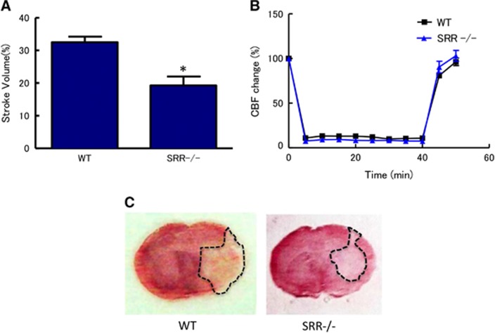
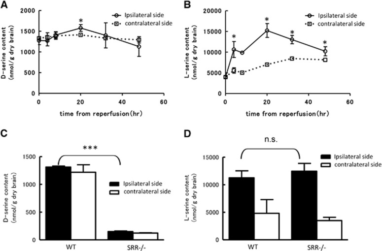
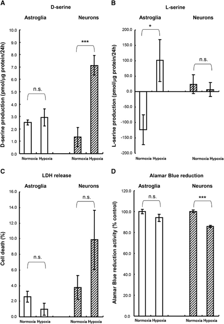
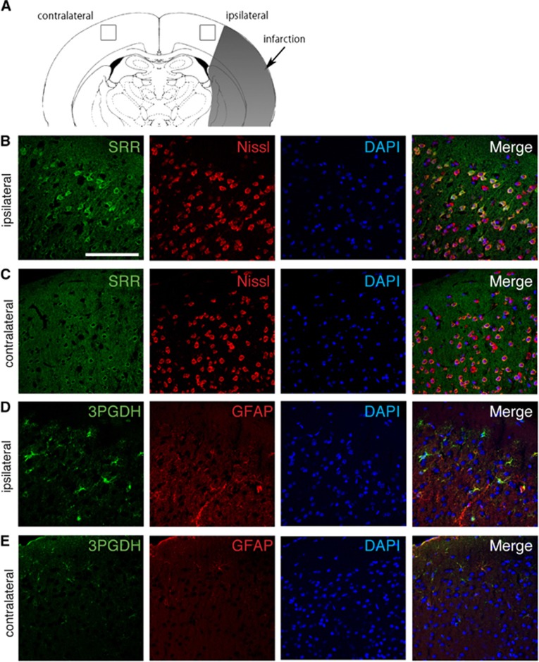
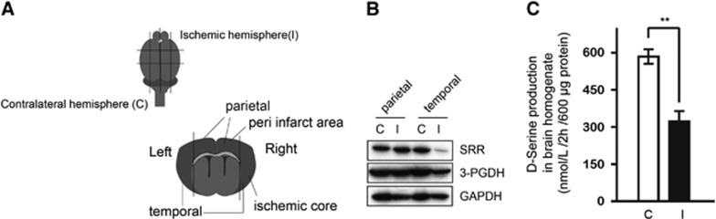
D-Serine is known to be essential for the activation of the N-methyl-D-aspartate (NMDA) receptor in the excitation of glutamatergic neurons, which have critical roles in long-term potentiation and memory formation. D-Serine is also thought to be involved in NMDA receptor-mediated neurotoxicity. The deletion of serine racemase (SRR), which synthesizes D-serine from L-serine, was recently reported to improve ischemic damage in mouse middle cerebral artery occlusion model. However, the cell type in which this phenomenon originates and the regulatory mechanism for D-/L-serine remain elusive. The D-/L-serine content in ischemic brain increased until 20 hours after recanalization and then leveled off gradually. The results of in vitro experiments using cultured cells suggested that D-serine is derived from neurons, while L-serine seems to be released from astroglia. Immunohistochemistry studies of brain tissue after cerebral ischemia showed that SRR is expressed in neurons, and 3-phosphoglycerate dehydrogenase (3-PGDH), which synthesizes L-serine from 3-phosphoglycerate, is located in astrocytes, supporting the results of the in vitro experiments. A western blot analysis showed that neither SRR nor 3-PGDH was upregulated after cerebral ischemia. Therefore, the increase in D-/L-serine was not related to an increase in SRR or 3-PGDH, but to an increase in the substrates of SRR and 3-PGDH.


Figures

Figure 1
Figure 1
Reduced infarct volumes in serine racemase (SRR) −/− mice. (A) Infarct volume was determined 24 hours after middle cerebral artery occlusion (MCAO) (n=7 to 8/group); *P<0.05 vs. wild type (WT) (Student's t-test). (B) Core blood flow during and after MCAO (n=7 to 8/group); not significant between WT and SRR−/− (analysis of variance, ANOVA). (C) Representative triphenyltetrazolium chloride-stained brain sections derived from animals analyzed in (A). CBF, cerebral blood flow.
Figure 2
Figure 2
Temporal profile of D-/L-serine content in the cortex after reperfusion. D-Serine (A) and L-serine (B) content in ipsilateral (ischemic) and contralateral hemisphere after middle cerebral artery occlusion (MCAO) in wild type (WT) mice (n=3−4/group). *P<0.05 vs. 0 hour (analysis of variance, ANOVA followed by Newman-Keuls test). D-Serine (C) and L-serine (D) content in WT and serine racemase (SRR) −/− mice at 20 hours after reperfusion (n=3 to 4/group). ***P<0.001, n.s., not significant vs. WT ipsilateral side (Student's t-test).
Figure 3
Figure 3
D-/L-Serine release to culture medium from neurons and astroglia during hypoxia. D-Serine (A) and L-serine (B) contents in the medium after 24 hours hypoxia (n=6). The Y axis shows the quantity of D- or L-serine in the culture medium during hypoxia. A negative value indicates the consumption of D- or L-serine from the culture medium during hypoxia. (C) Cell death during hypoxia was calculated based on lactate dehydrogenase (LDH) release (n=6). (D) Alamar Blue reduction activity at 24 hours after hypoxia (n=6). n.s., not significant; *P<0.05; ***P<0.001 vs. normoxia (Student's t-test).
Figure 4
Figure 4
Cellular localization of serine racemase (SRR) and 3-phosphoglycerate dehydrogenase (3-PGDH) in the brain after middle cerebral artery occlusion (MCAO). The squares in the ipsilateral and contralateral cortex show the lesion where the pictures were taken (A). Immunohistochemistry of SRR, 3-PGDH, Nissl, and glial fibrillary acidic protein (GFAP) in the peri-infarct area of the ipsilateral hemisphere (B, D) and the corresponding area in the contralateral hemisphere (C, E) after 20 hours of reperfusion. Scale bar=100 μm.
Figure 5
Figure 5
Expressions of serine racemase (SRR) and 3-phosphoglycerate dehydrogenase (3-PGDH) in the peri-infarct area and ischemic core. (A) Location where samples were obtained for the western blot. The parietal sample from the ischemic hemisphere contains tissue from the peri-infarct area, and the temporal sample contains tissue from the ischemic core. These tissues were compared with those from the corresponding area of the contralateral hemisphere. (B) Western blot for SRR and 3-PGDH in parietal (peri-infarct area) and temporal (ischemic core) samples at 20 hours after reperfusion in wild type (WT) mice. C: contralateral hemisphere, I: ischemic hemisphere. (C) D-Serine producing activity in brain homogenates at 20 hours after reperfusion in WT mice (n=4/group). The Y axis shows the amount of D-serine produced by 600 μg of protein for 2 hours. C, contralateral hemisphere; I, ischemic hemisphere. **P<0.01 vs ischemic hemisphere.

References

    1. Lee JM, Grabb MC, Zipfel GJ, Choi DW. Brain tissue responses to ischemia. J Clin Invest. 2000;106:723–731. - PMC - PubMed
    1. Itoh Y, Takaoka R, Ohira M, Abe T, Tanahashi N, Suzuki N. Reactive oxygen species generated by mitochondrial injury in human brain microvessel endothelial cells. Clin Hemorheol Microcirc. 2006;34:163–168. - PubMed
    1. Bonventre JV, Huang Z, Taheri MR, O'Leary E, Li E, Moskowitz MA, et al. Reduced fertility and postischaemic brain injury in mice deficient in cytosolic phospholipase A2. Nature. 1997;390:622–625. - PubMed
    1. Grotta J, Clark W, Coull B, Pettigrew LC, Mackay B, Goldstein LB, et al. Safety and tolerability of the glutamate antagonist CGS 19755 (Selfotel) in patients with acute ischemic stroke. Results of a phase IIa randomized trial. Stroke. 1995;26:602–605. - PubMed
    1. Davis SM, Albers GW, Diener HC, Lees KR, Norris J. Termination of acute stroke studies involving selfotel treatment. ASSIST steering committed. Lancet. 1997;349:32. - PubMed

Publication types

MeSH terms

LinkOut - more resources