. 2014 Oct 15;5(19):9349-61.
doi: 10.18632/oncotarget.2430.

Transcriptional coactivator CBP upregulates hTERT expression and tumor growth and predicts poor prognosis in human lung cancers

Affiliations

Transcriptional coactivator CBP upregulates hTERT expression and tumor growth and predicts poor prognosis in human lung cancers

Wei Guo et al. Oncotarget. .

Abstract

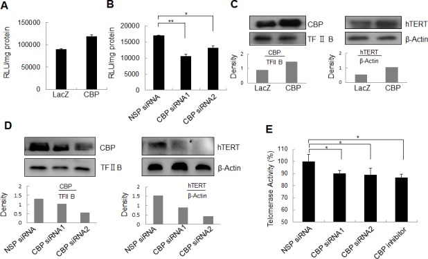
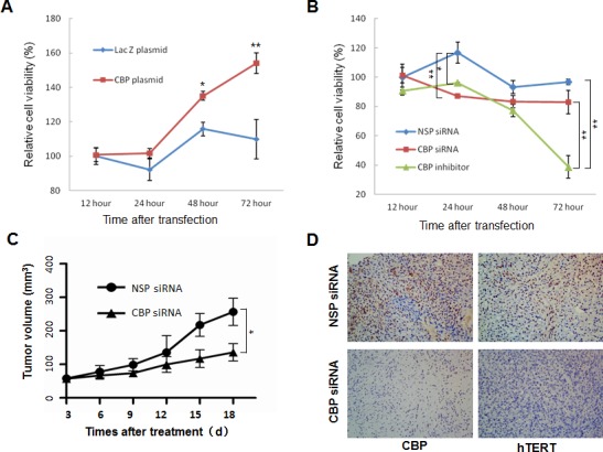
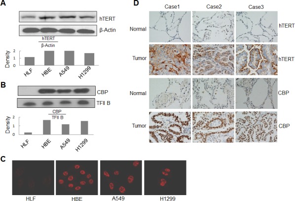
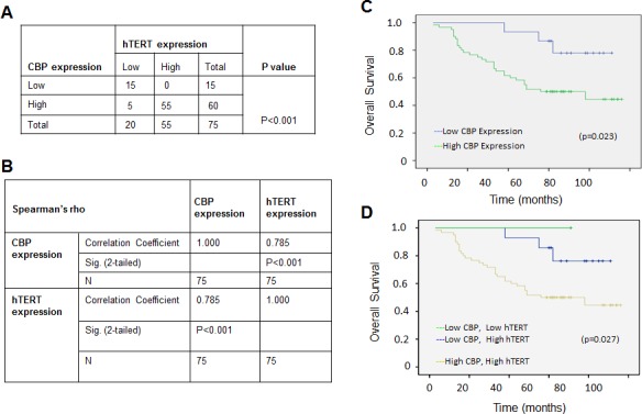
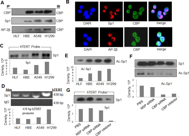
Upregulated expression and activation of human telomerase reverse transcriptase (hTERT) is a hallmarker of lung tumorigenesis. However, the mechanism underlying the aberrant hTERT activity in lung cancer cells remains poorly understood. In this study, we found the transcriptional co-activator CBP as a new hTERT promoter-binding protein that regulated hTERT expression and tumor growth in lung adenocarcinoma cells using a biotin-streptavidin-bead pulldown technique. Chromatin immunoprecipitation assay verified the immortalized cell and tumor cell-specific binding of CBP on hTERT promoter. Overexpression of exogenous CBP upregulated the expression of the hTERT promoter-driven luciferase and endogenous hTERT protein in lung cancer cells. Conversely, inhibition of CBP by CBP-specific siRNA or its chemical inhibitor repressed the expression of hTERT promoter-driven luciferase and endogenous hTERT protein as well as telomerase activity. Moreover, inhibition of CBP expression or activity also significantly reduced the proliferation of lung cancer cells in vitro and tumor growth in an xenograft mouse model in vivo. Immunohistochemical analysis of tissue microarrays of lung cancers revealed a positive correlation between CBP and hTERT. Importantly, the patients with high CBP and hTERT expression had a significantly shorter overall survival. Furthermore, CBP was found to interact with and acetylate transactivator Sp1 in lung cancer cells. Inhibition of CBP by CBP-specific siRNA or its chemical inhibitor significantly inhibited Sp1 acetylation and its binding to the hTERT promoter. Collectively, our results indicate that CBP contributes to the upregulation of hTERT expression and tumor growth, and overexpression of CBP predicts poor prognosis in human lung cancers.


Conflict of interest statement

Disclosure of conflict of interest

The authors declare no conflict of interest.

Figures

Figure 1
Figure 1. Identification of CBP as a hTERT promoter-binding protein in lung cancer cells
(A) Streptavidin-agarose bead pulldown assay with hTERT promoter (-378 to +60) as probes was done in human normal lung cells and lung cancer cells. The pulled down proteins were tested by immunoblot using antibodies against CBP. (B) Chromatin immunoprecipitation assay was done with normal lung cells and lung adenocarcinoma cells using antibodies against CBP. PCR products were separated on 1% agarose gels. The last lane represents the IgG control. The displayed gels were representative of 2-3 independent experiments. Densitometric analysis was used to analyze quantitatively the binding activity of CBP protein on hTERT promoter.
Figure 2
Figure 2. The effect of CBP on hTERT promoter activity, hTERT protein expression and telomerase activity
(A) Lung cancer cells were co-transfected with the plasmids of hTERT promoter driven-luciferase and pCDNA3.1-CBP or pCDNA3.1-Lac Z for 48 h followed by a dual-luciferase assay. The relative luciferase intensity per mg protein was calculated in the treated cells. (B) Lung cancer cells were co-transfected with hTERT promoter driven-luciferase and CBP siRNA or control siRNA for 48 h followed by a dual-luciferase assay. The relative luciferase intensity per mg protein was calculated in the treated cells. (C) Up-regulation of hTERT protein expression by the overexpression of CBP. H1299 cells were treated with pcDNA3.1-CBP for 48 h, and the expression of CBP itself in the nucleus and hTERT protein in the cytoplasm were analyzed by Western blot in lung cancer cells. (D, E) Downregulation of hTERT protein expression by the knockdown of CBP expression. H1299 cells were transfected with a CBP-specific siRNA for 48 h, the expression of CBP itself in the nucleus and hTERT protein in the cytoplasm were analyzed by Western blot (D), and the telomerase activity was assessed (E). All of the measurements represent the means ± SE of three independent experiments. *, P < 0.05, significant differences between treatment groups and DMSO control groups.
Figure 3
Figure 3. The effect of CBP on lung tumor growth in vitro and in vivo
(A) H1299 cells were transfected with CBP overexpression vector pcDNA3.1-CBP. At different time points after transfection, cell viability was measured by MTT assay. (B) H1299 cells were treated with CBP specific siRNA or inhibitor. At different time points after treatment, cell viability was measured by MTT assay. The mean and SE obtained from three independent experiments are plotted (*, P < 0.05,**, P < 0.01). (C) The nude mice containing xenografts of human lung cancer were intratumorally treated with non-specific control siRNA or CBP-specific siRNA, and the tumor volumes ± SE were calculated at different days after treatment. (N=5; *, P < 0.05). (D) Immunohistochemistry of CBP and hTERT from tumor xenografts in nonspecific control siRNA- and CBP-specific siRNA-treated nude mice (400× magnification).
Figure 4
Figure 4. Overexpression of CBP and hTERT in lung cancer cells and tumor tissues
(A) Western blot analysis of hTERT expression from cytoplasmic lysate in human lung normal and cancer cells. β-Actin was used as control. (B) Western blot analysis of CBP expression from nuclear lysate in human lung normal and cancer cells. TFIIB was used as control. (C) The expression and distribution of CBP in human lung normal and cancer cells through immunofluorescent analysis. (D) The expression of hTERT and CBP protein in tumor tissues of patients with lung adenocarcinomas and corresponding adjacent normal lung tissues by immunohistochemistry analysis (magnification, ×200).
Figure 5
Figure 5. The positive correlation between CBP and hTERT protein expression in lung adenocarcinoma specimens, and a comparatively poor prognosis indicated by the higher expression of CBP and hTERT
(A) The correlation between CBP and hTERT protein in lung adenocarcinoma tissues (P < 0.001, χ2 tests). (B) The correlation between CBP and hTERT protein in lung cancer tissues. (P<0.01, 2-tailed test). (C) Kaplan–Meier analysis of overall survival of lung cancer patients with different CBP expression (p<0.05, log-rank test). (D) Kaplan–Meier analysis of overall survival of lung cancer patients with different CBP and hTERT expression (p<0.05, log-rank test).
Figure 6
Figure 6. The interaction of CBP with Sp1 and AP-2 and the acetylationt of Sp1 by CBP in lung cancer cells
(A) The nuclear extracts of human lung normal and cancer cells were prepared for immunoprecipitation using an antibody against Sp1 or AP-2β and then evaluated by immunoblot using antibody against CBP. (B) Human lung cancer H1299 cells grown on chamber slides were cultivated for 24 h, and the subcellular localization and the colocalization of CBP with Sp1 or AP-2β were examined by confocal microscopy analysis with a confocal microscope. More than 100 cells were inspected per experiment, and cells with typical morphology were presented. (C) Streptavidin-agarose bead pulldown assay with hTERT promoter (-378 to +60) as probes was done. Sp1 was tested in the pulled down proteins by immunoblot using antibody against Sp1. (D) Chromatin immunoprecipitation assays were done using antibody against Sp1. PCR products of hTERT promoter (-378 to +60) were separated on 1% agarose gels. The last lane represents the IgG control. (E) Immunoprecipitation was performed using antibody against Sp1. The acetylated Sp1 was determined by immunoblot using the antibody against acetylation. (F) Immunoprecipitation was performed in human lung cancer cells (H1299) treated by non-specific siRNA or CBP specific siRNA or inhibitor using antibody against Sp1. The acetylated Sp1 was tested by immunoblot using antibody against acetylation. (G) Streptavidin-agarose bead pulldown assay with hTERT promoter (-378 to +60) as probes was done in lung cancer cells (H1299) treated by non-specific siRNA or CBP specific siRNA or CBP-specific inhibitor. The level of Sp1 in the pulled down proteins was determined by immunoblot. Densitometric analysis was used to analyze quantitatively the binding activity and acetylation level of Sp1 proteins.

Similar articles

Cited by

References

    1. Shay JW, Wright WE. Role of telomeres and telomerase in cancer. Seminars in cancer biology. 2011;21(6):349–353. - PMC - PubMed
    1. Ouellette MM, Wright WE, Shay JW. Targeting telomerase-expressing cancer cells. Journal of cellular and molecular medicine. 2011;15(7):1433–1442. - PMC - PubMed
    1. Stewart SA, Bertuch AA. The role of telomeres and telomerase in cancer research. Cancer research. 2010;70(19):7365–7371. - PMC - PubMed
    1. Jaskelioff M, Muller FL, Paik JH, Thomas E, Jiang S, Adams AC, Sahin E, Kost-Alimova M, Protopopov A, Cadinanos J, Horner JW, Maratos-Flier E, Depinho RA. Telomerase reactivation reverses tissue degeneration in aged telomerase-deficient mice. Nature. 2011;469(7328):102–106. - PMC - PubMed
    1. Tomas-Loba A, Flores I, Fernandez-Marcos PJ, Cayuela ML, Maraver A, Tejera A, Borras C, Matheu A, Klatt P, Flores JM, Vina J, Serrano M, Blasco MA. Telomerase reverse transcriptase delays aging in cancer-resistant mice. Cell. 2008;135(4):609–622. - PubMed

Publication types

MeSH terms