. 2014 Oct 29;42(19):12189-99.
doi: 10.1093/nar/gku878. Epub 2014 Oct 7.

Rio1 mediates ATP-dependent final maturation of 40S ribosomal subunits

Affiliations

Rio1 mediates ATP-dependent final maturation of 40S ribosomal subunits

Tomasz W Turowski et al. Nucleic Acids Res. .

Abstract

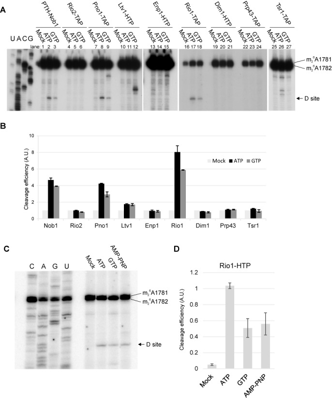
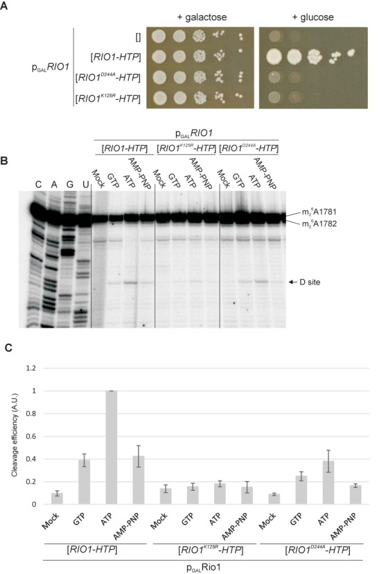
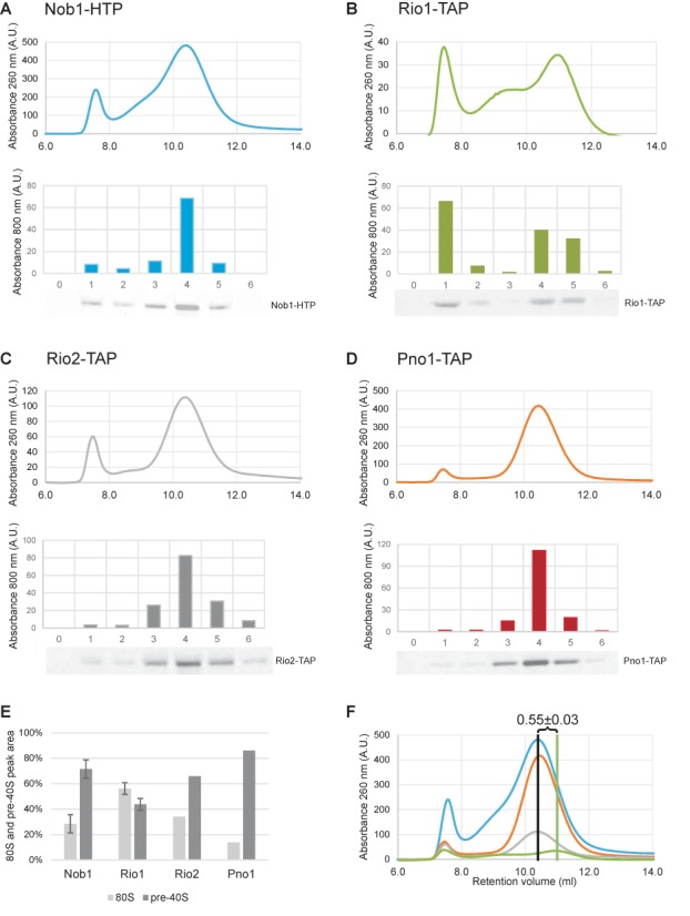
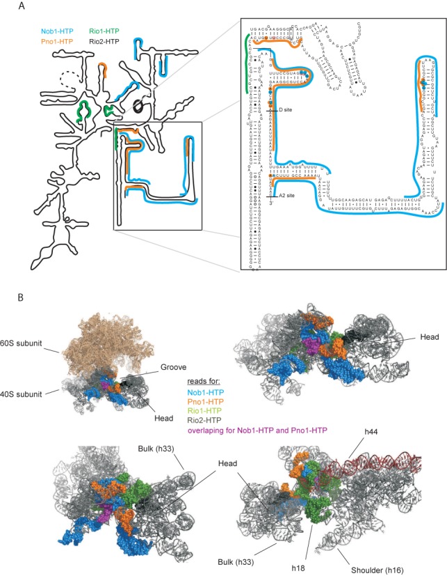
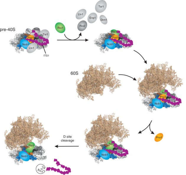
During the last step in 40S ribosome subunit biogenesis, the PIN-domain endonuclease Nob1 cleaves the 20S pre-rRNA at site D, to form the mature 18S rRNAs. Here we report that cleavage occurs in particles that have largely been stripped of previously characterized pre-40S components, but retain the endonuclease Nob1, its binding partner Pno1 (Dim2) and the atypical ATPase Rio1. Within the Rio1-associated pre-40S particles, in vitro pre-rRNA cleavage was strongly stimulated by ATP and required nucleotide binding by Rio1. In vivo binding sites for Rio1, Pno1 and Nob1 were mapped by UV cross-linking in actively growing cells. Nob1 and Pno1 bind overlapping regions within the internal transcribed spacer 1, and both bind directly over cleavage site D. Binding sites for Rio1 were within the core of the 18S rRNA, overlapping tRNA interaction sites and distinct from the related kinase Rio2. Site D cleavage occurs within pre-40S-60S complexes and Rio1-associated particles efficiently assemble into these complexes, whereas Pno1 appeared to be depleted relative to Nob1. We speculate that Rio1-mediated dissociation of Pno1 from cleavage site D is the trigger for final 18S rRNA maturation.


Figures

Figure 1.
Figure 1.
Pre-ribosomes associated with Rio1 or Pno1 undergo efficient cleavage. (A) Primer extension analyses of site D cleavage in pre-40S particles purified by association with the indicated bait proteins. The gels show the relative cleavage efficiencies in the absence of added nucleotides (mock) or with addition on 1 mM ATP or GTP. (B) Quantitation of cleavage efficiency. Cleavage analyses as in panel A were performed in three biological replicates. Cleavage efficiency was determined by the ratio of the primer extension stops site D and at 18S+1781/1782, and is expressed relative to the mock-treated sample incubated in the absence of added ATP or GTP. Error bars show ±1 SD. (C) Comparison of cleavage in Rio1-associated pre-ribosomes, in the presence of ATP, GTP or non-hydrolysable AMP-PNP each at 1 mM. (D) Quantitation of cleavage efficiency. Cleavage efficiency was calculated as in B except that ratio is expressed relative to the ATP-treated sample and error bars show standard error.
Figure 2.
Figure 2.
The ATP binding ability of Rio1 is required for growth and site D cleavage. (A) Growth of PGAL::RIO1 strain on permissive galactose medium or repressive glucose medium, without plasmid (empty brackets) or complemented by plasmids expressing functional Rio1-HTP or Rio1 with the D244A or K125R mutations. (B) Comparison of cleavage in pre-ribosomes associated with Rio1, Rio1K125R and Rio1D244A, in the presence of ATP, GTP or non-hydrolysable AMP-PNP each at 1 mM. (C) Quantitation of cleavage efficiencies. Cleavage efficiency was expressed relative to the ATP-treated sample for functional Rio1. Error bars show standard error.
Figure 3.
Figure 3.
Rio1-associated pre-ribosomes form 80S complexes. Pre-40S particles purified using Nob1-HTP (blue), Rio1-TAP (green), Rio2-TAP (gray) and Pno1-TAP (orange) were analyzed by size exclusion chromatography (top of panels A–D). Comparison to a calibration curve (not shown) revealed that the two major peaks correspond to 80S particles and 40S particles as previously reported (8). The distribution of tagged proteins in chosen fractions was assessed by western blot and quantified using a Licor Odyssey system (bottom of panels A–D). 80S and pre-40S peak areas were quantified and plotted as percent of its sum (E). Results of different experiments were superimposed presenting difference in retention volume of pre-40S peaks.
Figure 4.
Figure 4.
Mass-spectrometry of proteins associated with pre-ribosomal complexes. Fractions 1–5, previously tested by western blotting (Figure 3), were analyzed by mass spectrometry. Plots show ratios of summed-up peptide intensities for Nob1, Pno1, Rio1 and Rio2, Dim1, Enp1, Ltv1 and Tsr1, expressed relative to 40S subunit ribosomal protein Rps4A.
Figure 5.
Figure 5.
Pre-40S rRNA binding sites identified for Nob1, Pno1 and Rio1. (A) Mapped reads for Nob1-HTP (blue), Pno1-HTP (orange) and Rio1-HTP (green) in vivo on a simplified 20S pre-rRNA secondary structure. Inset: Sequence and secondary structure of the 3′ region of the 18S rRNA and 5′ region of ITS1. Nucleotides identified as direct protein contact sites are highlighted. (B) 18S rRNA derived from a crystal structure of the yeast 80S ribosome (PDB ID 3U5B and 3U5D). Sequences identified in CRAC analyses described here are indicated in space-filling mode and colored to indicate binding proteins: Nob1-HTP (blue), Pno1-HTP (orange) and Rio1-HTP (green). Reads common to Nob1-HTP and Pno-HTP, in the 3′ region of 18S rRNA are marked with purple. The crystal structure lacks the ITS1 regions that are bound by Nob1-HTP and Pno1-HTP.
Figure 6.
Figure 6.
Model for late pre-40S processing. The starting point is the previously characterized, major cytoplasmic pre-40S particle. As the particle matures, individual factors pre-40S assembly factors dissociate; Ltv1, Enp1, Tsr1, Dim1 and Rio2. However, Nob1 and Pno1 are retained and Rio1 binds to the particle. Following association with the mature 80S subunit, Pno1 is lost from site D, which is then cleaved by Nob1. As indicated schematically, the long stem structure of the 5′ region of ITS1 is predicted to fold back and to be held in close proximity to the body of the late pre-40S particle. Following D site cleavage, ITS1 is rapidly released and is degraded by the cytoplasmic 5′ exonuclease Xrn1.

References

    1. Fatica A., Tollervey D., Dlakic M. The PIN domain of Nob1p is required for 20S pre-rRNA cleavage at site D. RNA. 2004;10:1698–1701. - PMC - PubMed
    1. Pertschy B., Schneider C., Gnadig M., Schafer T., Tollervey D., Hurt E. RNA helicase Prp43 and its co-factor Pfa1 promote 20 to 18 S rRNA processing catalyzed by the endonuclease Nob1. J. Biol. Chem. 2009;284:35079–35091. - PMC - PubMed
    1. Glavan F., Behm-Ansmant I., Izaurralde E., Conti E. Structures of the PIN domains of SMG6 and SMG5 reveal a nuclease within the mRNA surveillance complex. EMBO J. 2006;25:5117–5125. - PMC - PubMed
    1. Schaeffer D., Tsanova B., Barbas A., Reis F.P., Dastidar E.G., Sanchez-Rotunno M., Arraiano C.M., van Hoof A. The exosome contains domains with specific endoribonuclease, exoribonuclease and cytoplasmic mRNA decay activities. Nat. Struct. Mol. Biol. 2009;16:56–62. - PMC - PubMed
    1. Schneider C., Leung E., Brown J., Tollervey D. The N-terminal PIN domain of the exosome subunit Rrp44 harbors endonuclease activity and tethers Rrp44 to the yeast core exosome. Nucleic Acids Res. 2009;37:1127–1140. - PMC - PubMed

Publication types

MeSH terms