

MHC class I loss is a frequent mechanism of immune escape in papillary thyroid cancer that is reversed by interferon and selumetinib treatment in vitro
- PMID: 25294906
- PMCID: PMC4252612
- DOI: 10.1158/1078-0432.CCR-14-0879
MHC class I loss is a frequent mechanism of immune escape in papillary thyroid cancer that is reversed by interferon and selumetinib treatment in vitro
Abstract
Purpose: To evaluate MHC class I expression on papillary thyroid cancer (PTC) and analyze changes in MHC expression and associated immune activation with current and experimental treatments for thyroid cancer using in vitro PTC cell lines.
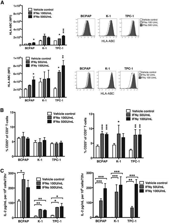
Experimental design: MHC class I expression and assessment of tumor-infiltrating leukocyte populations were evaluated by immunohistochemistry. PTC cell lines were analyzed for HLA-ABC expression by flow cytometry following tyrosine kinase inhibitor, IFNα or IFNγ, or radiation treatment. Functional changes in antigenicity were assessed by coculture of allogeneic donor peripheral blood leukocytes (PBL) with pretreated or untreated PTC cell lines and measurement of T-cell activation and cytokine production.
Results: Both MHC class I and β2-microglobulin expression was reduced or absent in 76% of PTC specimens and was associated with reduced tumor-infiltrating immune cells, including effector (CD3(+), CD8(+), CD16(+)) and suppressor (FoxP3(+)) populations. Treatment of PTC cell lines with the MEK1/2 inhibitor selumetinib or IFN increased HLA-ABC expression. This phenotypic change was associated with increased T-cell activation (%CD25(+) of CD3(+)) and IL2 production by PBL cocultured with treated PTC cell lines. Additive effects were seen with combination selumetinib and IFN treatment.
Conclusions: MHC class I expression loss is frequent in human PTC specimens and represents a significant mechanism of immune escape. Increased antigenicity following selumetinib and IFN treatment warrants further study for immunotherapy of progressive PTC.
©2014 American Association for Cancer Research.
Conflict of interest statement
All other authors have no conflicts of interest to declare.
Figures


Similar articles
-
IL-17A increases MHC class I expression and promotes T cell activation in papillary thyroid cancer patients with coexistent Hashimoto's thyroiditis.Diagn Pathol. 2019 Jun 4;14(1):52. doi: 10.1186/s13000-019-0832-2. Diagn Pathol. 2019. PMID: 31159823 Free PMC article.
-
Inhibition of BRAF Sensitizes Thyroid Carcinoma to Immunotherapy by Enhancing tsMHCII-mediated Immune Recognition.J Clin Endocrinol Metab. 2021 Jan 1;106(1):91-107. doi: 10.1210/clinem/dgaa656. J Clin Endocrinol Metab. 2021. PMID: 32936899
-
IL-10 Restores MHC Class I Expression and Interferes With Immunity in Papillary Thyroid Cancer With Hashimoto Thyroiditis.Endocrinology. 2020 Oct 1;161(10):bqaa062. doi: 10.1210/endocr/bqaa062. Endocrinology. 2020. PMID: 32348468 Free PMC article.
-
Evidence that one subset of anaplastic thyroid carcinomas are derived from papillary carcinomas due to BRAF and p53 mutations.Cancer. 2005 Jun 1;103(11):2261-8. doi: 10.1002/cncr.21073. Cancer. 2005. PMID: 15880523 Review.
-
BRAF mutations in papillary thyroid carcinoma and emerging targeted therapies (review).Mol Med Rep. 2012 Oct;6(4):687-94. doi: 10.3892/mmr.2012.1016. Epub 2012 Aug 2. Mol Med Rep. 2012. PMID: 22858857 Review.
Cited by
-
T cell receptor mimic antibodies for cancer therapy.Oncoimmunology. 2015 Jun 1;5(1):e1049803. doi: 10.1080/2162402X.2015.1049803. eCollection 2016. Oncoimmunology. 2015. PMID: 26942058 Free PMC article. Review.
-
The Escape of Cancer from T Cell-Mediated Immune Surveillance: HLA Class I Loss and Tumor Tissue Architecture.Vaccines (Basel). 2017 Feb 27;5(1):7. doi: 10.3390/vaccines5010007. Vaccines (Basel). 2017. PMID: 28264447 Free PMC article. Review.
-
Clinical and Molecular Correlates of NLRC5 Expression in Patients With Melanoma.Front Bioeng Biotechnol. 2021 Jul 9;9:690186. doi: 10.3389/fbioe.2021.690186. eCollection 2021. Front Bioeng Biotechnol. 2021. PMID: 34307322 Free PMC article.
-
Emerging roles of circular RNAs in regulating the hallmarks of thyroid cancer.Cancer Gene Ther. 2024 Apr;31(4):507-516. doi: 10.1038/s41417-024-00736-0. Epub 2024 Feb 5. Cancer Gene Ther. 2024. PMID: 38316961 Free PMC article. Review.
-
Immune Microenvironment of Thyroid Cancer.J Cancer. 2020 Jun 8;11(16):4884-4896. doi: 10.7150/jca.44506. eCollection 2020. J Cancer. 2020. PMID: 32626535 Free PMC article. Review.
References
-
- Siegel R, Naishadham D, Jemal A. Cancer statistics, 2013. CA Cancer J Clin. 2013;63:11–30. - PubMed
-
- Chen AY, Jemal A, Ward EM. Increasing incidence of differentiated thyroid cancer in the United States, 1988-2005. Cancer. 2009;115:3801–7. - PubMed
-
- Hay ID, Thompson GB, Grant CS, Bergstralh EJ, Dvorak CE, Gorman CA, et al. Papillary thyroid carcinoma managed at the Mayo Clinic during six decades (1940-1999): temporal trends in initial therapy and long-term outcome in 2444 consecutively treated patients. World J Surg. 2003;26:879–85. - PubMed
-
- American Thyroid Association (ATA) Guidelines Taskforce on Thyroid Nodules and Differentiated Thyroid Cancer. Cooper DS, Doherty GM, Haugen BR, Kloos RT, Lee SL, Mandel SJ, et al. Revised American Thyroid Association management guidelines for patients with thyroid nodules and differentiated thyroid cancer. Thyroid. 2009;19:1167–1214. - PubMed
Publication types
MeSH terms
Substances
Grants and funding
LinkOut - more resources
Full Text Sources
Other Literature Sources
Medical
Research Materials
Miscellaneous
