. 2014 Oct;141(20):4006-17.
doi: 10.1242/dev.111310.

Subcellular localisations of the CPTI collection of YFP-tagged proteins in Drosophila embryos

Affiliations

Subcellular localisations of the CPTI collection of YFP-tagged proteins in Drosophila embryos

Claire M Lye et al. Development. 2014 Oct.

Abstract

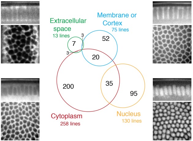
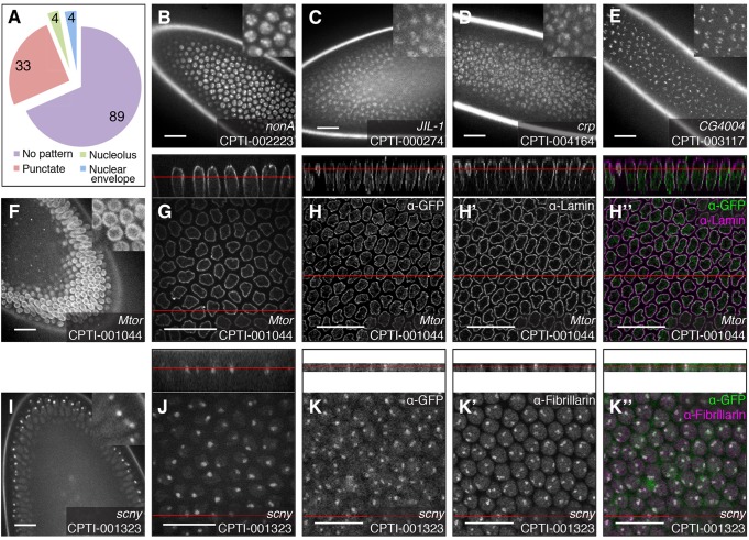
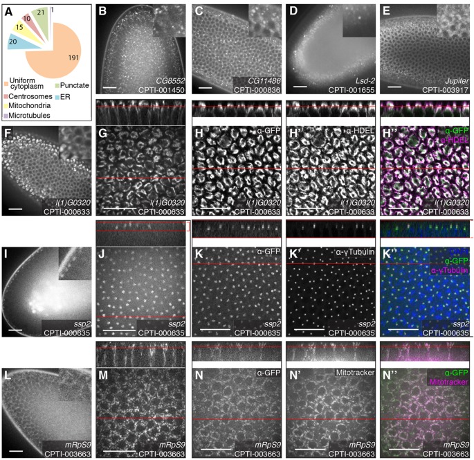
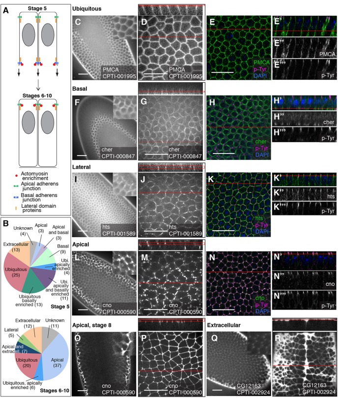
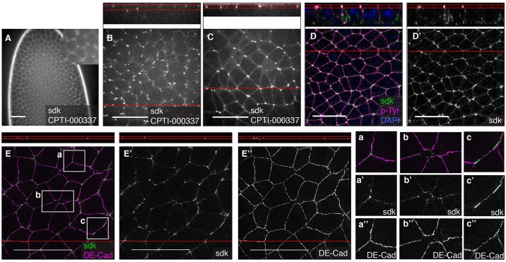
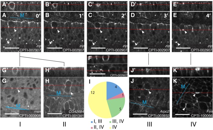
A key challenge in the post-genomic area is to identify the function of the genes discovered, with many still uncharacterised in all metazoans. A first step is transcription pattern characterisation, for which we now have near whole-genome coverage in Drosophila. However, we have much more limited information about the expression and subcellular localisation of the corresponding proteins. The Cambridge Protein Trap Consortium generated, via piggyBac transposition, over 600 novel YFP-trap proteins tagging just under 400 Drosophila loci. Here, we characterise the subcellular localisations and expression patterns of these insertions, called the CPTI lines, in Drosophila embryos. We have systematically analysed subcellular localisations at cellularisation (stage 5) and recorded expression patterns at stage 5, at mid-embryogenesis (stage 11) and at late embryogenesis (stages 15-17). At stage 5, 31% of the nuclear lines (41) and 26% of the cytoplasmic lines (67) show discrete localisations that provide clues on the function of the protein and markers for organelles or regions, including nucleoli, the nuclear envelope, nuclear speckles, centrosomes, mitochondria, the endoplasmic reticulum, Golgi, lysosomes and peroxisomes. We characterised the membranous/cortical lines (102) throughout stage 5 to 10 during epithelial morphogenesis, documenting their apico-basal position and identifying those secreted in the extracellular space. We identified the tricellular vertices as a specialized membrane domain marked by the integral membrane protein Sidekick. Finally, we categorised the localisation of the membranous/cortical proteins during cytokinesis.

Keywords: Epithelium; GFP; Morphogenesis; Protein trap.


Figures

Fig. 1.
Fig. 1.
Subcellular localisation of the CPTI collection at cellularisation. Four-hundred and fifteen YFP-tagged lines are expressed at stage 5 during cellularisation. Nuclear, cytoplasmic, membranous or extracellular subcellular localisations of the proteins were identified at this stage by examining confocal sections showing the arrangement of cells in the apico-basal plane (top panels) and in the plane of the tissue (bottom panels). The Venn diagram shows the number of lines localising in a given compartment, including those detected in two compartments.
Fig. 2.
Fig. 2.
Examples of nuclear localisation patterns of the CPTI lines at cellularisation. (A) Distribution of nuclear patterns at stage 5. No pattern indicates a uniform distribution in the nucleoplasm. (B-E) Example of punctate patterns observed during screening in live embryos: CPTI-002223, inserted in nonA; CPTI-000274, inserted in JIL-1; CPTI-004164, inserted in crp; CPTI-003117, inserted in GC4004. Localisation of CPTI-001044 (inserted in Megator) in the nuclear membrane during screening (F) and at higher magnification (G) in live embryos. Fixed embryos stained for GFP (H), Lamin (H′) and merge showing the two stainings together (H″). The top panels show a cross-section taken through the z-stack. Localisation of CPTI-001323 (inserted in scrawny) in the nucleoli during screening (I) and at higher magnification (J) in live embryos. Fixed embryos stained for GFP (K), Fibrillarin (K′) and merge (K″). Red lines indicate the position of the face view in the z-section, and vice versa. Scale bars: 20 μm.
Fig. 3.
Fig. 3.
Example of cytoplasmic localisation patterns of the CPTI lines at cellularisation. (A) Distribution of cytoplasmic patterns at stage 5. (B-D) Examples of punctate localisation in the cytoplasm: CPTI-001450 in CG8552 marks the Golgi apparatus and ER-to-Golgi transport vesicles (B); CPTI-000836 in CG11486 marks P-bodies (C); and CPTI-001655 in Lsd-2 labels the lipid droplets at the basal end of the cells. (E) CPTI-003917 inserted in Jupiter labels the microtubule cytoskeleton. (F-H″) The localisation of CPTI-000633 in l(1)G0320 to the endoplasmic reticulum appears as a dense perinuclear pattern that is especially strong above the apical side of the nucleus, in live tissue during screening (F) and at high magnification (G). (H-H″) Fixed embryo stained for GFP (H) and HDEL, an ER marker (H′) and merge (H″). (I-K″) CPTI-000635 in ssp2 localises to the centrosomes, recognizable as a pair of puncta on opposite sides of the nucleus, in the apical part of each cell. Live tissue during screening (I) and at high magnification (J). Fixed embryo stained for GFP (K) and the centrosome marker γ Tubulin (K′), and merge with DAPI in blue (K″). (L-N″) Localisation to the mitochondria of CPTI-003663, inserted in Mitochondrial ribosomal protein S9, gives the cytoplasm a granular appearance. Live tissue during screening (L) and at high magnification (M). Live embryo permeabilised and treated with Mitotracker dye to mark the mitochondria: GFP (N), Mitotracker (N′) and merge (N″). Red lines indicate the position of the face view in the z-section, and vice versa. Scale bars: 20 μm.
Fig. 4.
Fig. 4.
Examples of apico-basal localisations of membranous/cortical and extracellular YFP-tagged proteins. (A) The position of adherens junctions, lateral proteins and actomyosin cytoskeleton at stage 5 (cellularisation) and stages 6 to 10 (early morphogenesis). At stage 5, a transient basal junction forms. (B) Distribution of the lines along the apico-basal domains of the plasma membrane and presence in the extracellular space, at stage 5 and stages 6 to 10. (C-E‴) Example of ubiquitous membranous localisation: CPTI-001995, inserted in the plasma membrane calcium ATPase (PMCA). Live embryos at stage 5 during screening (C) and at high magnification (D). Co-staining of fixed stage 5 embryos with anti-GFP and the adherens junction marker p-Tyr, with nuclei labelled by DAPI in blue: merge face view (E) and z section (E′); GFP staining (E″) and p-Tyr staining (E‴). PMCA-YFP is distributed throughout the plasma membrane. (F-H‴) Example of basal localisation at stage 5, at the cellularisation front: CPTI-000847, inserted in Cheerio. Live embryos at stage 5 during screening (F) and at high magnification (G). Co-staining of fixed stage 5 embryos: merge GFP and p-Tyr stainings for face view (H) and z section (H′); GFP only (H″) and p-Tyr only (H‴). In face views, Cheerio-YFP localises in a ring-like pattern typical of the actomyosin-rich cellularisation front. (I-K‴) Example of lateral localisation at stage 5: CPTI-001589, inserted in hu li tai shao (hts). Live embryos at stage 5 during screening (I) and at high magnification (J). Co-staining of fixed stage 5 embryos: merge GFP and p-Tyr stainings for face view (K) and z section (K′); GFP only (K″) and p-Tyr only (K‴). In z sections, Hts-YFP localises just below and above the apical and basal junctions at stage 5, which is typical of the localisation of lateral proteins at cellularisation (see A). (L-N‴) Example of apical localisation at stage 5: CPTI-000590, inserted in canoe (cno). Live embryos at stage 5 during screening (L) and at high magnification (M). Co-staining of fixed stage 5 embryos: merge GFP and p-Tyr stainings for face view (N) and z section (N′); GFP only (N″) and p-Tyr only (N‴). At stage 5, most but not all the Cno-YFP signal has reached an apical position and colocalises with p-Tyr. Low (O) and high (P) magnification in live embryos at stage 8: all Cno-YFP is now apical and forms a continuous belt at the apical junctional domain. (Q,R) Example of extracellular localisation: CPTI-002924, inserted in GC12163. Live embryos during screening at stage 5 (Q) and stage 8 (R). The YFP signal pools in gaps between the apical ends of the cells (Q,R) and in the groove formed by the ventral midline (top to bottom groove in R). In all images, red lines indicate the position of the z section in the face view, and vice versa. When face views are projections of several z planes, two red lines in the z section indicate the bottom and top-most planes used for the projection. Scale bars: 20 μm.
Fig. 5.
Fig. 5.
Localisation of Sidekick at tricellular vertices. (A,B) In live embryos at stage 5, CPTI-000337 inserted in Sidekick localises in spots that become progressively enriched apically, at cell vertices. (C) In live embryos at stage 8, Sidekick-YFP is localised apically and marks cell vertices between three or more cells. (D,D′) In fixed embryos of same stage, co-staining with p-Tyr confirms the apical junctional position of Sidekick-YFP. (E-E″) Superresolution imaging of fixed embryos at stage 8 stained for DE-Cadherin and YFP shows that DE-Cadherin and Sidekick tend to localise in complementary domains, with DE-Cadherin mainly at bicellular contacts (E,E″,a″,b″) and Sidekick (E,E′) at tricellular (a′) or multicellular contacts (b′). Occasionally, Sidekick forms plaques at bicellular contacts where Cadherin is less enriched (c′,c″). Top panels show side views from the reconstruction of the z planes at the position of the red line in the main images. Scale bars: 20 μm.
Fig. 6.
Fig. 6.
Localisation patterns during cytokinesis. (A-E) The localisation of CPTI-002907, an insertion in the gene zipper coding for Myosin II Heavy Chain, reveals the closure of the cytokinesis ring, from its formation (A) to its closure with the formation of the midbody (D,E). Side views from the reconstruction of the z planes show the asymmetric apico-basal shape of the cytokinesis ring (A′) and its closure from basal to apical (B′-E′). (F) An image taken by light sheet imaging, confirming the asymmetric shape of the cytokinesis ring. Membranous/cortical protein localisations at cytokinesis were classified as follows. Categories I and II correspond to the beginning of cytokinesis, I for localisation at the ring (G,G′) and II for localisation at the apical cortex (H,H′). Category III corresponds to proteins localising immediately adjacent to the midbody (J,J′) and category IV proteins localise a little later at the new membrane between daughter cells, on either side of the midbody (K,K′). The pie chart in I shows the number of insertions in each category. Cytokinesis was imaged in stage 9 or 10 embryos. Top panels show side views from the reconstruction of the z planes at the position of the red line in the main images. M, ventral midline. Scale bars: 20 μm.

References

    1. Aleksic, J., Lazic, R., Müller, I., Russell, S. R. and Adryan, B. (2009). Biases in Drosophila melanogaster protein trap screens. BMC Genomics 10, 249 10.1186/1471-2164-10-249 - DOI - PMC - PubMed
    1. Alonso, J., Rodriguez, J. M., Baena-López, L. A. and Santarén, J. F. (2005). Characterization of the Drosophila melanogaster mitochondrial proteome. J. Proteome Res. 4, 1636-1645 10.1021/pr050130c - DOI - PubMed
    1. Andrew, D. J. and Baker, B. S. (2008). Expression of the Drosophila secreted cuticle protein 73 (dsc73) requires Shavenbaby. Dev. Dyn. 237, 1198-1206 10.1002/dvdy.21512 - DOI - PMC - PubMed
    1. Antao, J. M., Mason, J. M., Dejardin, J. and Kingston, R. E. (2012). Protein landscape at Drosophila melanogaster telomere-associated sequence repeats. Mol. Cell. Biol. 32, 2170-2182 10.1128/MCB.00010-12 - DOI - PMC - PubMed
    1. Bobinnec, Y., Morin, X. and Debec, A. (2006). Shaggy/GSK-3beta kinase localizes to the centrosome and to specialized cytoskeletal structures in Drosophila. Cell Motil. Cytoskeleton 63, 313-320 10.1002/cm.20128 - DOI - PubMed

Publication types

MeSH terms

LinkOut - more resources