. 2014:2014:475919.
doi: 10.1155/2014/475919. Epub 2014 Sep 9.

Insulin-like growth factor-I induces arginase activity in Leishmania amazonensis amastigote-infected macrophages through a cytokine-independent mechanism

Affiliations

Insulin-like growth factor-I induces arginase activity in Leishmania amazonensis amastigote-infected macrophages through a cytokine-independent mechanism

Celia Maria Vieira Vendrame et al. Mediators Inflamm. 2014.

Abstract

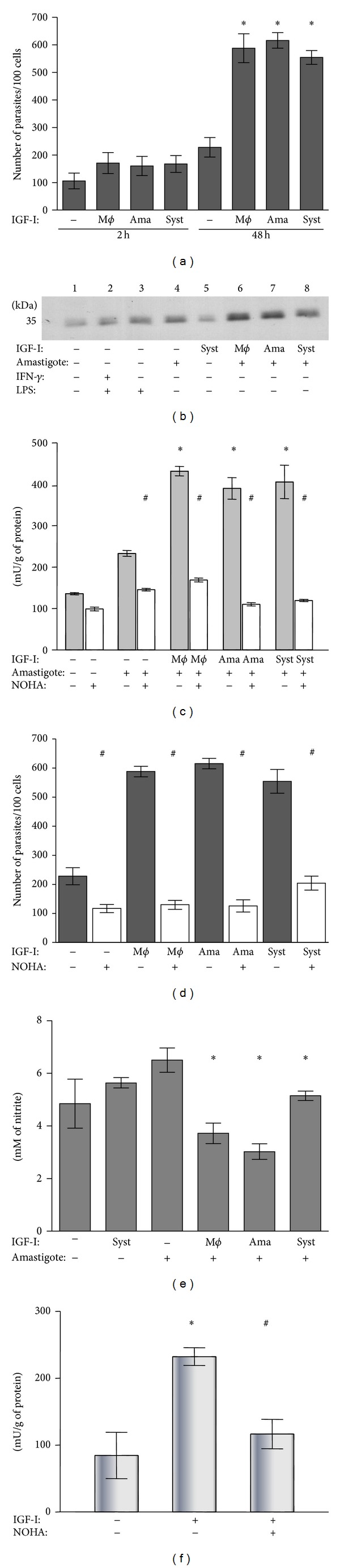
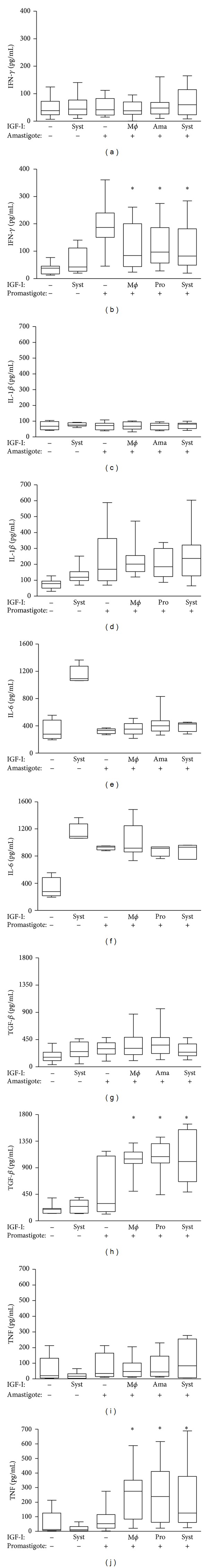
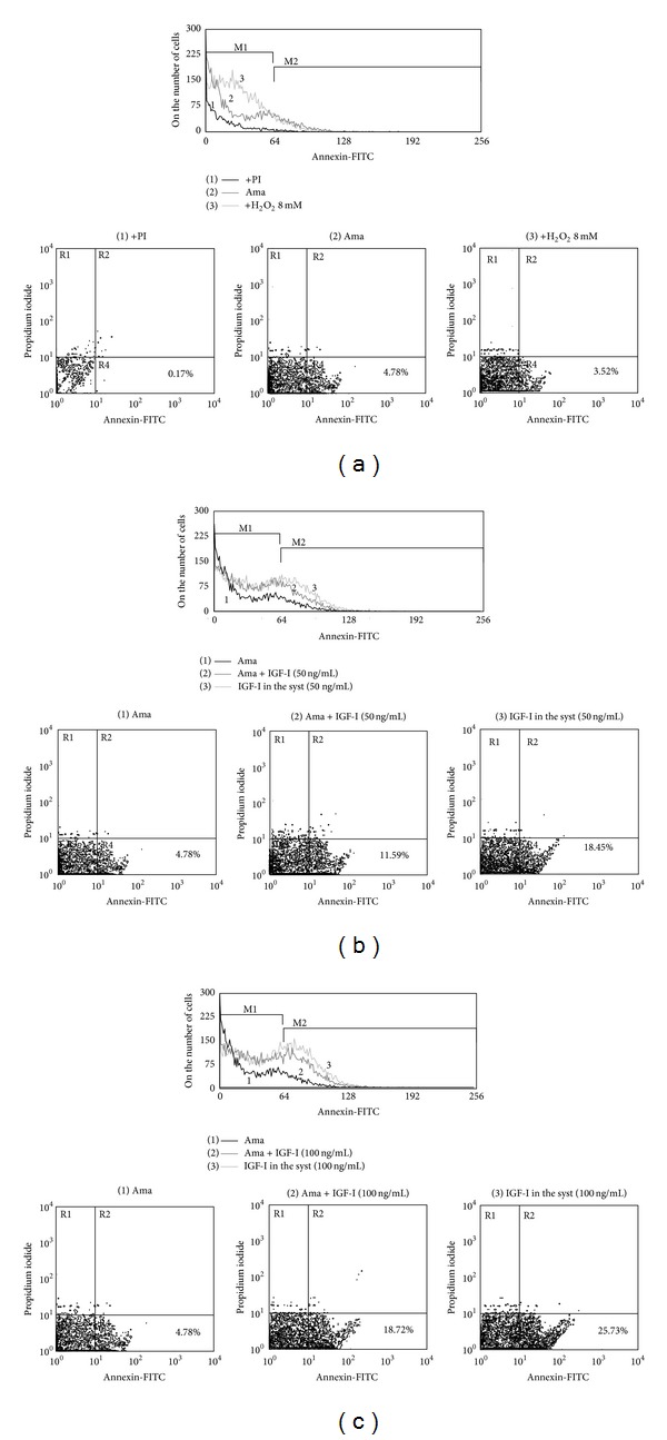
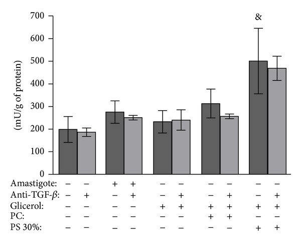
Leishmania (Leishmania) amazonensis exhibits peculiarities in its interactions with hosts. Because amastigotes are the primary form associated with the progression of infection, we studied the effect of insulin-like growth factor (IGF)-I on interactions between L. (L.) amazonensis amastigotes and macrophages. Upon stimulation of infected macrophages with IGF-I, we observed decreased nitric oxide production but increased arginase expression and activity, which lead to increased parasitism. However, stimulation of amastigote-infected macrophages with IGF-I did not result in altered cytokine levels compared to unstimulated controls. Because IGF-I is present in tissue fluids and also within macrophages, we examined the possible effect of this factor on phosphatidylserine (PS) exposure on amastigotes, seen previously in tissue-derived amastigotes leading to increased parasitism. Stimulation with IGF-I induced PS exposure on amastigotes but not on promastigotes. Using a PS-liposome instead of amastigotes, we observed that the PS-liposome but not the control phosphatidylcholine-liposome led to increased arginase activity in macrophages, and this process was not blocked by anti-TGF-β antibodies. Our results suggest that in L. (L.) amazonensis amastigote-infected macrophages, IGF-I induces arginase activity directly in amastigotes and in macrophages through the induction of PS exposure on amastigotes in the latter, which could lead to the alternative activation of macrophages through cytokine-independent mechanisms.


Figures

Figure 1
Figure 1
Effect of IGF-I on the Leishmania amastigote-macrophage interaction. (a) Parasite burden evaluated after 2 and 48 h of culture. (b) Arginase expression analyzed in infected macrophages at 24 h with western blotting using an anti-arginase-I antibody. (c) Arginase activity evaluated in the interaction measuring urea production with and without the arginase inhibitor NOHA. (d) Effect of inhibition by NOHA on the parasite burden in Leishmania amastigote-infected macrophages. (e) Measurement of nitrite production in the culture supernatant by the Griess method. (f) Effect of IGF-I on the arginase activity of Leishmania amastigotes. BALB/c peritoneal macrophages were infected with amastigotes of Leishmania (L.) amazonensis. Amastigotes (ama) or macrophages (mϕ) were prestimulated for 5 min with IGF-I (50 ng/mL) before interaction or the factors were maintained in the culture system (syst) throughout the experiment period. For further details see Material and Methods Section. *P < 0.05 compared with amastigote-infected macrophage without IGF-I stimulation (ANOVA and Student Newman-Keuls tests). # P < 0.05 compared with culture without NOHA (ANOVA and Student Newman-Keuls tests).
Figure 2
Figure 2
Effect of IGF-I on cytokine production by BALB/c peritoneal macrophages infected with amastigotes or promastigotes of L. (L.) amazonensis. Cytokine levels were determined in the culture supernatant by ELISA: IFN-γ: (a) and (b); IL1-β: (c) and (d); IL-6: (e) and (f); TGF-β: (g) and (h); TNF: (i) and (j). Boxes represent the median values and the 25th and 75th percentiles (3 experiments, n = 6). Mϕ = macrophage, ama = amastigote, pro = promastigote and Syst = System. *P < 0.05 compared with promastigote-infected macrophage without IGF-I stimulation. (Kruskal-Wallis test with the Student Newman-Keuls contrast post-test).
Figure 3
Figure 3
Effect of IGF-I on Annexin V binding to L. (L.) amazonensis after IGF-I stimulation. Annexin V-FITC binding to parasites by flow cytometry. (a) Controls. (1) Negative control-amastigotes without Annexin V-FITC label; only with propidium iodide (PI) stain; (2) Unstimulated amastigotes. (3) Positive control-amastigotes after 8 mM H2O2 stimulation. (b) IGF-I (50 ng/mL) stimulation and (c) IGF-I (100 ng/mL) stimulation. (1) Unstimulated amastigotes. (2) Amastigotes prestimulated for 5 min and maintained in culture without IGF-I for 24 h. (3) Amastigotes maintained in culture (syst) with IGF-I for 24 h. The results are representative of 3 independent experiments. Data were collected in a BD FACScalibur and analyzed by CellQuest Pro (BD Biosciences). A total of 10,000 events were harvested from each sample.
Figure 4
Figure 4
Effect of negatively charged lipid phosphatidylserine (PS)-liposomes on arginase activity of BALB/c macrophages. Peritoneal macrophages were infected with phosphatidylserine- (PS-) liposomes, phosphatidylcholine- (PC-) liposome or glycerol. Amastigotes of Leishmania (L.) amazonensis were used as controls. In parallel, an anti-TGF-β antibody was used in all interaction conditions. Macrophages were lysed and the arginase activity was determined by measuring the urea level. Assays were run in triplicate. The data are presented as the mean ± standard deviation of enzyme activity units (amount of enzyme that catalyzes the formation of 1 μmol urea/min). The results are representative of two similar experiments. & P < 0.05 compared with control without liposome (ANOVA and Student Newman-Keuls tests).

References

    1. Desjeux P. Leishmaniasis: current situation and new perspectives. Comparative Immunology, Microbiology and Infectious Diseases. 2004;27(5):305–318. - PubMed
    1. Goto H, Lauletta Lindoso JA. Cutaneous and mucocutaneous leishmaniasis. Infectious Disease Clinics of North America. 2012;26(2):293–307. - PubMed
    1. Cunningham AC. Parasitic adaptive mechanisms in infection by Leishmania. Experimental and Molecular Pathology. 2002;72(2):132–141. - PubMed
    1. Da-Cruz AM, Bittar R, Mattos M, et al. T-cell-mediated immune responses in patients with cutaneous or mucosal leishmaniasis: long-term evaluation after therapy. Clinical and Diagnostic Laboratory Immunology. 2002;9(2):251–256. - PMC - PubMed
    1. Silveira FT, Lainson R, de Castro Gomes CM, Laurenti MD, Corbett CEP. Immunopathogenic competences of Leishmania (V.) braziliensis and L. (L.) amazonensis in American cutaneous leishmaniasis. Parasite Immunology. 2009;31(8):423–431. - PubMed

Publication types

LinkOut - more resources