. 2014:2014:917535.
doi: 10.1155/2014/917535. Epub 2014 Sep 10.

A Novel Rat Model of Orthodontic Tooth Movement Using Temporary Skeletal Anchorage Devices: 3D Finite Element Analysis and In Vivo Validation

Affiliations

A Novel Rat Model of Orthodontic Tooth Movement Using Temporary Skeletal Anchorage Devices: 3D Finite Element Analysis and In Vivo Validation

Neelambar Kaipatur et al. Int J Dent. 2014.

Abstract

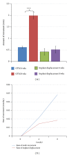
The aim of this animal study was to develop a model of orthodontic tooth movement using a microimplant as a TSAD in rodents. A finite element model of the TSAD in alveolar bone was built using μCT images of rat maxilla to determine the von Mises stresses and displacement in the alveolar bone surrounding the TSAD. For in vivo validation of the FE model, Sprague-Dawley rats (n = 25) were used and a Stryker 1.2 × 3 mm microimplant was inserted in the right maxilla and used to protract the right first permanent molar using a NiTi closed coil spring. Tooth movement measurements were taken at baseline, 4 and 8 weeks. At 8 weeks, animals were euthanized and tissues were analyzed by histology and EPMA. FE modeling showed maximum von Mises stress of 45 Mpa near the apex of TSAD but the average von Mises stress was under 25 Mpa. Appreciable tooth movement of 0.62 ± 0.04 mm at 4 weeks and 1.99 ± 0.14 mm at 8 weeks was obtained. Histological and EPMA results demonstrated no active bone remodeling around the TSAD at 8 weeks depicting good secondary stability. This study provided evidence that protracted tooth movement is achieved in small animals using TSADs.


Figures

Figure 1
Figure 1
Computer modeling of TSAD placement into the rat maxilla 3D model of hemimaxillae in (a) sagittal view and (b). Transverse view (c). 3D model of the hemimaxillae showing the direction of force applied during FE analysis (d), (e). Fine mesh of the TSAD and the surrounding maxillary bone.
Figure 2
Figure 2
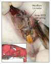
Orthodontic appliance in the right maxilla for tooth movement using TSAD and NiTi closed coil spring. Inset: μCT 3D rendered cross-section model of rat maxilla with appliance.
Figure 3
Figure 3
Displacement (panel above) and von Mises stresses (panel below) at (a). 0 gms, (b). 30 gms (c). 60 gms and (d). 140 gms of force. The warmer colors depict increase in the amount and distribution of the stress and displacement and shows stresses concentrated at the apex of the TSAD and at the coronal contact area between the TSAD and the bone.
Figure 4
Figure 4
Von Mises stress localization in the cortical bone surrounding the TSAD. Dark blue color represents minimal von Mises stress and red color represents maximum von Mises stress localization around the TSAD.
Figure 5
Figure 5
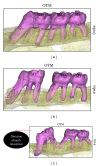
Three-dimensional microcomputed tomography—rendered images showing the amount of tooth movement of the right first permanent molar at 4 and 8 weeks. (M1, right permanent first molar; M2, right permanent second molar; M3, right permanent third molar).
Figure 6
Figure 6
(a) Mean (±S.E) amount of orthodontic tooth movement and implant displacement measured at 4 and 8 weeks. (Significance level: *P < 0.05). (b) Mean rate of orthodontic tooth movement and implant displacement measured at 0, 4, and 8 weeks.
Figure 7
Figure 7
Sagittal (a) and cross-sectional (e) backscattered images and electron microprobe mapping of calcium ((b) and (f)); phosphorus ((c) and (g)); strontium ((d) and (h)) composition of the alveolar bone surrounding the micro-TSAD. No evidence of recognizable difference in the calcium or phosphorus levels of bone immediately around the micro-TSAD and the surrounding preexisting bone (). ((d) and (h)) show no strontium deposition in the alveolar bone immediately surrounding the micro-TSAD. Lack of strontium deposition confirms no active bone remodeling and excellent micro-TSAD stability. ∗TSAD cavity. Scale bars = 1 mm in ((a)–(h)).
Figure 8
Figure 8
Electron microprobe mapping of strontium ((a) and (d)) calcium ((b) and (e)); phosphorus ((c) and (f)) composition of the alveolar bone surrounding the upper left (control) and right first permanent molar (OTM), respectively. Panel (d) shows increased strontium deposition (∗) surrounding the roots of right first permanent molar where OTM occurred. Evidence of increased strontium deposition indicates increased bone remodeling on the tension side of OTM. No difference in the Ca ((b) and (e)) and P ((c) and (f)) composition could be seen between control and OTM side. OTM-orthodontic tooth movement, ⟵ indicates direction of tooth movement. Scale bars = 2 mm in ((a)–(c)); 5 mm in ((d)–(f)).
Figure 9
Figure 9
Histologic hematoxylin and eosin stained paraffin sections of the maxillary left (a) and right (b) first permanent molar area. While normal bone remodeling occurs on the control side with no gap between the first and second molar (a), increased separation between the first and second permanent molars (tooth movement) and increased bone remodeling is evident on the tension side of OTM (b). (c) Higher magnification of boxed area in (b) with stretched PDL fibers (Inset). (d) Alveolar bone surrounding the micro-TSAD, (arrows) with part of TSAD inadvertently into PDL space surrounding the tooth root. ∗ denotes TSAD space; scale bars = 1 mm.

References

    1. Krishnan V, Davidovitch Z. Cellular, molecular, and tissue-level reactions to orthodontic force. American Journal of Orthodontics and Dentofacial Orthopedics. 2006;129(4):469.e1–469.e32. - PubMed
    1. Karras JC, Miller JR, Hodges JS, Beyer JP, Larson BE. Effect of alendronate on orthodontic tooth movement in rats. The American Journal of Orthodontics and Dentofacial Orthopedics. 2009;136(6):843–847. - PubMed
    1. Gonzales C, Hotokezaka H, Yoshimatsu M, Yozgatian JH, Darendeliler MA, Yoshida N. Force magnitude and duration effects on amount of tooth movement and root resorption in the rat molar. Angle Orthodontist. 2008;78(3):502–509. - PubMed
    1. Dunn MD, Park CH, Kostenuik PJ, Kapila S, Giannobile WV. Local delivery of osteoprotegerin inhibits mechanically mediated bone modeling in orthodontic tooth movement. Bone. 2007;41(3):446–455. - PMC - PubMed
    1. Drevenek M, Volk J, Sprogar S, Drevenek G. Orthodontic force decreases the eruption rate of rat incisors. European Journal of Orthodontics. 2009;31(1):46–50. - PubMed

LinkOut - more resources