

Primary hepatic malignant fibrous histiocytoma mimicking hepatocellular carcinoma: A report of two cases
- PMID: 25295102
- PMCID: PMC4186583
- DOI: 10.3892/ol.2014.2483
Primary hepatic malignant fibrous histiocytoma mimicking hepatocellular carcinoma: A report of two cases
Abstract
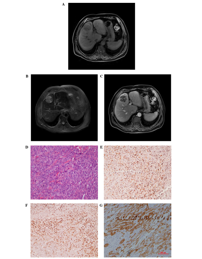
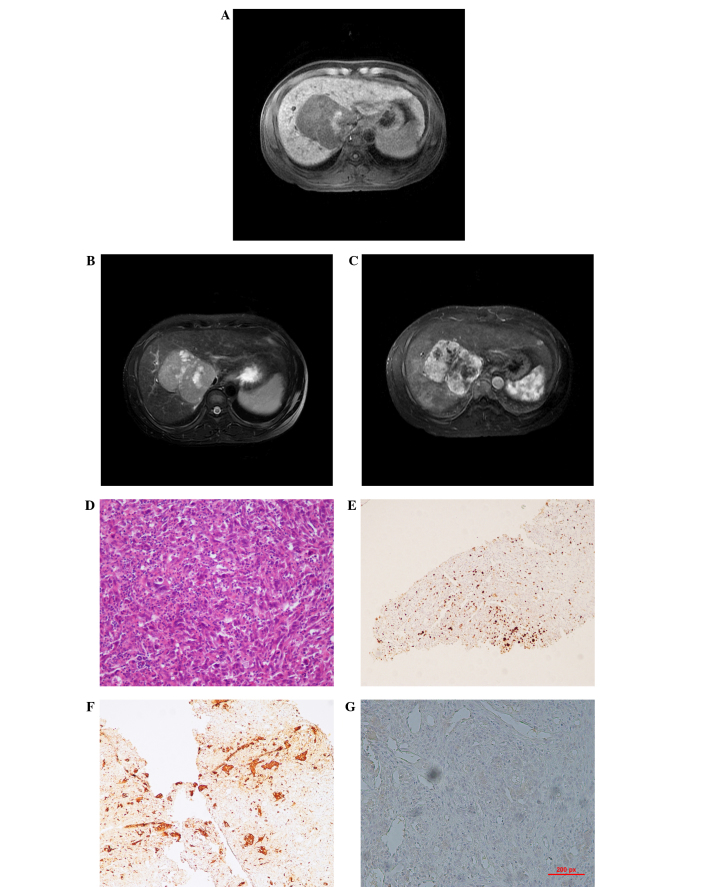
Malignant fibrous histiocytoma (MFH) is a tumor that occurs throughout the body as a relatively uncommon entity. The current study presents two cases of primary malignant fibrous histiocytoma of the liver. The first case was of a 67-year-old male who exhibited no symptoms or abnormal physical signs, and in whom the lesion was found by ultrasound examination during a routine physical examination. The second case was of a 35-year-old male who presented with persistent malaise, weight loss and intermittent right upper quadrant pain. The presence of liver cirrhosis due to hepatitis B virus, which was identified 10 years previously, and the clinical appearance caused MFH to appear as hepatocellular carcinoma at the time of the initial diagnosis. Abdominal magnetic resonance imaging scans were the main tools of diagnosis, but the MFH mimicked hepatocellular carcinoma due to the similar morphological characteristics, the rare occurrence of MFH and the underlying diseases of the liver. The first patient underwent a complete resection and recovered well, while the second patient underwent palliative treatment due to the large size of the tumor and the obstructive emboli in the portal vein. The diagnoses of the tumors were confirmed as MFH by histopathology and immunohistochemistry.
Keywords: histology; magnetic resonance imaging; malignant fibrous histiocytoma.
Figures
Similar articles
-
Simultaneous occurrence of malignant fibrous histiocytoma and hepatocellular carcinoma in cirrhotic liver: A case report.World J Hepatol. 2011 Sep 27;3(9):256-61. doi: 10.4254/wjh.v3.i9.256. World J Hepatol. 2011. PMID: 21969879 Free PMC article.
-
Clinical characteristics of the primary hepatic malignant fibrous histiocytoma in China: case report and review of the literature.World J Surg Oncol. 2012 Jan 5;10:2. doi: 10.1186/1477-7819-10-2. World J Surg Oncol. 2012. PMID: 22221822 Free PMC article. Review.
-
A rare case of primary malignant fibrous histiocytoma: a sarcoma of the kidney.BMC Urol. 2019 Jun 4;19(1):45. doi: 10.1186/s12894-019-0471-7. BMC Urol. 2019. PMID: 31164132 Free PMC article.
-
Malignant fibrous histiocytoma: an uncommon sarcoma with pathological fracture of mandible.J Maxillofac Oral Surg. 2015 Mar;14(Suppl 1):283-7. doi: 10.1007/s12663-013-0491-x. Epub 2013 Mar 27. J Maxillofac Oral Surg. 2015. PMID: 25838711 Free PMC article.
-
Primary malignant fibrous histiocytoma of the jejunum: report of a case and review of subject.J R Coll Surg Edinb. 1997 Oct;42(5):355-8. J R Coll Surg Edinb. 1997. PMID: 9354075 Review.
Cited by
-
MRI and CT findings of a primary malignant fibrous hystiocitoma presenting as a huge glissonian mass; imaging findings with surgical and histological correlations.BJR Case Rep. 2018 Aug 2;5(1):20180055. doi: 10.1259/bjrcr.20180055. eCollection 2019 Feb. BJR Case Rep. 2018. PMID: 31131128 Free PMC article.
-
Clinical and imaging characteristics of primary hepatic sarcomatoid carcinoma and sarcoma: a comparative study.BMC Cancer. 2020 Oct 9;20(1):977. doi: 10.1186/s12885-020-07475-z. BMC Cancer. 2020. PMID: 33036589 Free PMC article.
-
miR-143 down-regulates TLR2 expression in hepatoma cells and inhibits hepatoma cell proliferation and invasion.Int J Clin Exp Pathol. 2015 Oct 1;8(10):12738-47. eCollection 2015. Int J Clin Exp Pathol. 2015. PMID: 26722463 Free PMC article.
-
Primary malignant fibrous histiocytoma of the colon: A case report and review of the literature.Mol Clin Oncol. 2016 Jun;4(6):1006-1008. doi: 10.3892/mco.2016.849. Epub 2016 Apr 6. Mol Clin Oncol. 2016. PMID: 27284436 Free PMC article.
References
-
- Weiss SW, Enzinger FM. Malignant fibrous histiocytoma: an analysis of 200 cases. Cancer. 1978;41:2250–2266. - PubMed
-
- Kearney MM, Soule EH, Ivins JC. Malignant fibrous histiocytoma: a retrospective study of 167 cases. Cancer. 1980;45:167–178. - PubMed
-
- Yu JS, Kim KW, Kim CS, et al. Primary malignant fibrous histiocytoma of the liver: imaging features of five surgically confirmed cases. Abdom Imaging. 1999;24:386–391. - PubMed
-
- Li YR, Akbari E, Tretiakova MS, et al. Primary hepatic malignant fibrous histiocytoma: clinicopathologic characteristics and prognostic value of ezrin expression. Am J Surg Pathol. 2008;32:1144–1158. - PubMed
LinkOut - more resources
Full Text Sources
Other Literature Sources