Review
. 2014:2014:697923.
doi: 10.1155/2014/697923. Epub 2014 Sep 11.

Aptamers as both drugs and drug-carriers

Affiliations
Review

Aptamers as both drugs and drug-carriers

Md Ashrafuzzaman. Biomed Res Int. 2014.

Abstract

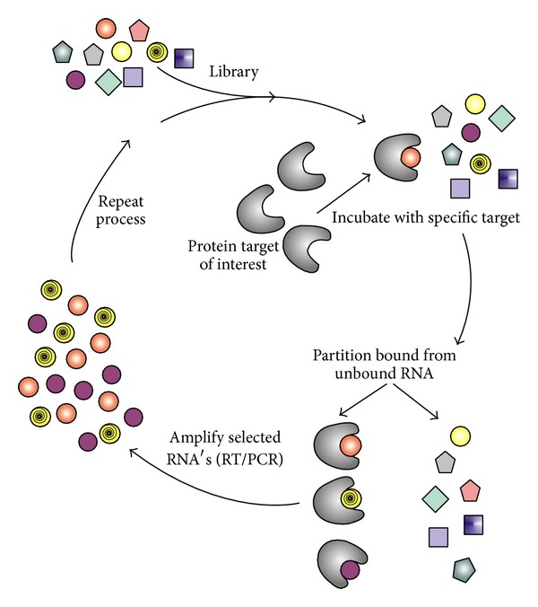
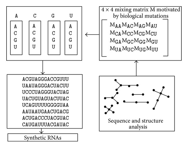
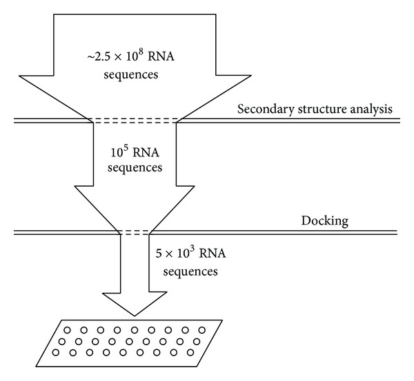
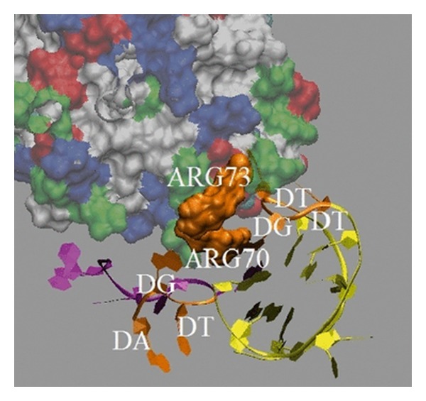
Aptamers are short nucleic acid oligos. They may serve as both drugs and drug-carriers. Their use as diagnostic tools is also evident. They can be generated using various experimental, theoretical, and computational techniques. The systematic evolution of ligands by exponential enrichment which uses iterative screening of nucleic acid libraries is a popular experimental technique. Theory inspired methodology entropy-based seed-and-grow strategy that designs aptamer templates to bind specifically to targets is another one. Aptamers are predicted to be highly useful in producing general drugs and theranostic drugs occasionally for certain diseases like cancer, Alzheimer's disease, and so on. They bind to various targets like lipids, nucleic acids, proteins, small organic compounds, and even entire organisms. Aptamers may also serve as drug-carriers or nanoparticles helping drugs to get released in specific target regions. Due to better target specific physical binding properties aptamers cause less off-target toxicity effects. Therefore, search for aptamer based drugs, drug-carriers, and even diagnostic tools is expanding fast. The biophysical properties in relation to the target specific binding phenomena of aptamers, energetics behind the aptamer transport of drugs, and the consequent biological implications will be discussed. This review will open up avenues leading to novel drug discovery and drug delivery.


Figures

Figure 1
Figure 1
Schematic of SELEX. This figure is taken with due permission from [36].
Figure 2
Figure 2
Modeling the RNA pool generation process using mixing matrices and analysis of pool structural distributions using tree graphs. This has been redrawn in light of figure presented in [41]. The mixing matrix applied to any starting sequence specifies the mutation rates for all nucleotide bases. The matrix elements of each row represent nucleotide base (A, C, G, and U) composition in a vialor synthesis port. Mixing matrices and starting sequences can be optimized to yield target structured pools.
Figure 3
Figure 3
Reduction in size of the RNA sequence space for experimental screening and selection of RNA aptamers by in silico approach. The secondary structure of more than 2.5 × 10⁸ RNA sequences was analyzed to select 105 sequences for the RNA 3D structure library. The high-throughput virtual screening of the developed library selected 103-104 sequences suitable for the experimental screening and verification.
Figure 4
Figure 4
Binding modes of ME and SELEX thrombin aptamer are presented here (taken with due permission from [11]). The tertiary structure of the ME aptamer that binds to thrombin protein is shown in purple, and its binding location at 1 ns of the simulation was generated using visual molecular dynamics [8]. The structure of the SELEX aptamer colored by yellow and binding location at 1 ns of the simulation are shown for comparison. Both form nonbonded interactions with Arg70 and Arg73. The TGA and TGT loop from the ME and SELEX aptamers and residues Arg70 and Arg73 are shown in orange. The properties of residues in thrombin are color labeled. The acid residues are shown in red. The polar and nonpolar residues are shown in green and white, respectively. The remaining residues are represented in blue.
Figure 5
Figure 5
Surface representations of two ME aptamer structures at 5 ns are presented; A is shown in green, C in pink, and G in green. Panels (a) and (b) show the binding modes between two ME aptamers (PS-aptamer I and PS-aptamer II, resp.) and PS. Panel (c) shows the binding mode between PS-aptamer II and PC. The figure was rendered in VMD [43]. This figure is taken with due permission from [11].
Figure 6
Figure 6
(a) Fluorescence (FL) measured in relative fluorescence units (RFU) versus aptamer concentration. Left panel: the selective binding of two designed DNA aptamers with liposomes containing PS. Right panel: the low, nonspecific binding of the designed DNA aptamer with liposomes containing only PC. The DNA concentration shown here is the actual concentration used in lipids/cholesterol in HEPES buffer. PS-aptamer I/SIAp1: AAAAGA, PS-aptamer II/SIAp4: AAAGAC [11, 38]. The data clearly show that PS-aptamer II/SIApIV binds to PS better than PS-aptamer I. This figure is taken with due permission from [11]. (b) The dissociation constant of SIAp4. Fluorescence versus aptamer (SIAp3: TAAAGA and SIAp4) concentrations. Only PS binding is measured. This figure is taken from [38].
Figure 7
Figure 7
PS externalization [44, 45] in the apoptotic cell membrane. Early in the apoptotic process there is a rapid redistribution and exposure of phosphatidylserine (PS) on the cell surface mediated by the enzyme scramblase. Due to perhaps specific lipid properties, for example, especially intrinsic curvature, the PS concentration varies between inner and outer leaflets on lipid monolayers in a membrane. PS is normally restricted to the inner leaflet of the lipid bilayer by an ATPdependent enzyme called flippase (translocase). Flippase, in concert with a second ATP-dependent enzyme, floppase, that pumps cationic phospholipids such as phosphatidylcholine (PC) and sphingomyelin to the cell surface, maintains an asymmetric distribution of different phospholipids between the inner and outer leaflets of the plasma membrane. This figure is redrawn [46] in light of the model diagram and description of the general apoptosis process presented in [44, 45].
Figure 8
Figure 8
The pharmacokinetics of aptamers conjugated to different molecular mass pegs (taken from [47]).
Figure 9
Figure 9
Model diagram on an aptamer/hairpin DNA-gold nanoparticle (apt/hp-Au NP) conjugate for targeted delivery of drugs. This model diagram has been redrawn in light of the figure presented in [48].
Figure 10
Figure 10
Nanoparticles with nonlipid interacting criteria, for example, long DNA aptamers [11], naturally diffuse through the type of toroidal pores where the opening region inside the pore consists of no pore inducing agents. The absence of pore inducing agents in the opening regions helps nanoparticles experience no considerable direct interactions with the agents. We have possibly observed such a type of pore induced by chemotherapy drugs. Beside chemotherapy drugs, a possible search for a better set of agents, for example, any natural or synthetic antimicrobial peptides or biomolecules which might create the type of pore as mentioned here, should be a meaningful investment. This will ensure a combination therapy at low cytotoxicity.

References

    1. Banerjee A, Luduena RF. Kinetics of colchicine binding to purified β-tubulin isotypes from bovine brain. The Journal of Biological Chemistry. 1992;267(19):13335–13339. - PubMed
    1. Tseng C-Y, Mane JY, Winter P, et al. Quantitative analysis of the effect of tubulin isotype expression on sensitivity of cancer cell lines to a set of novel colchicine derivatives. Molecular Cancer. 2010;9, article 131:1–19. - PMC - PubMed
    1. Gan PP, McCarroll JA, Byrne FL, Garner J, Kavallaris M. Specific β-tubulin isotypes can functionally enhance or diminish epothilone B sensitivity in non-small cell lung cancer cells. PLoS ONE. 2011;6(6):1–8.e21717 - PMC - PubMed
    1. Haga T, Kurokawa M. Microtubule formation from two components separated by gel filtration of a tubulin preparation. Biochimica et Biophysica Acta: General Subjects. 1975;392(2):335–345. - PubMed
    1. Holmes FA, Kudelka AP, Kavanagh JJ, Huber MH, Ajani JA, Valero V. Current status of clinical trials with paclitaxel and docutaxel. In: Georg GI, Chen TC, Ojima I, Vyas DM, editors. Taxane Anticancer Agents: Basic Science and Current Status. Vol. 583. Washington, DC, USA: American Chemical Society; 1995. pp. 31–57. (ACS Symposium Series).

Publication types