Review
. 2014:2014:263625.
doi: 10.1155/2014/263625. Epub 2014 Sep 10.

Cytokine-mediated bone destruction in rheumatoid arthritis

Affiliations
Review

Cytokine-mediated bone destruction in rheumatoid arthritis

Seung Min Jung et al. J Immunol Res. 2014.

Abstract

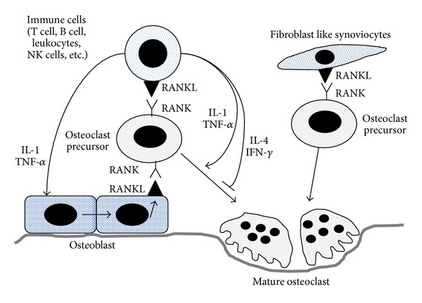
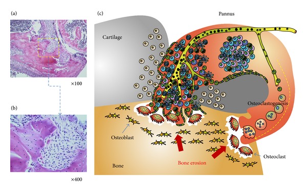
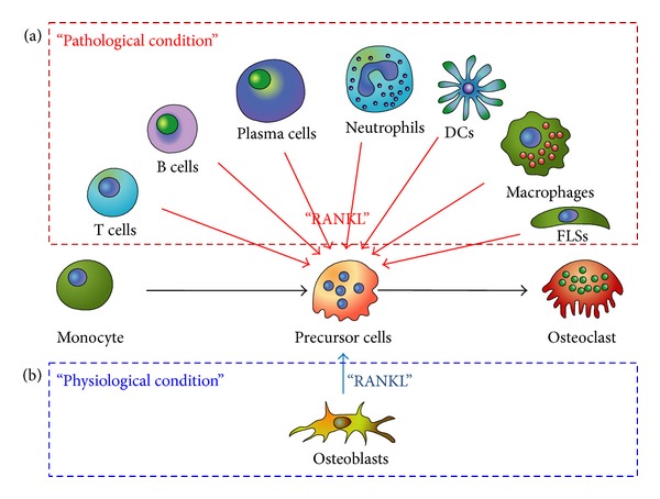
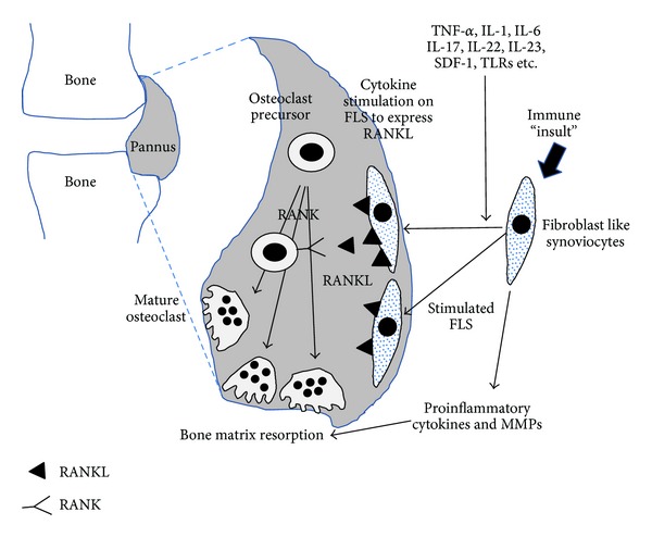
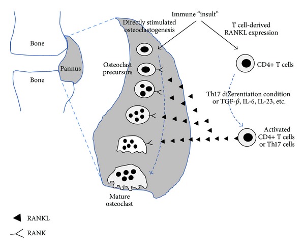
Bone homeostasis, which involves formation and resorption, is an important process for maintaining adequate bone mass in humans. Rheumatoid arthritis (RA) is an autoimmune disease characterized by inflammation and bone loss, leading to joint destruction and deformity, and is a representative disease of disrupted bone homeostasis. The bone loss and joint destruction are mediated by immunological insults by proinflammatory cytokines and various immune cells. The connection between bone and immunity has been intensely studied and comprises the emerging field of osteoimmunology. Osteoimmunology is an interdisciplinary science investigating the interplay between the skeletal and the immune systems. The main contributors in osteoimmunology are the bone effector cells, such as osteoclasts or osteoblasts, and the immune cells, particularly lymphocytes and monocytes. Physiologically, osteoclasts originate from immune cells, and immune cells regulate osteoblasts and vice versa. Pathological conditions such as RA might affect these interactions, thereby altering bone homeostasis, resulting in the unfavorable outcome of bone destruction. In this review, we describe the osteoclastogenic roles of the proinflammatory cytokines and immune cells that are important in the pathophysiology of RA.


Figures

Figure 1
Figure 1
Osteoblast-derived RANKL binds to RANK on monocytes to differentiate them into mature osteoclasts. Osteoblast-derived RANKL plays important role in generating osteoclast in physiological condition. However, immune cell and FLS-derived RANKL play pathogenic role in RA. Proinflammatory cytokines such as IL-1 and TNFα effectively stimulate osteoblast to express RANKL. FLS-derived RANKL enhances osteoclastogenesis in RA joints. RANK: receptor activator of the nuclear factor kappa B; RANKL: receptor activator of the nuclear factor kappa B ligand; FLS: fibroblast like synoviocyte; RA: rheumatoid arthritis; IL-1: Interleukin-1; TNFα: tumor necrosis factor-alpha.
Figure 2
Figure 2
(a) Bone is destroyed by a proliferative and invasive synovium, which is called pannus. It originates from adjacent synovial tissue and invades the cartilages and bones. (b) Magnified view of the pannus-bone interface. The pannus-bone interface is lined with mature osteoclasts (arrows). Various inflammatory cells and stromal cells comprise the invading pannus. (c) Schematic depiction of the pannus-cartilage-bone structure. Inflammatory cells such as B cells, T cells, macrophages, monocytes, and fibroblast-like synoviocytes accumulate in the pannus. For metabolic support, intensive angiogenesis is usually followed. Excessive provision of RANKL from the accumulated cells in the pannus enhances osteoclastogenesis, resulting in the erosion of bone at the pannus-bone interface.
Figure 3
Figure 3
(a) Monocytes are differentiated into mature osteoclasts by the aid of RANKL. In pathological conditions such as inflammation, cancer, and hypermetabolism, various cells extraordinarily provide RANKL to the monocytes, resulting in overweighed osteoclastogenesis. In this condition, the osteoclasts outnumber the osteoblasts, disrupting the bone homeostasis. Bone erosion or osteoporosis is the major outcome of disrupted homeostasis. (b) In normal physiological conditions, a few cells, predominantly osteoblasts, express RANKL. A similar number of osteoclasts and osteoblasts maintain the bone mass by homeostatic equilibrium.
Figure 4
Figure 4
FLS could express RANKL in response to various stimuli. FLS-expressed RANKL enhances osteoclastogenesis and results in bone erosion in RA. Inflammatory and immune stimulation induce the FLS to produce proinflammatory cytokines and matrix metalloproteinase. These cytokines and enzymes aid osteoclasts to destroy the bone matrix.
Figure 5
Figure 5
T cells are activated to produce RANKL or osteoclastogenic cytokines by various stimuli. RANKL and activated T cell-cytokines have the potential to induce osteoclastogenesis. With T cells, the outnumbered osteoclasts destroy the bone in RA.

References

    1. Nakashima T, Kobayashi Y, Yamasaki S, et al. Protein expression and functional difference of membrane-bound and soluble receptor activator of NF-κB ligand: modulation of the expression by osteotropic factors and cytokines. Biochemical and Biophysical Research Communications. 2000;275(3):768–775. - PubMed
    1. Takayanagi H, Kim S, Taniguchi T. Signaling crosstalk between RANKL and interferons in osteoclast differentiation. Arthritis Research & Therapy. 2002;4(supplement 3):S227–S232. - PMC - PubMed
    1. Kotake S, Nanke Y, Yago T, Kawamoto M, Yamanaka H. Human osteoclastogenic T cells and human osteoclastology. Arthritis and Rheumatism. 2009;60(11):3158–3163. - PubMed
    1. Gao Y, Grassi F, Ryan MR, et al. IFN-γ stimulates osteoclast formation and bone loss in vivo via antigen-driven T cell activation. The Journal of Clinical Investigation. 2007;117(1):122–132. - PMC - PubMed
    1. Janssens K, ten Dijke P, Janssens S, van Hul W. Transforming growth factor-β1 to the bone. Endocrine Reviews. 2005;26(6):743–774. - PubMed

Publication types

MeSH terms