. 2014 Oct 8;9(10):e109201.
doi: 10.1371/journal.pone.0109201. eCollection 2014.

Antinematode activity of Violacein and the role of the insulin/IGF-1 pathway in controlling violacein sensitivity in Caenorhabditis elegans

Affiliations

Antinematode activity of Violacein and the role of the insulin/IGF-1 pathway in controlling violacein sensitivity in Caenorhabditis elegans

Francesco Ballestriero et al. PLoS One. .

Erratum in

Abstract

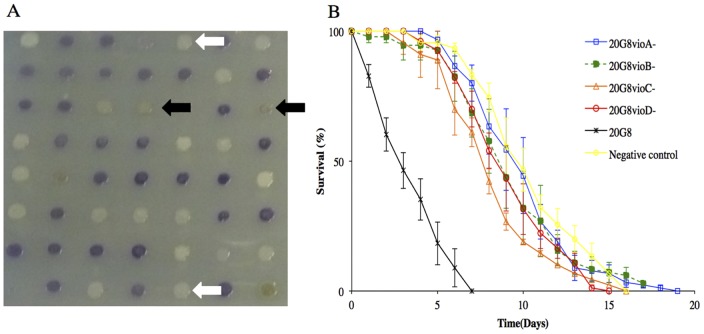
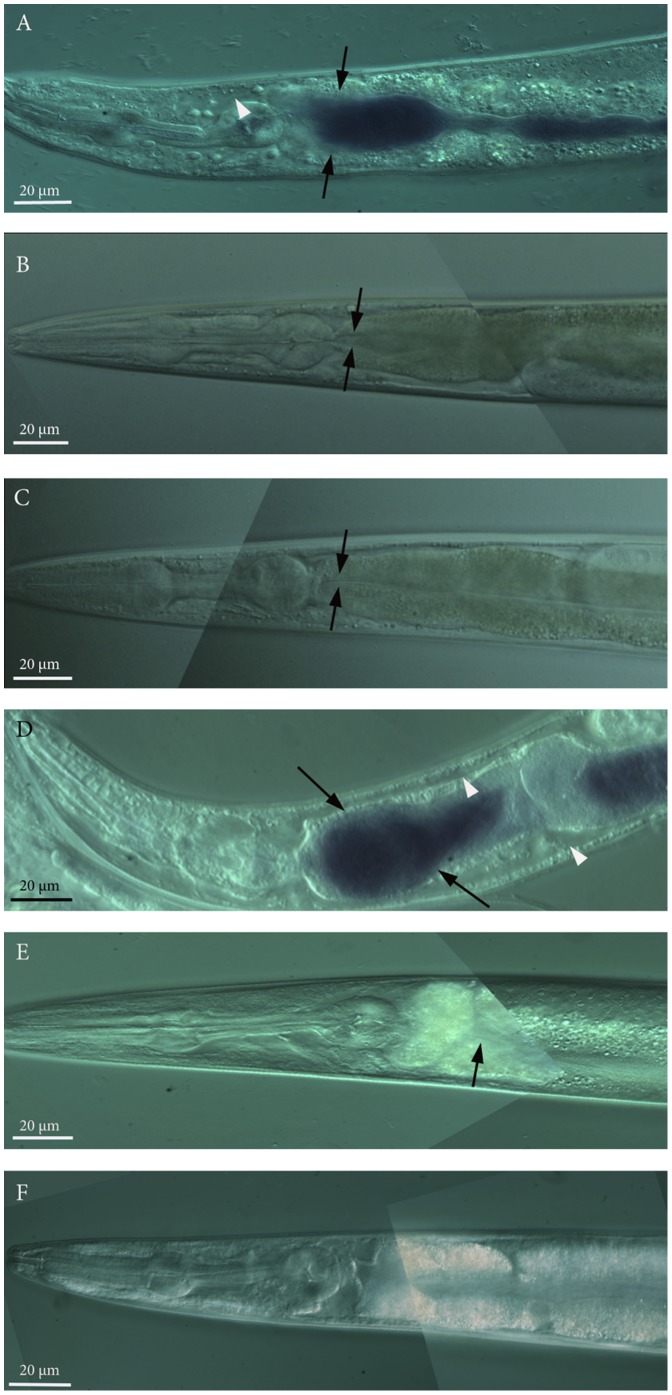
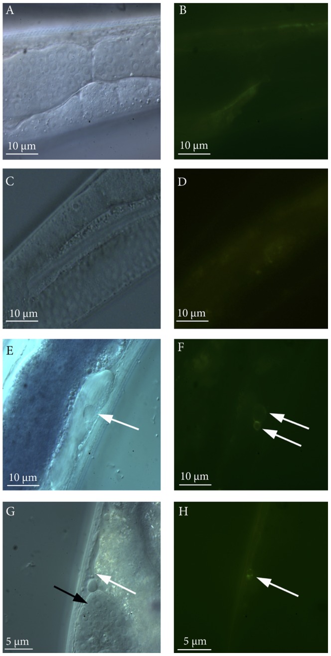
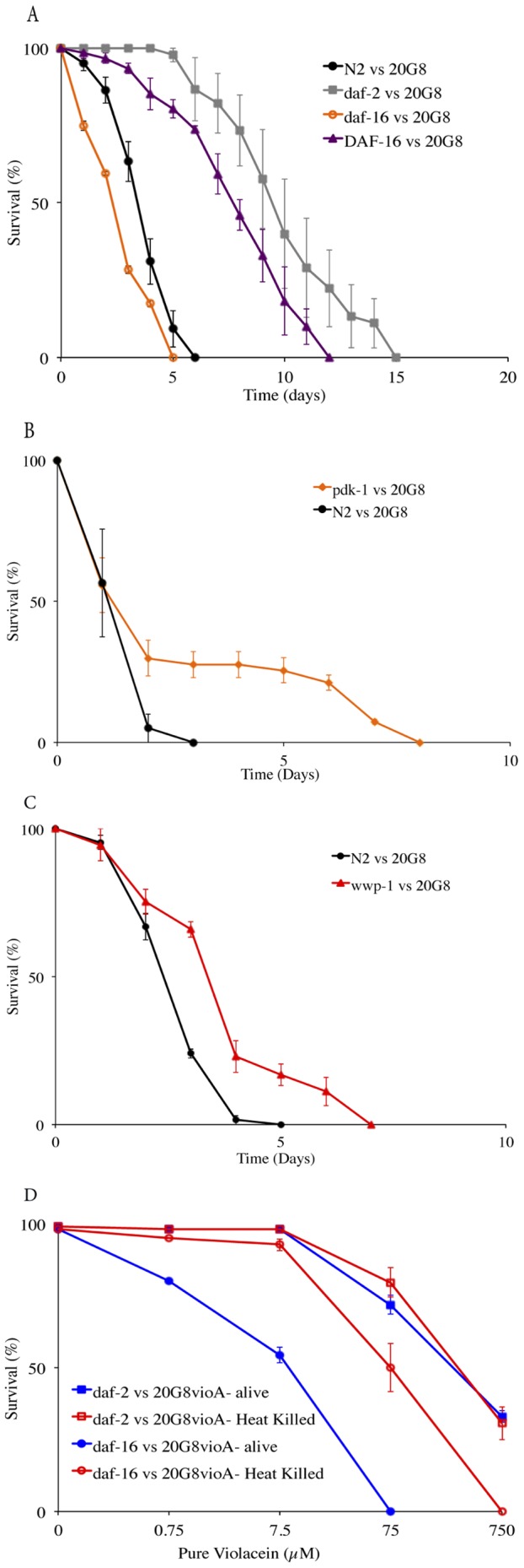
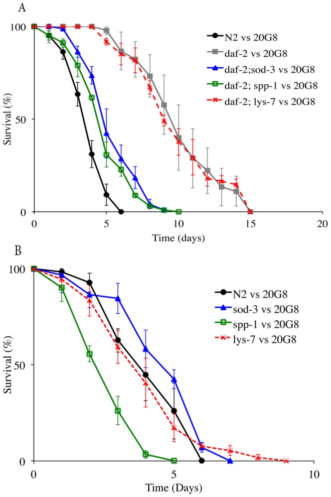
The purple pigment violacein is well known for its numerous biological activities including antibacterial, antiviral, antiprotozoan, and antitumor effects. In the current study we identify violacein as the antinematode agent produced by the marine bacterium Microbulbifer sp. D250, thereby extending the target range of this small molecule. Heterologous expression of the violacein biosynthetic pathway in E. coli and experiments using pure violacein demonstrated that this secondary metabolite facilitates bacterial accumulation in the nematode intestine, which is accompanied by tissue damage and apoptosis. Nematodes such as Caenorhabditis elegans utilise a well-defined innate immune system to defend against pathogens. Using C. elegans as a model we demonstrate the DAF-2/DAF-16 insulin/IGF-1 signalling (IIS) component of the innate immune pathway modulates sensitivity to violacein-mediated killing. Further analysis shows that resistance to violacein can occur due to a loss of DAF-2 function and/or an increased function of DAF-16 controlled genes involved in antimicrobial production (spp-1) and detoxification (sod-3). These data suggest that violacein is a novel candidate antinematode agent and that the IIS pathway is also involved in the defence against metabolites from non-pathogenic bacteria.


Conflict of interest statement

Competing Interests: The authors have declared that no competing interests exist.

Figures

Figure 1
Figure 1. Characterization of the transposon mutant library of violacein producing clone 20G8.
(A) Several mutants exhibit changes in violet pigmentation typical of the presence of violacein: 20G8vioA and 20G8vioB mutants have lost pigmentation (white arrows). 20G8vioC and 20G8vioD mutants express green-grey pigmentation (black arrows). (B) Nematode killing assay (N2 animals vs bacterial strains 20G8 clone and 20G8vioABCD mutants). Survival kinetics of nematodes fed with either the 20G8 clone or the 20G8vioABCD mutants deficient in violacein production. The killing phenotype of the four mutant clones is significantly reduced (p<0.0001) when compared to the wild type clone 20G8. A randomly chosen clone from the library with no activity is used as a negative control. Each data point represents means ± the standard error of three replicate plates. p values were calculated on the pooled data of all of the plates in each experiment by using the log-rank (Mantel–Cox) method.
Figure 2
Figure 2. Nematode killing assay and dose response assay (N2 animal vs the alive and heat killed 20G8 clone and D250 strains).
(A) Killing kinetics of Microbulbifer sp. D250 and dV2 mutant deficient in violacein production. Negative control OP50 is a non-pathogenic strain of E. coli. (B) Dose response of C. elegans to pure violacein added to 20G8vioA mutant bacteria alive and heat killed. Each point on the graph represents the average survival of worms after seven days exposure to violacein (C) Kinetics of nematode killing when nematodes are fed either the live or heat killed 20G8 clone or 20G8vioA mutant. Each data point represents means ± the standard error of three replicate plates. p values were calculated on the pooled data of all of the plates in each experiment by using the log-rank (Mantel–Cox) method.
Figure 3
Figure 3. Visualization of bacterial accumulation in the nematode intestine by the E. coli clones (wild type animals, the daf-2 and daf-16 null mutant animals and the DAF-16 overexpressing nematodes vs the E. coli clone 20G8 and 20G8vioA and 20G8vioC mutant clones).
All images present C. elegans anterior to the left and show the pharynx and first part of the intestinal lumen by differential interference contrast (DIC) microscopy. Accumulation by 20G8 cells in the nematodes intestine (arrows in panels A and D) and extensive enlargement of extracellular regions (white arrowheads panels A and D) in N2 wild type (panel A) and daf-16 mutant (panel D) animals. No change in phenotype was observed in N2 nematodes fed with violacein deficient mutant clones 20G8vioA (negative control-panel B) and 20G8vioC (panel C). Similarly, no bacterial cells or enlargement of extracellular regions were detected in daf-2 and DAF-16 over-expressing mutant nematodes exposed to the violacein producing clone 20G8 (arrows in panel E and F respectively). Each panel was assembled from multiple photomicrographs taken with the same magnification and same acquisition settings.
Figure 4
Figure 4. Apoptosis analysis in CED-1::GFP transgenic nematode strain CU1546.
Visualization of GFP apoptosis marker inside the nematodes exposed to the violacein deficient mutant clones (20G8vioA panels A and B; 20G8vioC panels C and D) or violacein producing clone (20G8, panels E and F and G and H). Images show a section of the intestine and the gonads of C. elegans by DIC (panels A, C, E, G) and by epifluorescence microscopy (panels B, D, F, H). A ring-shaped GFP signal was detected in somatic cells of CU1546 nematodes exposed to 20G8 clone (two white arrows in panel F and white arrow in panel H) but not to 20G8vioA and 20G8vioC mutants (panel B and D). The ring-shaped GFP signal is associated with the expression of CED-1 receptor on the cell wall of engulfing cells only when phagocytosis of apoptotic cells is taking place. The ring-shaped GFP signal is apoptosis-specific and is visible in somatic cells of the nematodes (white arrows in E and G), the germ line (gonad) of the nematode is shown in panel G (black arrow). A non-specific fluorescence signal is visible in panels B and D.
Figure 5
Figure 5. Nematode killing assay and dose response assay (wild type animals and daf-2, daf-16, pdk-1, wwp-1 and DAF-16 mutant nematodes vs 20G8 clone).
The survival of nematodes fed with 20G8 clone was measured for (A) daf-2, daf-16 and DAF-16 mutant animals. (B) pdk-1 mutant animals (C) wwp-1 mutant animals (D) Dose response of C. elegans daf-2 and daf-16 strains to pure violacein added to 20G8vioA mutant bacteria alive and heat killed. Each data point represents means ± the standard error of three replicate plates p values were calculated on the pooled data of all of the plates in each experiment by using a Students t-test on the number of surviving nematodes at day seven.
Figure 6
Figure 6. Nematode killing assay (wild type animals and daf-2, daf-2;spp-1, daf-2;lys-7, daf-2;sod-3, spp-1, lys-7, sod-3 mutant nematodes vs the 20G8 clone).
(A) The survival of the nematode was tested using C. elegans double mutants daf-2;sod-3, daf-2;spp-1, daf-2;lys-7 and (B) the single mutant animals sod-3, spp-1, and lys-7. Each data point represents means ± the standard error of three replicate plates. p values were calculated on the pooled data of all of the plates done in each experiment by using the log-rank (Mantel–Cox) method and the values are provide in the text.

References

    1. Holden-Dye L, Walker RJ (2007) Anthelmintic drugs. WormBook 2: 1–13. - PMC - PubMed
    1. Kaplan RM (2004) Drug resistance in nematodes of veterinary importance: a status report. Trends Parasitol 20: 477–481. - PubMed
    1. Prichard RK, Geary TG (2008) Drug discovery: Fresh hope to can the worms. Nature 452: 157–158. - PubMed
    1. Jones AK, Buckingham SD, Sattelle DB (2005) Chemistry-to-gene screens in Caenorhabditis elegans . Nat Rev Drug Discov 4: 321–330. - PubMed
    1. Sifri CD, Begun J, Ausubel FM (2005) The worm has turned-microbial virulence modeled in Caenorhabditis elegans . Trends Microbiol 13: 119–127. - PubMed

MeSH terms