

Establishment of Myotis myotis cell lines--model for investigation of host-pathogen interaction in a natural host for emerging viruses
- PMID: 25295526
- PMCID: PMC4190323
- DOI: 10.1371/journal.pone.0109795
Establishment of Myotis myotis cell lines--model for investigation of host-pathogen interaction in a natural host for emerging viruses
Abstract
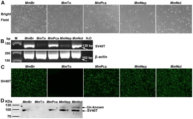
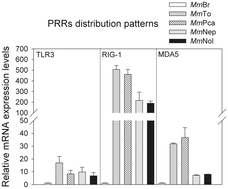
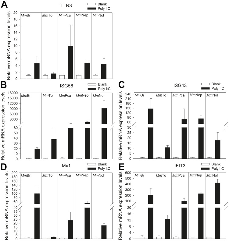
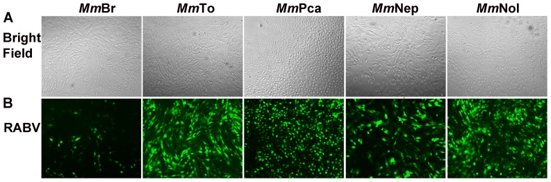
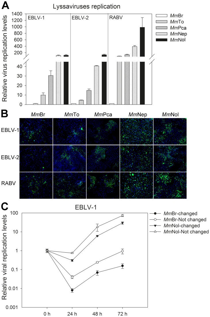
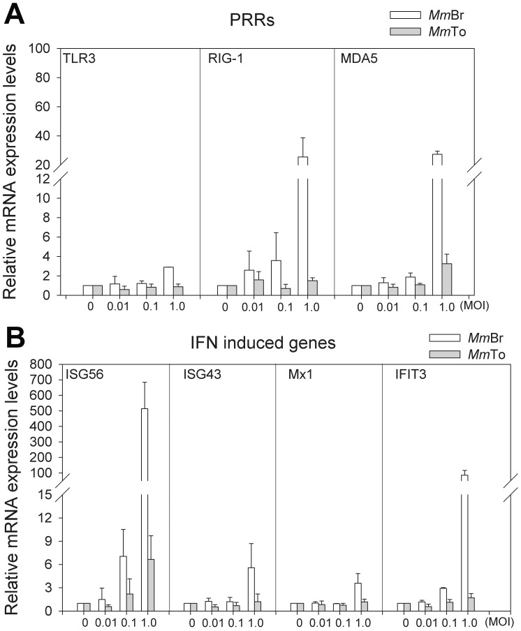
Bats are found to be the natural reservoirs for many emerging viruses. In most cases, severe clinical signs caused by such virus infections are normally not seen in bats. This indicates differences in the virus-host interactions and underlines the necessity to develop natural host related models to study these phenomena. Due to the strict protection of European bat species, immortalized cell lines are the only alternative to investigate the innate anti-virus immune mechanisms. Here, we report about the establishment and functional characterization of Myotis myotis derived cell lines from different tissues: brain (MmBr), tonsil (MmTo), peritoneal cavity (MmPca), nasal epithelium (MmNep) and nervus olfactorius (MmNol) after immortalization by SV 40 large T antigen. The usefulness of these cell lines to study antiviral responses has been confirmed by analysis of their susceptibility to lyssavirus infection and the mRNA patterns of immune-relevant genes after poly I:C stimulation. Performed experiments indicated varying susceptibility to lyssavirus infection with MmBr being considerably less susceptible than the other cell lines. Further investigation demonstrated a strong activation of interferon mediated antiviral response in MmBr contributing to its resistance. The pattern recognition receptors: RIG-I and MDA5 were highly up-regulated during rabies virus infection in MmBr, suggesting their involvement in promotion of antiviral responses. The presence of CD14 and CD68 in MmBr suggested MmBr cells are microglia-like cells which play a key role in host defense against infections in the central nervous system (CNS). Thus the expression pattern of MmBr combined with the observed limitation of lyssavirus replication underpin a protective mechanism of the CNS controlling the lyssavirus infection. Overall, the established cell lines are important tools to analyze antiviral innate immunity in M. myotis against neurotropic virus infections and present a valuable tool for a broad spectrum of future investigations in cellular biology of M. myotis.
Conflict of interest statement
Figures
Similar articles
-
Transcriptomic responses of bat cells to European bat lyssavirus 1 infection under conditions simulating euthermia and hibernation.BMC Immunol. 2023 Apr 21;24(1):7. doi: 10.1186/s12865-023-00542-7. BMC Immunol. 2023. PMID: 37085747 Free PMC article.
-
Accelerated viral dynamics in bat cell lines, with implications for zoonotic emergence.Elife. 2020 Feb 3;9:e48401. doi: 10.7554/eLife.48401. Elife. 2020. PMID: 32011232 Free PMC article.
-
Evolution and Antiviral Specificities of Interferon-Induced Mx Proteins of Bats against Ebola, Influenza, and Other RNA Viruses.J Virol. 2017 Jul 12;91(15):e00361-17. doi: 10.1128/JVI.00361-17. Print 2017 Aug 1. J Virol. 2017. PMID: 28490593 Free PMC article.
-
Tools to study pathogen-host interactions in bats.Virus Res. 2018 Mar 15;248:5-12. doi: 10.1016/j.virusres.2018.02.013. Epub 2018 Feb 15. Virus Res. 2018. PMID: 29454637 Free PMC article. Review.
-
Exploring the Role of Innate Lymphocytes in the Immune System of Bats and Virus-Host Interactions.Viruses. 2022 Jan 14;14(1):150. doi: 10.3390/v14010150. Viruses. 2022. PMID: 35062356 Free PMC article. Review.
Cited by
-
Temperature sensitivity of bat antibodies links metabolic state of bats with antigen-recognition diversity.Nat Commun. 2024 Jul 13;15(1):5878. doi: 10.1038/s41467-024-50316-x. Nat Commun. 2024. PMID: 38997292 Free PMC article.
-
Utility of primary cells to examine NPC1 receptor expression in Mops condylurus, a potential Ebola virus reservoir.PLoS Negl Trop Dis. 2020 Jan 21;14(1):e0007952. doi: 10.1371/journal.pntd.0007952. eCollection 2020 Jan. PLoS Negl Trop Dis. 2020. PMID: 31961874 Free PMC article.
-
Transcriptomic responses of bat cells to European bat lyssavirus 1 infection under conditions simulating euthermia and hibernation.BMC Immunol. 2023 Apr 21;24(1):7. doi: 10.1186/s12865-023-00542-7. BMC Immunol. 2023. PMID: 37085747 Free PMC article.
-
Generation and Characterization of Eptesicus fuscus (Big brown bat) kidney cell lines immortalized using the Myotis polyomavirus large T-antigen.J Virol Methods. 2016 Nov;237:166-173. doi: 10.1016/j.jviromet.2016.09.008. Epub 2016 Sep 14. J Virol Methods. 2016. PMID: 27639955 Free PMC article.
-
Extensive longevity and DNA virus-driven adaptation in nearctic Myotis bats.bioRxiv [Preprint]. 2024 Nov 27:2024.10.10.617725. doi: 10.1101/2024.10.10.617725. bioRxiv. 2024. PMID: 39416019 Free PMC article. Preprint.
References
-
- Simmons NB, Seymour KL, Habersetzer J, Gunnell GF (2008) Primitive Early Eocene bat from Wyoming and the evolution of flight and echolocation. Nature 451: 818–821. - PubMed
Publication types
MeSH terms
LinkOut - more resources
Full Text Sources
Other Literature Sources
Research Materials
