. 2014 Nov;124(11):4989-5001.
doi: 10.1172/JCI76042. Epub 2014 Oct 8.

TRPM2 mediates ischemic kidney injury and oxidant stress through RAC1

TRPM2 mediates ischemic kidney injury and oxidant stress through RAC1

Guofeng Gao et al. J Clin Invest. 2014 Nov.

Abstract

Ischemia is a leading cause of acute kidney injury. Kidney ischemia is associated with loss of cellular ion homeostasis; however, the pathways that underlie ion homeostasis dysfunction are poorly understood. Here, we evaluated the nonselective cation channel transient receptor potential melastatin 2 (TRPM2) in a murine model of kidney ischemia/reperfusion (I/R) injury. TRPM2-deficient mice were resistant to ischemic injury, as reflected by improved kidney function, reduced histologic damage, suppression of proapoptotic pathways, and reduced inflammation. Moreover, pharmacologic TRPM2 inhibition was also protective against I/R injury. TRPM2 was localized mainly in kidney proximal tubule epithelial cells, and studies in chimeric mice indicated that the effects of TRPM2 are due to expression in parenchymal cells rather than hematopoietic cells. TRPM2-deficient mice had less oxidative stress and lower levels of NADPH oxidase activity after ischemia. While RAC1 is a component of the NADPH oxidase complex, its relation to TRPM2 and kidney ischemic injury is unknown. Following kidney ischemia, TRPM2 promoted RAC1 activation, with active RAC1 physically interacting with TRPM2 and increasing TRPM2 expression at the cell membrane. Finally, inhibition of RAC1 reduced oxidant stress and ischemic injury in vivo. These results demonstrate that TRPM2-dependent RAC1 activation increases oxidant stress and suggest that therapeutic approaches targeting TRPM2 and/or RAC1 may be effective in reducing ischemic kidney injury.


Figures

Figure 8
Figure 8. Pharmacological blockade of RAC1 protects against ischemic AKI.
(A) WT (circles) or MDA (squares) mice were pretreated with RAC1 inhibitor NSC23766 (NSC) (black symbols) or vehicle (Veh) (white symbols) and subjected to bilateral kidney ischemia. BUN and serum creatinine levels prior to (BL) and 24 hours after I/R.(B) Western blot analysis of kidney lysates from WT and Trpm2-KO mice 24 hours after I/R using Abs against cleaved PARP, BCL-2, caspase 3, and GAPDH. (C) Densitometric analysis of Western blotting of kidney lysates using anti–4-HNE. (D) TBARs (MDA) in kidney tissues from Trpm2-KO and WT mice. (E) NADPH oxidase activity in WT mice subjected to sham or I/R in the presence or absence of NSC23766. *P < 0.05; **P < 0.01; ***P < 0.001.
Figure 7
Figure 7. H2O2 increases membrane expression of TRPM2 by activation of RAC1.
HEK293 cells were transiently transfected with V5-TRPM2 and either Myc-RAC1-WT, Myc-RAC1-CA, or Myc-RAC1-DN and then treated with H2O2 or vehicle. After H2O2 treatment, cell surface biotinylation was performed. Biotinylated proteins were isolated using streptavidin beads followed by Western blotting with anti-V5 Ab to identify biotinylated TRPM2 (top panel). Total TRPM2 and RAC1 content in the cell lysate is shown in the lower panels.
Figure 6
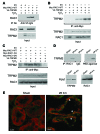
Figure 6. TRPM2 interacts with active RAC1.
(A) HEK293 cells were transiently transfected with TRPM2 with an N-terminal V5 Tag and RAC1 with an N-terminal Myc Tag. Top panel: IP of TRPM2 using anti-V5 and immunoblot of RAC1 using anti-Myc Ab. Bottom panels: Equal expression of transfected Myc-RAC1 and V5-TRPM2 in the cell lysates. (B) Transfected cells were treated with H2O2 prior to lysis. Lysates were subjected to IP of RAC1 using anti-Myc Ab followed by immunoblotting for TRPM2. (C) HEK293 cells were transiently transfected with V5-TRPM2 and either Myc-RAC1-WT, Myc-RAC1-CA, or Myc-RAC1-DN. After H2O2 treatment, cells were lysed, followed by IP with anti-Myc Ab and immunoblotting with anti-V5 Ab. EV, empty vector. (D) Pull-down of active RAC1 from kidneys subjected to sham surgery or I/R using a RAC1 Ab (left) or PBD agarose (right), with immunoblotting for TRPM2. (E) Merged confocal images of TRPM2 (red) and RAC1 (green) in kidneys subjected to sham or I/R surgery. Arrowheads denote areas of overlap (yellow). Scale bars: 10 μm.
Figure 5
Figure 5. Reduction of oxidative stress and RAC1 activation in Trpm2-KO mice.
(A) Western blot analysis of 4-HNE adducts in kidney lysates from Trpm2-KO and WT mice subjected to bilateral kidney ischemia. The same gel used in Figure 4C was reprobed with a 4-HNE Ab. (B) NADPH oxidase activity in kidney lysates from WT and Trpm2-KO mice subjected to bilateral kidney ischemia followed by 6 hours of reperfusion. ***P < 0.001. (C) RAC1 activity in kidney lysates from WT and Trpm2-KO mice subjected to ischemia or sham surgery. (D) RAC1 activity in primary kidney PTCs after H2O2 treatment.
Figure 4
Figure 4. Reduction of epithelial cell apoptosis in Trpm2-KO mice.
(A) TUNEL staining of kidney sections from Trpm2-KO and WT mice subjected to bilateral kidney ischemia or sham surgery. (B) Quantitation of apoptotic cells. ***P < 0.001. Scale bars: 50 μm. (C) Western blot analysis of kidney lysates from Trpm2-KO and WT mice subjected to bilateral kidney ischemia or sham surgery to detect pro- and antiapoptotic proteins. (D) Western blot analysis of PARP cleavage as a measure of apoptosis.
Figure 3
Figure 3. Parenchymal TRPM2 mediates ischemic AKI.
(A) Chimeric mice were generated and subjected to kidney ischemia or sham surgery. Blood collected 24 hours after I/R was analyzed for BUN and serum creatinine. *P < 0.001 versus WT-WT. (B) PAS-stained kidney tissue sections. Scale bars: 50 μm. (C) Cell viability of primary cultured PTCs from WT or KO mice subjected to hypoxia/reoxygenation in vitro. *P < 0.01 versus normoxia.
Figure 2
Figure 2. Deletion or pharmacologic inhibition of TRPM2 reduces ischemic AKI.
(A) Trpm2-KO and WT mice were subjected to 28 minutes of bilateral kidney ischemia or sham surgery. Blood collected 24 hours after surgery was analyzed for BUN and serum creatinine as measures of kidney function. (B) Serum creatinine in WT and Trpm2-KO mice were pretreated with 2-APB or vehicle and then subjected to bilateral kidney ischemia. (C) PAS-stained kidney tissue sections from Trpm2-KO and WT mice subjected to kidney ischemia or sham surgery. Scale bars: 50 μm. (D) FACS analysis of neutrophils in kidneys from Trpm2-KO and WT mice subjected to kidney ischemia or sham surgery. Numbers denote the percentage of CD45 cells that were also Ly6G+. (E) Quantification of tubular injury, NGAL expression, and neutrophil infiltration. *P < 0.05; **P < 0.01.
Figure 1
Figure 1. Localization of TRPM2 in the kidney.
Top row: Kidney sections were labeled with a TRPM2 Ab (red), DAPI (blue), and LTL (green), a marker of proximal tubules. Bottom row: Confocal images of TRPM2 (red) and DAPI-stained sections from kidneys harvested after sham surgery or 6 and 24 hours after I/R. Sections from TRPM2-deficient mice (top right) or sections stained without incubation with the TRPM2 Ab (bottom right) served as negative controls. Scale bars: 50 μm (top panels), 10 μm (bottom panels).

Similar articles

Cited by

References

    1. Nash K, Hafeez A, Hou S. Hospital-acquired renal insufficiency. Am J Kidney Dis. 2002;39(5):930–936. doi: 10.1053/ajkd.2002.32766. - DOI - PubMed
    1. Kosieradzki M, RowiDski W. Ischemia/reperfusion injury in kidney transplantation: mechanisms and prevention. Transplant Proc. 2008;40(10):3279–3288. doi: 10.1016/j.transproceed.2008.10.004. - DOI - PubMed
    1. Mason J, Beck F, Dorge A, Rick R, Thurau K. Intracellular electrolyte composition following renal ischemia. Kidney Int. 1981;20(1):61–70. doi: 10.1038/ki.1981.105. - DOI - PubMed
    1. Beck FX, Ohno A, Dorge A, Thurau K. Ischemia-induced changes in cell element composition and osmolyte contents of outer medulla. Kidney Int. 1995;48(2):449–457. doi: 10.1038/ki.1995.313. - DOI - PubMed
    1. Leaf A. Cell swelling: A factor in ischemic tissue injury. Circulation. 1973;48(3):455–458. doi: 10.1161/01.CIR.48.3.455. - DOI - PubMed

Publication types

MeSH terms