. 2014 Nov;124(11):4965-75.
doi: 10.1172/JCI75660. Epub 2014 Oct 8.

Skin exposure promotes a Th2-dependent sensitization to peanut allergens

Skin exposure promotes a Th2-dependent sensitization to peanut allergens

Leticia Tordesillas et al. J Clin Invest. 2014 Nov.

Abstract

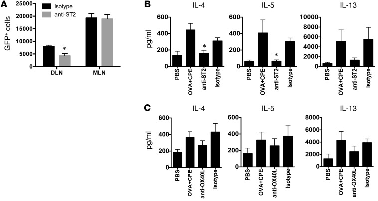
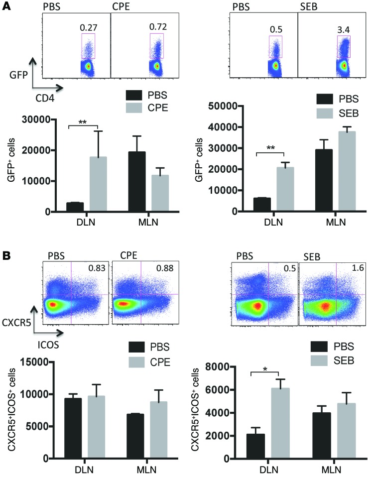
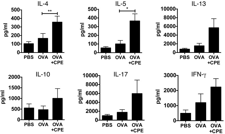
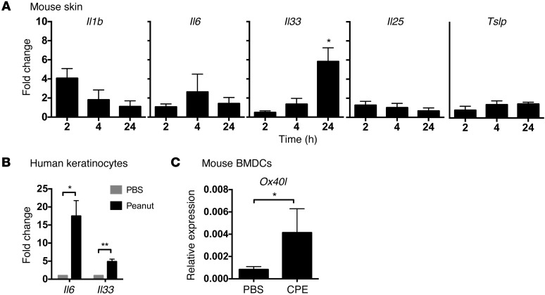
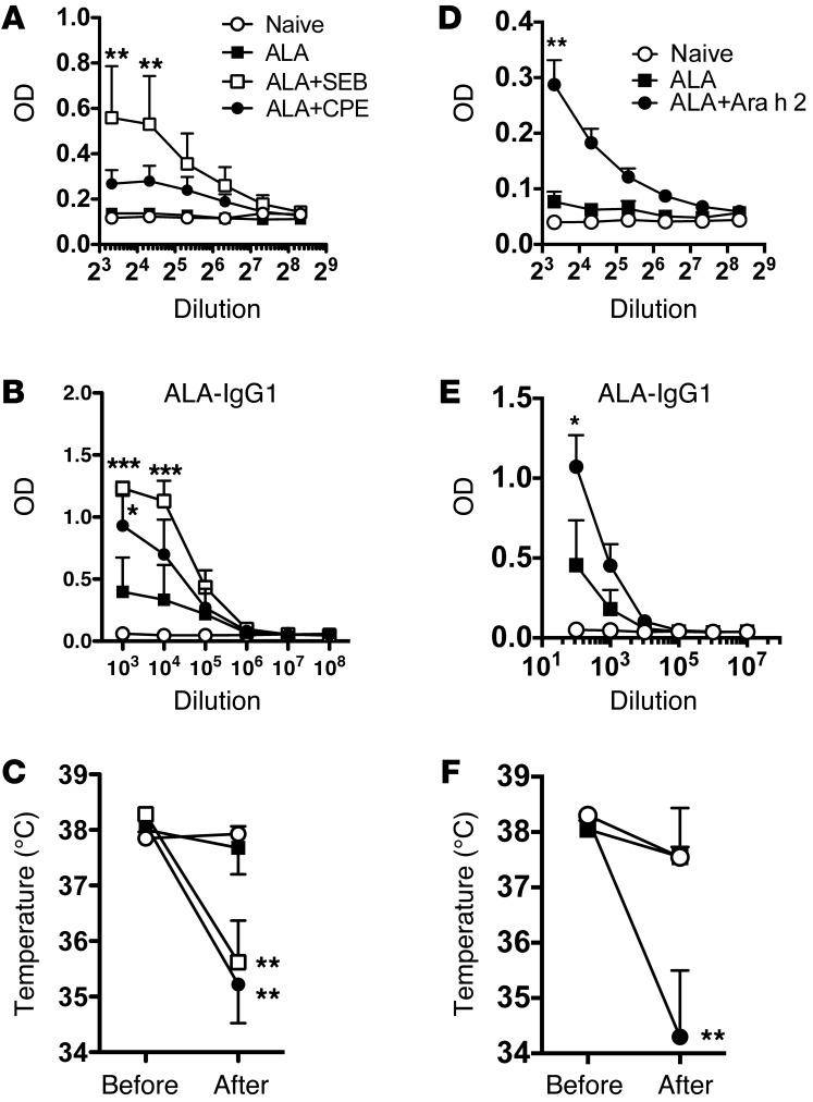
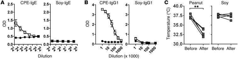
Sensitization to foods often occurs in infancy, without a known prior oral exposure, suggesting that alternative exposure routes contribute to food allergy. Here, we tested the hypothesis that peanut proteins activate innate immune pathways in the skin that promote sensitization. We exposed mice to peanut protein extract on undamaged areas of skin and observed that repeated topical exposure to peanut allergens led to sensitization and anaphylaxis upon rechallenge. In mice, this epicutaneous peanut exposure induced sensitization to the peanut components Ara h 1 and Ara h 2, which is also observed in human peanut allergy. Both crude peanut extract and Ara h 2 alone served as adjuvants, as both induced a bystander sensitization that was similar to that induced by the atopic dermatitis-associated staphylococcal enterotoxin B. In cultured human keratinocytes and in murine skin, peanut extract directly induced cytokine expression. Moreover, topical peanut extract application induced an alteration dependent on the IL-33 receptor ST2 in skin-draining DCs, resulting in Th2 cytokine production from T cells. Together, our data support the hypothesis that peanuts are allergenic due to inherent adjuvant activity and suggest that skin exposure to food allergens contributes to sensitization to foods in early life.


Figures

Figure 7
Figure 7. Peanut induces Th2 polarization through ST2-mediated effects on skin-draining DCs.
4get mice were pretreated with either isotype control or neutralizing ST2 antibodies before epicutaneous exposure to CPE. One week after exposure, draining lymph nodes and mesenteric lymph nodes were harvested, and (A) IL-4 reporter activity was quantified. Summary data (mean ± SEM) showing number of cells per million CD4+ T cells are shown. n = 3 mice per condition. *P < 0.05. (B) BALB/c mice were pretreated with isotype control or anti-ST2 antibody prior to exposure to PBS or OVA and CPE. DCs from skin-draining lymph nodes were cocultured with DO11.10 T cells, and cytokine output was measured. Data are mean ± SEM of 3 independent experiments. *P < 0.05, compared with isotype control. (C) DCs from skin-draining lymph nodes were isolated after exposure to OVA and CPE and cocultured with DO11.10 T cells in the presence of anti-OX40L or isotype control antibody. Data are mean ± SEM of 4 independent experiments.
Figure 6
Figure 6. Contribution of IL-1, IL-6, and IL-33 to Th2 and Tfh induction by SEB.
4get mice were pretreated with either isotype control or (A and B) neutralizing IL-6 antibodies plus IL-1 receptor antagonist or (C and D) neutralizing anti-ST2 antibody before epicutaneous exposure to SEB. Draining lymph nodes and mesenteric lymph nodes were harvested 1 week later. Summary plots of (A and C) IL-4+ cells or (B and D) CXCR5+ICOS+ cells per million CD4+ T cells are shown. Data are mean ± SEM of 3 mice per condition. *P < 0.05, **P < 0.01.
Figure 5
Figure 5. Effect of CPE or SEB on Th2 and Tfh cells.
4get mice were epicutaneously exposed to PBS, peanut (CPE), or SEB. One week later, skin-draining lymph nodes (DLN) and mesenteric lymph nodes (MLN) were assessed for (A) IL-4 reporter expression or (B) Tfh markers CXCR5 and ICOS. Representative plots of IL-4+ or CXCR5+ICOS+ cells of total CD4+ cells in skin-draining lymph nodes are shown above summary plots of the mean ± SEM number of cells normalized per million CD4+ T cells for 3 mice per condition. *P < 0.05, **P < 0.01.
Figure 4
Figure 4. Effect of in vivo exposure to CPE on priming by DCs.
DCs were purified from the skin-draining lymph nodes of BALB/c mice epicutaneously exposed to PBS, OVA, or OVA and CPE and cocultured with DO11.10 CD4+ T cells for 3 days, followed by restimulation with CD3/CD28. Data are mean ± SEM of 7 independent experiments, with each experiment consisting of 3 pooled mice per group. *P < 0.05, **P < 0.01.
Figure 3
Figure 3. Innate response to epicutaneous peanut exposure.
(A) Mice were exposed to peanut or PBS as control on the ear, followed by RNA isolation and RT-PCR for cytokines. (B) Human keratinocytes or (C) mouse bone marrow DCs were exposed in vitro to CPE or PBS as control. Fold change is shown with respect to PBS. Relative expression refers to data normalized to the housekeeping gene. Data are mean ± SEM of a least 3 different experiments. *P < 0.05, **P < 0.01.
Figure 2
Figure 2. Peanut and Ara h 2 induce bystander sensitization, demonstrating adjuvant activity.
(AC) Mice were exposed to 0.1 mg ALA on the ear, alone, or in the presence of 10 μg SEB or 1 mg CPE once a week for 6 weeks. ALA-specific (A) IgE and (B) IgG1 were measured in serum prior to oral challenge with ALA. (C) Body temperature was measured at baseline and 30 minutes after challenge. n = 5 mice per group. (DF) The experiment was repeated with exposure to ALA in the presence or absence of 0.1 mg purified Ara h 2. n = 4 per group. *P < 0.05, **P < 0.01, ***P < 0.001, vs. ALA alone.
Figure 1
Figure 1. Adjuvant-independent sensitization to peanut through epicutaneous exposure.
Mice were topically exposed to 1 mg CPE or soy extract weekly for 6 weeks. Antigen-specific (A) IgE and (B) IgG1 were measured prior to allergen challenge. Black circles represent values in naive mice, and gray rectangles represent mice exposed to the extract. Mice were challenged with 0.1 mg CPE by intraperitoneal challenge. Body temperature prior to challenge and 30 minutes after the last challenge are shown in C. **P < 0.01.

References

    1. Sicherer SH, Sampson HA. Food allergy: epidemiology, pathogenesis, diagnosis, and treatment. J Allergy Clin Immunol. 2014;133(2):291–307. doi: 10.1016/j.jaci.2013.11.020. - DOI - PubMed
    1. Sicherer SH, Munoz-Furlong A, Godbold JH, Sampson HA. US prevalence of self-reported peanut, tree nut, and sesame allergy: 11-year follow-up. J Allergy Clin Immunol. 2010;125(6):1322–1326. doi: 10.1016/j.jaci.2010.03.029. - DOI - PubMed
    1. Shreffler WG, et al. The major glycoprotein allergen from Arachis hypogaea, Ara h 1, is a ligand of dendritic cell-specific ICAM-grabbing nonintegrin and acts as a Th2 adjuvant in vitro. J Immunol. 2006;177(6):3677–3685. doi: 10.4049/jimmunol.177.6.3677. - DOI - PubMed
    1. Hsu SC, et al. Functional interaction of common allergens and a C-type lectin receptor, dendritic cell-specific ICAM3-grabbing non-integrin (DC-SIGN), on human dendritic cells. J Biol Chem. 2010;285(11):7903–7910. doi: 10.1074/jbc.M109.058370. - DOI - PMC - PubMed
    1. Trompette A, et al. Allergenicity resulting from functional mimicry of a Toll-like receptor complex protein. Nature. 2009;457(7229):585–588. doi: 10.1038/nature07548. - DOI - PMC - PubMed

Publication types

MeSH terms