. 2014 Oct 8;9(10):e109116.
doi: 10.1371/journal.pone.0109116. eCollection 2014.

Deficits in tactile learning in a mouse model of fragile X syndrome

Affiliations

Deficits in tactile learning in a mouse model of fragile X syndrome

Megan T Arnett et al. PLoS One. .

Abstract

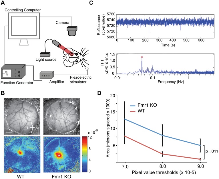
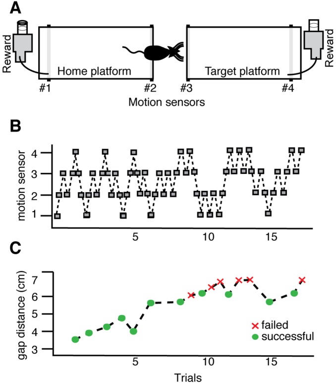
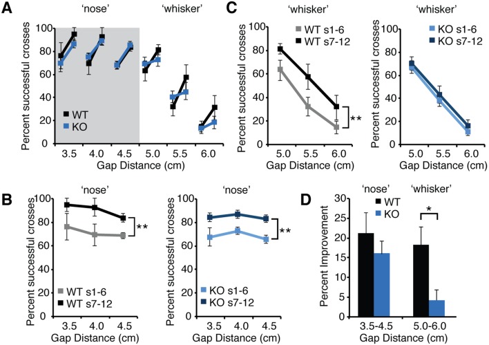
The fragile X mental retardation 1 mutant mouse (Fmr1 KO) recapitulates several of the neurologic deficits associated with Fragile X syndrome (FXS). As tactile hypersensitivity is a hallmark of FXS, we examined the sensory representation of individual whiskers in somatosensory barrel cortex of Fmr1 KO and wild-type (WT) mice and compared their performance in a whisker-dependent learning paradigm, the gap cross assay. Fmr1 KO mice exhibited elevated responses to stimulation of individual whiskers as measured by optical imaging of intrinsic signals. In the gap cross task, initial performance of Fmr1 KO mice was indistinguishable from WT controls. However, while WT mice improved significantly with experience at all gap distances, Fmr1 KO mice displayed significant and specific deficits in improvement at longer distances which rely solely on tactile information from whiskers. Thus, Fmr1 KO mice possess altered cortical responses to sensory input that correlates with a deficit in tactile learning.


Conflict of interest statement

Competing Interests: The authors have declared that no competing interests exits.

Figures

Figure 1
Figure 1. Fmr1 KO mice exhibit increased evoked activity in primary somatosensory cortex during whisker stimulation.
(A) Schematic showing experimental set up of intrinsic optical imaging over primary somatosensory cortex (black circle) during periodic whisker stimulation. (B) Pictures of the thin skull preparation and example images collected from a wild-type (WT) mouse (left) and Fmr1 KO mouse (right) mouse. Scale bar = 0.4 mm. Rostral (R), Caudal (C), Lateral (L) and Medial (M) coordinates are shown. (C) Representative examples of data collected during a typical imaging session. Above, a time series of pixel values for the cortical location indicated by the asterisk in the Fmr1 KO in panel B. Below, a fast-fourier transform (FFT) of the raw trace extracts the magnitude of the change in reflectance (ΔR/R) corresponding to the frequency of whisker stimulation (red square). (D) The number of pixels within the region of response with ΔR/R magnitudes greater than the threshold indicated on the abscissa for WT (n = 10) and Fmr1 KO (n = 10) mice. The response to whisker stimulation is elevated in Fmr1 KO mice (WT vs. KO, p = .011; 2-way ANOVA).
Figure 2
Figure 2. The Gap Cross task is a whisker-dependent sensory learning paradigm.
(A) Schematic of the gap cross learning task. Motion sensors positioned at four points along the 2 platforms (labeled #1–4) track the mouse as it moves from the starting platform across a given gap distance to the target platform. (B) Activation of each sensor (grey box) indicates the position of the mouse. (C) Successful crosses are defined as the movement of the mouse from the starting platform to the target platform (green circles). Failures are defined as trials in which the mouse approaches the edge of the home or target platform and returns to the back of the home platform (red crosses).
Figure 3
Figure 3. Fmr1 KO mice display normal learning on the gap cross assay at shorter gap distances but impaired learning at longer whisker-dependent distances.
(A) The percent successful crosses averaged across the first six sessions and subsequent six sessions across gap distances ranging from 3.0 cm to 6.0 cm for both wild-type mice (black lines, n = 6) and Fmr1 KO mice (blue lines, n = 9). For each distance, the line marker on the left is the average success rate of the first six sessions and the connected line marker on the right is the average success rate of the subsequent six sessions. Error bars represent standard error of the mean. (B) At shorter ‘nose’ distances, both wild-type (WT) and Fmr1 KO mice (KO) improve to a greater percentage of successful crosses between the average of the first six sessions (WT, grey, KO light blue) and the last six sessions (WT, black, KO dark blue). This improvement is statistically significant (WT, p = .007; n = 6; KO, p<.001, n = 9; WT; two-way ANOVA) (C) At whisker-dependent distances, wild-type (WT) improve between early sessions (grey line) and subsequent sessions (black line) despite the lower overall success rate at increasing gap distances. However, KO mice do not display significant improvement between early sessions (light blue line) and later sessions (dark blue line) (WT, p = .002, n = 6, KO, p = .14; n = 9, two-way ANOVA). (D) Average improvement for WT and KO mice at shorter ‘nose’ distances and longer ‘whisker’ distances. WT mice display significantly greater improvement at whisker-dependent distances that KO mice (p = .02, two-tailed t-test).

Similar articles

Cited by

References

    1. Turner G, Webb T, Wake S, Robinson H (1996) Prevalence of fragile X syndrome. Am J Med Genet 64: 196–197 doi:;10.1002/(SICI)1096-8628(19960712)64:1<196::AID-AJMG35>3.0.CO;2-G - DOI - PubMed
    1. O’Donnell WT, Warren ST (2002) A Decade of Molecular Studies of Fragile X Syndrome. Annu Rev Neurosci 25: 315–338 10.1146/annurev.neuro.25.112701.142909 - DOI - PubMed
    1. Garber K, Smith KT, Reines D, Warren ST (2006) Transcription, translation and fragile X syndrome. Current Opinion in Genetics & Development 16: 270–275 10.1016/j.gde.2006.04.010 - DOI - PubMed
    1. Pfeiffer BE, Huber KM (2009) The State of Synapses in Fragile X Syndrome. The Neuroscientist: a review journal bringing neurobiology, neurology and psychiatry 15: 549–567 10.1177/1073858409333075 - DOI - PMC - PubMed
    1. Miller LJ, McIntosh DN, McGrath J, Shyu V, Lampe M, et al. (1999) Electrodermal responses to sensory stimuli in individuals with fragile X syndrome: a preliminary report. Am J Med Genet 83: 268–279. - PubMed

Publication types

Substances