. 2014 Oct 7;107(7):1637-46.
doi: 10.1016/j.bpj.2014.07.075.

The myosin inhibitor blebbistatin stabilizes the super-relaxed state in skeletal muscle

Affiliations

The myosin inhibitor blebbistatin stabilizes the super-relaxed state in skeletal muscle

Clyde Wilson et al. Biophys J. .

Abstract

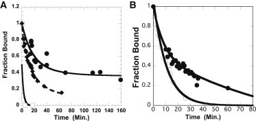
The super-relaxed state of myosin (SRX), in which the myosin ATPase activity is strongly inhibited, has been observed in a variety of muscle types. It has been proposed that myosin heads in this state are inhibited by binding to the core of the thick filament in a structure known as the interacting-heads motif. The myosin inhibitor blebbistatin has been shown in structural studies to stabilize the binding of myosin heads to the thick filament, and here we have utilized measurements of single ATP turnovers to show that blebbistatin also stabilizes the SRX in both fast and slow skeletal muscle, providing further support for the proposal that myosin heads in the SRX are also in the interacting-heads motif. We find that the SRX is stabilized using blebbistatin even in conditions that normally destabilize it, e.g., rigor ADP. Using blebbistatin we show that spin-labeled nucleotides bound to myosin have an oriented spectrum in the SRX in both slow and fast skeletal muscle. This is to our knowledge the first observation of oriented spin probes on the myosin motor domain in relaxed skeletal muscle fibers. The spectra for skeletal muscle with blebbistatin are similar to those observed in relaxed tarantula fibers in the absence of blebbistatin, demonstrating that the structure of the SRX is similar in different muscle types and in the presence and absence of blebbistatin. The mobility of spin probes attached to nucleotides bound to myosin shows that the conformation of the nucleotide site is closed in the SRX.


Figures

Figure 1
Figure 1
(A) Fiber fluorescence is shown during the chase phase for a slow-twitch fiber in the absence (lower trace) and presence (upper trace) of blebbistatin. The fiber was first incubated in 250 μM mantATP and subsequently chased with a solution containing 4 mM ATP. The decay of fluorescence was fit by a two-exponential decay function, as described previously (22). For the experiment with blebbistatin, blebbistatin was included in both the incubation and chase solutions. (B) The decay of fiber fluorescence during the chase phase in the presence of blebbistatin is contrasted for a fast- (lower trace) and slow-twitch (upper trace) muscle fiber. As can be seen, the effect of blebbistatin on the rate of release of nucleotides is greater in the slow-twitch muscle fiber than in the fast-twitch muscle fiber.
Figure 2
Figure 2
Decay of fiber fluorescence late in the chase phase is shown as a function of time. A muscle fiber was first incubated in mantATP and subsequently chased by ATP. The arrow marks the time where the solution was exchanged for one containing 4 mM ADP. All solutions were in the presence of blebbistatin. The two upper traces represent experiments performed with a slow-twitch fiber in 40 μM blebbistatin (solid circles) or in 20 μM blebbistatin (solid diamonds). The lower trace represents the experiment performed with a fast-twitch fiber in 40 μM blebbistatin (open squares). As can be seen, there are only modest changes in the rate of release of fluorescent nucleotides in ADP relative to ATP, showing the stability of the SRX in blebbistatin.
Figure 3
Figure 3
(A) EPR spectra of the spin-labeled analog of ATP, 2′3′SLATP, bound to slow-twitch muscle fibers in the presence of blebbistatin. The first derivative of the absorption is shown as a function of the magnetic field. The fibers were oriented with their long axis parallel to the magnetic field (blue) or perpendicular to the magnetic field (red), showing a strong dependence on the orientation. The three sharp peaks (P2–P4) in the central region of the spectrum arise from free nucleotides; the broader peaks at high (P5) and low field (P1) arise from nucleotides bound to specific sites in the fiber, almost entirely to myosin. (B) EPR spectra are shown for slow-twitch muscle fibers aligned parallel to the magnetic field in the presence of blebbistatin (blue) and for tarantula fibers in the absence of blebbistatin (red). The spectra show that the orientation of the spin-labeled nucleotides is very similar for these two fibers and conditions. The spectra from the two muscle types were likewise similar when the fibers were aligned perpendicular to the magnetic field. The center field is 349.1 mT for parallel spectra and 350.0 mT for perpendicular spectra. All spectra are 10 mT wide. Spectra were aligned on the central (P3) peak of unbound nucleotide. To see this figure in color, go online.
Figure 4
Figure 4
(A) The release of spin-labeled nucleotides from slow-twitch fibers and from myosin is shown as a function of time during a chase with unlabeled ATP. Data obtained from fibers are shown in the presence (solid circles) and absence (open squares) of blebbistatin. Solid diamonds represent data obtained from myosin isolated from slow-twitch fibers in the presence of blebbistatin. In the presence of blebbistatin, the release of nucleotides from fibers is slow, with a time constant similar to that seen with fluorescent nucleotides. A fraction of the probes, 40%, is released fairly quickly, with a time constant of 14 min. Another faction, 31%, is released more slowly, with a time constant of 88 min, and the remaining probes, 29%, are released even more slowly (too slowly to be measured in this assay). In the absence of blebbistatin, all nucleotides are released from the fibers before the first time point at 3 min. The major fraction of nucleotides, 83%, is released from myosin plus blebbistatin with a time constant of 16 min. (B) Release of spin-labeled nucleotides from fast-twitch fibers (solid circles) and from purified myosin (open circles), both in the presence of blebbistatin. Fits to the data show a single-exponential decay with a time constant of 10.6 min for myosin and a two-phase decay for fibers with a slow phase representing 66% of the initial intensity and with a time constant of 31 min.

References

    1. Huxley H.E., Faruqi A.R. Time-resolved x-ray diffraction studies on vertebrate striated muscle. Annu. Rev. Biophys. Bioeng. 1983;12:381–417. - PubMed
    1. Jung H.S., Komatsu S., Craig R. Head-head and head-tail interaction: a general mechanism for switching off myosin II activity in cells. Mol. Biol. Cell. 2008;19:3234–3242. - PMC - PubMed
    1. Xu S., Offer G., Yu L.C. Temperature and ligand dependence of conformation and helical order in myosin filaments. Biochemistry. 2003;42:390–401. - PubMed
    1. Craig R., Woodhead J.L. Structure and function of myosin filaments. Curr. Opin. Struct. Biol. 2006;16:204–212. - PubMed
    1. Alamo L., Wriggers W., Padrón R. Three-dimensional reconstruction of tarantula myosin filaments suggests how phosphorylation may regulate myosin activity. J. Mol. Biol. 2008;384:780–797. - PMC - PubMed

Publication types

LinkOut - more resources