. 2014 Dec;5(6):731-42.
doi: 10.1007/s13244-014-0357-x. Epub 2014 Oct 9.

Imaging adults on extracorporeal membrane oxygenation (ECMO)

Affiliations

Imaging adults on extracorporeal membrane oxygenation (ECMO)

Steven Lee et al. Insights Imaging. 2014 Dec.

Abstract

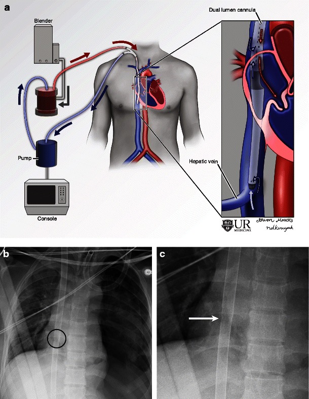
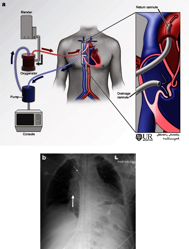
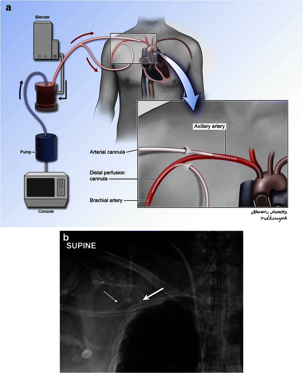
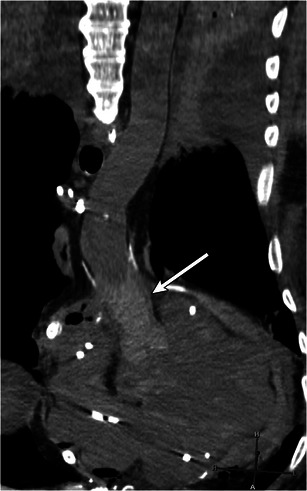
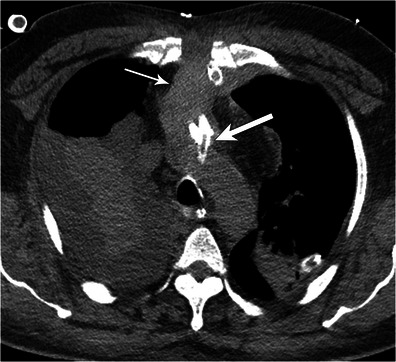
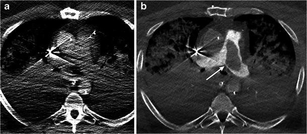
Extracorporeal membrane oxygenation (ECMO) is increasingly being used in adults following failure to wean from cardiopulmonary bypass, after cardiac surgery or in cases of severe respiratory failure. Knowledge of the different types of ECMO circuits, expected locations of cannulas and imaging appearance of complications is essential for accurate imaging interpretation and diagnosis. Commonly encountered complications are malposition of cannulas, adjacent or distal haemorrhage, stroke, stasis thrombus in access vessels, and distal emboli. This article will describe the imaging appearance of different ECMO circuits in adults as well as commonly encountered complications. If a CT (computed tomography) angiogram is being performed on these patients to evaluate for pulmonary embolism, the scan may be suboptimal from siphoning off of the contrast by the ECMO. In such cases, an optimal image can be obtained by lowering the flow rate of the ECMO circuit or by disabling the circuit for the duration of image acquisition. Key Points • Femoroatrial VV ECMO: femoral vein drainage cannula and right atrial return cannula. • Femorofemoral VV ECMO: return and drainage cannulas placed in femoral veins. • Dual-lumen single cannula VV ECMO: via the right IJ/Femoral vein with the tip in the IVC/SVC. • Peripheral VA ECMO: peripheral venous drainage cannula and peripheral arterial return cannula. • Central VA ECMO: direct right atrial drainage cannula and aortic return cannula.


Figures

Fig. 1
Fig. 1
Illustration (a) and radiograph (b) of femoroatrial VV ECMO demonstrate a right femoral drainage cannula and right atrial return cannula. Frontal radiographs show the expected locations of the right atrial return cannula (b, arrow) and right femoral drainage cannula (c, arrow). While the Murray score depends on several factors, this patient would receive a 4 for the chest radiograph element as there is four-quadrant infiltration
Fig. 2
Fig. 2
Illustration (a) and radiograph (b) of dual lumen, single cannula VV ECMO demonstrate the drainage cannula tip in the IVC and a sidehole in the right atrium, which is used for blood return. A magnified view of the right atrium and cannula (c) demonstrates the atrial opening identified by the slight discontinuity of the cannula (arrow)
Fig. 3
Fig. 3
Illustration (a) and radiograph (b) of mediastinal (central) VA ECMO. Note the drainage cannula in the right atrium (thick arrow) and the return cannula in the ascending aorta (thin arrow)
Fig. 4
Fig. 4
Sagittal reformat from a CT demonstrating a left ventricular drain (arrow) terminating in the left ventricle. This drain aids in decompression when left ventricular function is poor
Fig. 5
Fig. 5
Illustration (a) and radiograph (b) demonstrating peripheral VA ECMO. The drainage cannula is advanced in the distal IVC to the level of the diaphragm. The arterial return cannula is not advanced very far. Radiograph (b) demonstrating the arterial cannula (black arrow) in the proximal femoral artery with multiple surgical clips used for anchoring. The full extent of the venous drainage cannula (white arrow) can be better assessed with a chest radiograph (not shown)
Fig. 6
Fig. 6
Illustration (a) and radiograph (b) demonstrating a distal arterial perfusion cannula (thin arrow) in a patient on peripheral VA ECMO. The arterial return cannula (thick arrow) projects over the axillary artery and points proximally as compared to the perfusion cannula which points distally
Fig. 7
Fig. 7
Malpositioned cannulas. Chest radiograph of a 54-year-old male status post MI on peripheral VA ECMO a demonstrates the tip of a superior approach venous ECMO cannula projecting too inferiorly over the IVC (thin arrow). A chest radiograph from a different patient b shows the tip of an inferior approach venous cannula projecting over the lower SVC, much higher than the expected location of the right atrium
Fig. 8
Fig. 8
Arterial vascular obstruction. Spectral Doppler image of the right axillary artery (a) in a 42-year-old female on VA ECMO demonstrates diminished amplitude and prolonged systolic peaks of the arterial waveforms, classic for pulsus parvus et tardus. In a different patient, B flow ultrasound (b) shows complete occlusion of the radial artery (arrow) just distal to the bifurcation
Fig. 9
Fig. 9
Arterial stasis thrombus. Non-contrast reconstructed CT image in a 67-year-old male on VA ECMO after a failed heart transplant demonstrates intraluminal high attenuation material in the proximal tubular ascending aorta compatible with thrombus
Fig. 10
Fig. 10
Stroke in a 48-year-old male on VA ECMO with mental status change. CT of the head demonstrates a large hypodensity in the distribution of the right MCA compatible with subacute infarct
Fig. 11
Fig. 11
Haematoma. Axial CT in a 44-year-old male on mediastinal ECMO for heart failure after myocardial infarction shows mediastinal haematoma (thin arrow) tracking along the arterial cannula (thick arrow) up to the aortic arch
Fig. 12
Fig. 12
Pulmonary embolism. Axial CT image of a 44-year-old male on central VA ECMO a shows insufficient opacification of the pulmonary arteries using the standard protocol secondary to siphoning of IV contrast by ECMO. Diagnositc images are obtained in the same patient b after putting the circuit in a minimal flow state (500 ml/min) for 15 s with reinjection of IV contrast. Thrombus is seen in the main pulmonary artery (b, arrow)

References

    1. Allen S, et al. A review of the fundamental principles and evidence base in the use of extracorporeal membrane oxygenation (ECMO) in critically ill adult patients. J Intensive Care Med. 2011;26(1):13–26. doi: 10.1177/0885066610384061. - DOI - PubMed
    1. Organization ELS (2014) ELSO website, under registry information. [website] 2014 [cited 2014; Available from: http://www.elso.org/index.php?option=com_content&view=article&id=95&Item....
    1. Zapol WM, et al. Extracorporeal membrane oxygenation in severe acute respiratory failure. A randomized prospective study. JAMA. 1979;242(20):2193–2196. doi: 10.1001/jama.1979.03300200023016. - DOI - PubMed
    1. Maslach-Hubbard A, Bratton SL. Extracorporeal membrane oxygenation for pediatric respiratory failure: history, development and current status. World J Crit Care Med. 2013;2(4):29–39. doi: 10.5492/wjccm.v2.i4.29. - DOI - PMC - PubMed
    1. Fenik JC, Rais-Bahrami K. Neonatal cerebral oximetry monitoring during ECMO cannulation. J Perinatol. 2009;29(5):376–381. doi: 10.1038/jp.2008.231. - DOI - PubMed

LinkOut - more resources