. 2014 Nov;15(11):1210-8.
doi: 10.15252/embr.201438622. Epub 2014 Oct 8.

Iro/IRX transcription factors negatively regulate Dpp/TGF-β pathway activity during intestinal tumorigenesis

Affiliations

Iro/IRX transcription factors negatively regulate Dpp/TGF-β pathway activity during intestinal tumorigenesis

Òscar Martorell et al. EMBO Rep. 2014 Nov.

Abstract

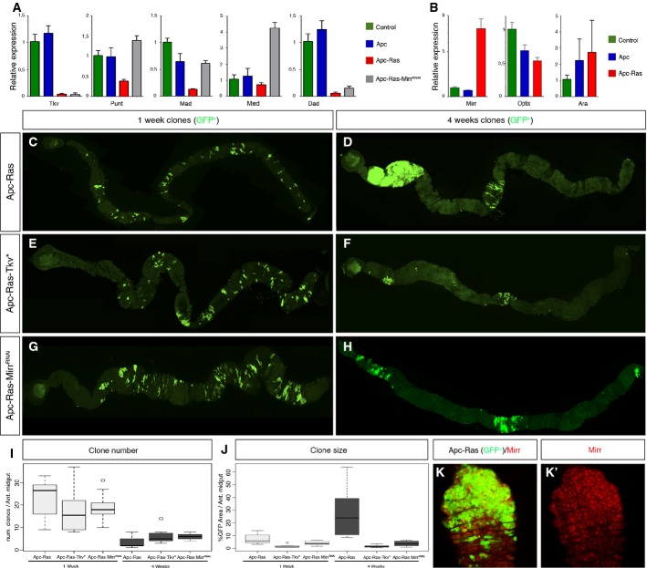
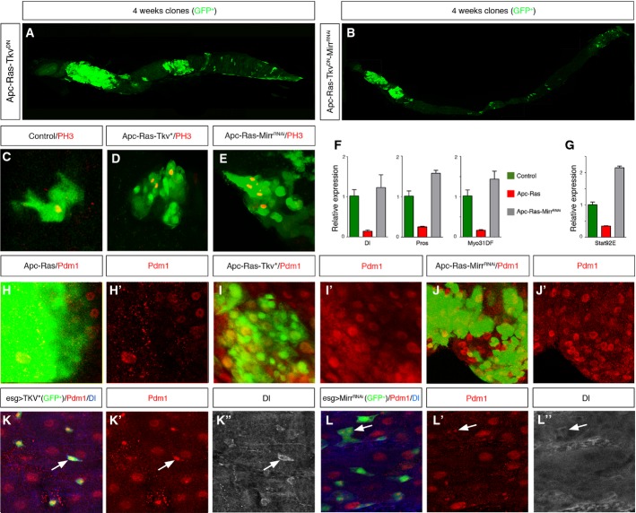
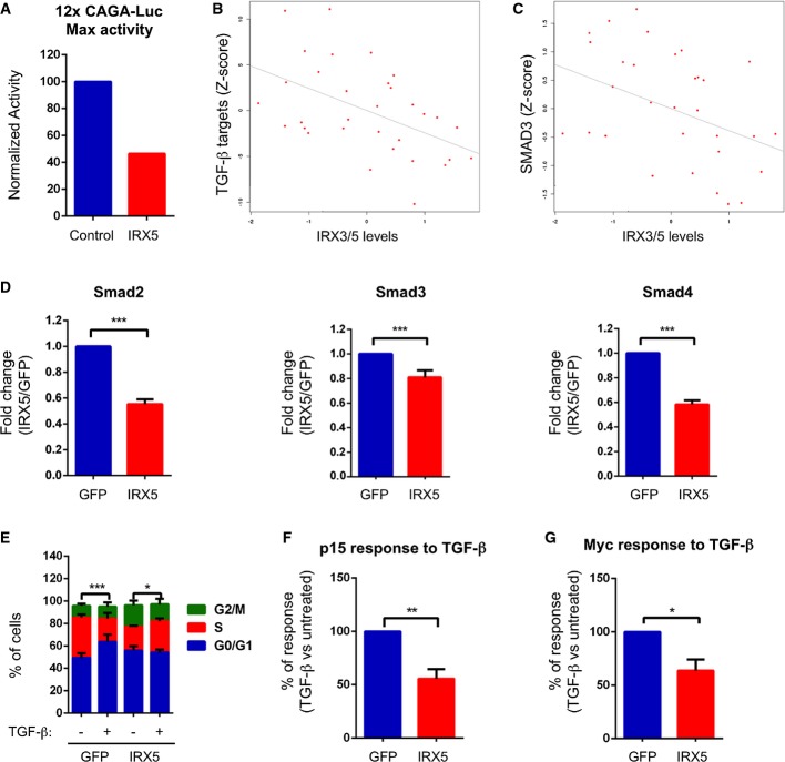
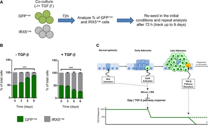
Activating mutations in Wnt and EGFR/Ras signaling pathways are common in colorectal cancer (CRC). Remarkably, clonal co-activation of these pathways in the adult Drosophila midgut induces "tumor-like" overgrowths. Here, we show that, in these clones and in CRC cell lines, Dpp/TGF-β acts as a tumor suppressor. Moreover, we discover that the Iroquois/IRX-family-protein Mirror downregulates the transcription of core components of the Dpp pathway, reducing its tumor suppressor activity. We also show that this genetic interaction is conserved in human CRC cells, where the Iro/IRX proteins IRX3 and IRX5 diminish the response to TGF-β. IRX3 and IRX5 are upregulated in human adenomas, and their levels correlate inversely with the gene expression signature of response to TGF-β. In addition, Irx5 expression confers a growth advantage in the presence of TGF-β, but is selected against in its absence. Together, our results identify a set of Iro/IRX proteins as conserved negative regulators of Dpp/TGF-β activity. We propose that during the characteristic adenoma-to-carcinoma transition of human CRC, the activity of IRX proteins could reduce the sensitivity to the cytostatic effect of TGF-β, conferring a growth advantage to tumor cells prior to the acquisition of mutations in TGF-β pathway components.

Keywords: Dpp; EGFR/Ras; TGF‐β; Wnt.


Figures

Figure 1
Figure 1. Dpp pathway suppresses Apc-Ras tumor growth and is regulated by Mirror
A qRT–PCRs of the main core components of the Dpp pathway (tkv, put, Mad, and Med) and the target gene Dad 4 weeks after clone induction. Note that their expression is lower in Apc-Ras clones compared to wild-type or Apc clones. B qRT–PCRs of Mirr, Optix, and Ara. Notice that Mirr is the only upregulated gene in Apc-Ras clones, but not in wild-type or Apc clones. Of note, qRT–PCR of Caup was negative in all conditions, and we discarded its expression in Apc-Ras clones by antibody staining (data not shown). C–H Adult midguts showing Apc-Ras (C, D), Apc-Ras-Tkv* (E, F), and Apc-Ras-MirrRNAi (G, H) clones marked by GFP (green), 1 and 4 weeks after induction. I Box-plot graph of the total number of clones in the anterior midgut. J Box-plot graph of the clone area (GFP+) per anterior midgut area. K Apc-Ras clones (green) stained with Mirr (red). Data information: Graphs in (A, B) show a representative experiment (mean ± SEM of relative expression, n = 3). Box-plots in (I, J) show data from Table 1. The length of the box represents the distance between the 25th and 75th percentiles, the interior horizontal line represents the group median, and the whiskers extend to the group minimum and maximum values. Source data are available online for this figure.
Figure 2
Figure 2. TkvDN is epistatic to Mirr
A, B Adult midguts showing Apc-Ras-TkvDN (A) and Apc-Ras-TkvDN-MirrRNAi (B) clones marked by GFP (green) 4 weeks after clone induction. Note that Dpp pathway inactivation blocks the tumor suppressor effect of MirrRNAi. C–E Wild-type (C), Apc-Ras-Tkv* (D), and Apc-Ras-MirrRNAi (E) GFP+ clones (green) stained with PH3 (red). Note the increased number of PH3+ cells in Apc-Ras-Tkv* and Apc-Ras-MirrRNAi clones compared to a wild-type clone of similar size. F, G qRT–PCRs of Dl, Pros, and Myo31DF (F) and stat92e (G) showing that Apc-Ras-MirrRNAi clones restore the expression of these markers to wild-type levels. Graphs show a representative experiment (mean ± SEM of relative expression, n = 3). H–J Apc-Ras (H), Apc-Ras-Tkv* (I), and Apc-Ras-MirrRNAi (J) GFP+ clones (green) marked with the EC marker Pdm1 (red). K, L Expression of activated Tkv* (K) or MirrRNAi (L) in progenitor cells induces the expression of the EC marker Pdm1 (red) in ISCs, marked by Dl (blue). Source data are available online for this figure.
Figure 3
Figure 3. Irx3 and Irx5 regulate the response to TGF-β in human cells
A Graph of a representative experiment showing maximum luciferase activity upon TGF-β treatment of cells transfected with an empty vector or the Irx5-expressing vector. B Negative correlation between TGF-β targets and IRX3/IRX5 expression in the cohort of adenoma samples from the GSE8671 dataset (Spearman’s correlation = −0.47507, P-value = 0.0065275, n = 32). C Negative correlation between SMAD3 and IRX3/IRX5 expression in the same dataset as in (B) (Spearman’s correlation = −0.43218, P-value = 0.014199). D qRT–PCRs of Smad2, Smad3, and Smad4 in LSTBRII cells expressing GFP or IRX5. Cells were induced with doxycycline for 24 h, and the levels of Smad2, Smad3, and Smad4 were assessed by qRT–PCR. Data are presented as mean fold change over GFP cells ± SEM (n = 4) (***P-value < 0.001, two-tailed t-test). E Cell cycle profile of LSTBRII cells expressing GFP or IRX5 in response to TGF-β. Cells were induced with doxycycline and treated with TGF-β for 72 h. Data are presented as % of cells in G0/G1, S, or G2/M from the total population ± SEM (n = 6) (***P-value < 0.001; *P-value < 0.05, two-way ANOVA). Comparisons refer to cells in S phase. F, G p15/Cdkn2b (F) or cMyc (G) response to TGF-β treatment of LSTBRII cells expressing GFP or IRX5. Cells were induced with doxycycline for 24 h and then treated with TGF-β for an additional 24 h. Results are expressed considering the transcriptional upregulation of p15/Cdkn2b (F) or the transcriptional downregulation of cMyc (G) of GFP cells (+TGF-β versus –TGF-β) as 100%. Data are presented as mean % of response ± SEM (n = 4) (*P-value < 0.05, **P-value < 0.01, two-tailed t-test). Source data are available online for this figure.
Figure 4
Figure 4. Irx proteins may play an essential role during the CRC adenoma-to-carcinoma transition
A Schematic representation of cell competition assay. The same number of doxycycline-induced GFP or IRX5 LSTBRII cells was co-cultured in the presence or absence of TGF-β. After 72 h, cells were analyzed by FACS to quantify the % of GFP and IRX5 cells in the culture. Cells were then re-seeded and kept in the same culture conditions as before (either with or without TGF-β). This analysis was repeated at 6 and 9 days from the initial seeding. B Cell competition assay of IRX5 and GFP LSTBRII cells. Data are presented as % of GFP and IRX5 cells ± SEM (n = 3) (***P-value < 0.001, two-way ANOVA). C Proposed model in which expression of IRX genes in early adenomas would reduce the ability of tumor cells to respond to TGF-β, precluding its tumor suppressor activity. This reduced response would allow tumor cells to survive long enough to accumulate additional mutations. Source data are available online for this figure.

References

    1. Fearon ER, Vogelstein B. A genetic model for colorectal tumorigenesis. Cell. 1990;61:759–767. - PubMed
    1. Morin PJ, Morin PJ, Sparks AB, Sparks AB, Korinek V, Korinek V, Barker N, Barker N, Clevers H, Clevers H, et al. Activation of beta-catenin-Tcf signaling in colon cancer by mutations in beta-catenin or APC. Science. 1997;275:1787–1790. - PubMed
    1. Korinek V, Barker N, Morin PJ, van Wichen D, de Weger R, Kinzler KW, Vogelstein B, Clevers H. Constitutive transcriptional activation by a beta-catenin-Tcf complex in APC-/- colon carcinoma. Science. 1997;275:1784–1787. - PubMed
    1. van de Wetering M, Sancho E, Verweij C, de Lau W, Oving I, Hurlstone A, van der Horn K, Batlle E, Coudreuse D, Haramis AP, et al. The beta-catenin/TCF-4 complex imposes a crypt progenitor phenotype on colorectal cancer cells. Cell. 2002;111:241–250. - PubMed
    1. Batlle E, Henderson JT, Beghtel H, van den Born MMW, Sancho E, Huls G, Meeldijk J, Robertson J, van de Wetering M, Pawson T, et al. Beta-catenin and TCF mediate cell positioning in the intestinal epithelium by controlling the expression of EphB/ephrinB. Cell. 2002;111:251–263. - PubMed

Publication types

MeSH terms