. 2014 Dec 5;115(12):1007-16.
doi: 10.1161/CIRCRESAHA.115.304216. Epub 2014 Oct 8.

Abi3bp regulates cardiac progenitor cell proliferation and differentiation

Affiliations

Abi3bp regulates cardiac progenitor cell proliferation and differentiation

Conrad P Hodgkinson et al. Circ Res. .

Abstract

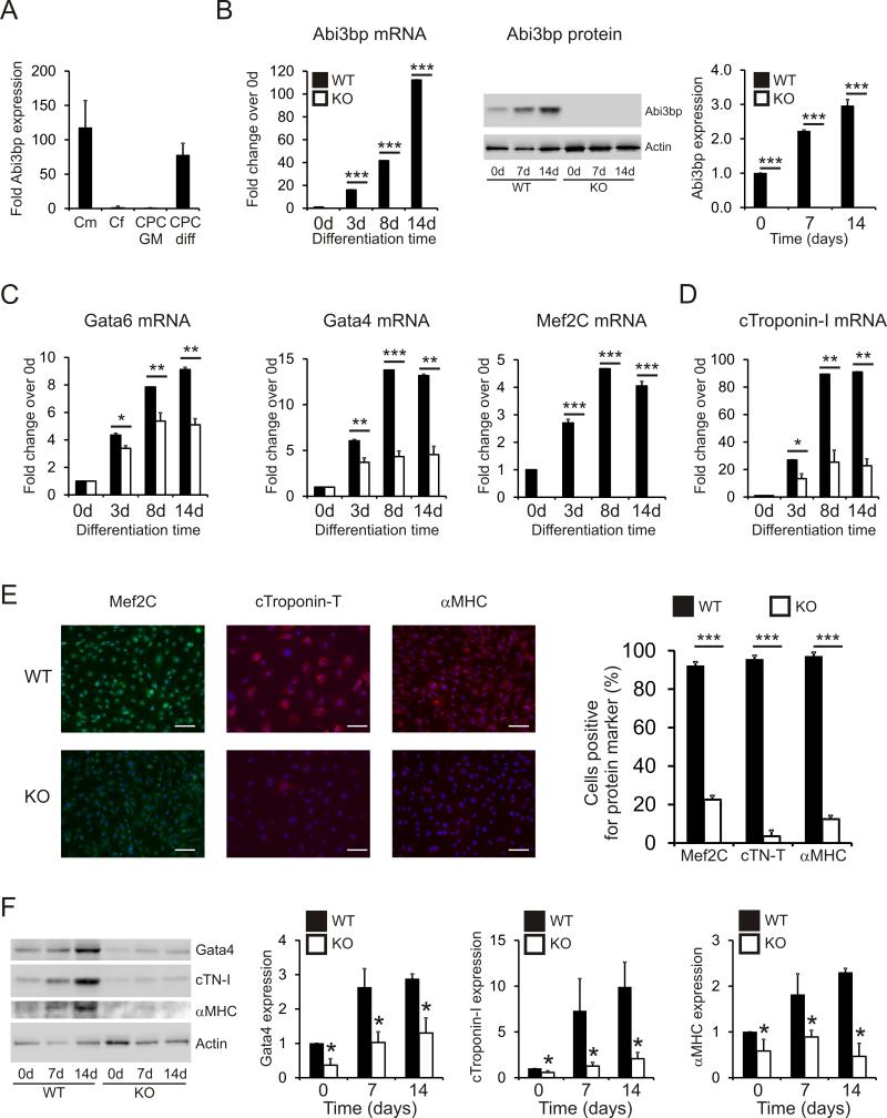
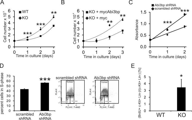
Rationale: Cardiac progenitor cells (CPCs) are thought to differentiate into the major cell types of the heart: cardiomyocytes, smooth muscle cells, and endothelial cells. We have recently identified ABI family, member 3 (NESH) binding protein (Abi3bp) as a protein important for mesenchymal stem cell biology. Because CPCs share several characteristics with mesenchymal stem cells, we hypothesized that Abi3bp would similarly affect CPC differentiation and proliferation.

Objective: To determine whether Abi3bp regulates CPC proliferation and differentiation.

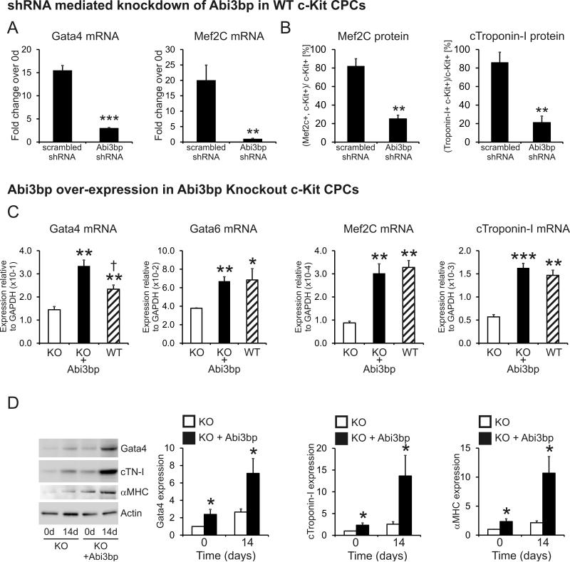
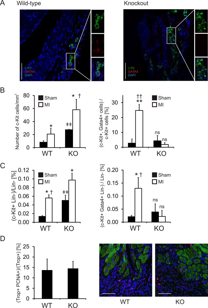
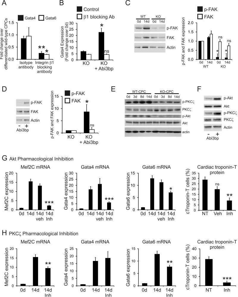
Methods and results: In vivo, genetic ablation of the Abi3bp gene inhibited CPC differentiation, whereas CPC number and proliferative capacity were increased. This correlated with adverse recovery after myocardial infarction. In vitro, CPCs, either isolated from Abi3bp knockout mice or expressing an Abi3bp shRNA construct, displayed a higher proliferative capacity and, under differentiating conditions, reduced expression of both early and late cardiomyocyte markers. Abi3bp controlled CPC differentiation via integrin-β1, protein kinase C-ζ, and v-akt murine thymoma viral oncogene homolog.

Conclusions: We have identified Abi3bp as a protein important for CPC differentiation and proliferation.

Keywords: extracellular matrix; integrin-β.


Figures

Figure 1
Figure 1. Abi3bp knockout inhibits CPC differentiation
(A) Abi3bp expression in cardiomyocytes [Cm], cardiac fibroblasts [Cf], and CPCs grown in either growth media [CPC GM] or differentiation media [CPC diff] was determined by qPCR. N=3. Data is shown as a fold expression with c-Kit+ CPCs grown in CPC-maintenance media taken to be 1. (B) Wild-type and Abi3bp knockout CPCs were cultured in CPC-differentiation media for up to 14 days. Expression of Abi3bp was determined by qPCR and immunoblotting. Expression in day 0 wild-type CPCs was taken to be 1. N=3. ***P≤0.001. (C-D) Wild-type and Abi3bp knockout CPCs were cultured in CPC-differentiation media for up to 14 days. Expression of Gata4, Gata6, Mef2C (C), and cardiac troponin-I (D) was determined by qPCR at the indicated time-points. Expression in day 0 wild-type CPCs was taken to be 1. N=3. ***P≤0.001. (E) Wild-type and Abi3bp knockout CPCs were cultured for 14 days in CPC-differentiation media. The cells were subsequently stained with Mef2C, cardiac troponin-T, or αMHC antibodies. DAPI was used to stain nuclei. Scale bar 100 microns. N=4. (F) Protein extracts (7.5μg) from wild-type and Abi3bp knockout CPCs cultured in CPC-differentiation media for 0, 7 and 14 days were probed for the indicated proteins. Actin was used as a loading control. Intensities were normalized to the loading control; normalized intensity of wild-type cells at day 0 was taken to be 1. N=3. *P≤0.05.
Figure 2
Figure 2. Re-expression of the Abi3bp in Abi3bp knockout CPCs recapitulates the wild-type phenotype
(A) Wild-type CPCs, expressing either a scrambled control or an Abi3bp targeting shRNA, were cultured for 14 days in CPC-differentiation media. Gata4 and Mef2C expression was determined by qPCR. Expression of cells at day 0 was taken to be 1. N=3. Comparisons made between scrambled and Abi3bp shRNA expressing cells **P≤0.01, ***P≤0.001. (B) Following culturing in CPC-differentiation media for 14 days the percentage of CPCs positive for Mef2C or cardiac troponin-I was determined by flow cytometry. N=3. Comparisons made between scrambled and Abi3bp shRNA expressing cells, **P≤0.01, ***P≤0.001. (C) Wild-type and Abi3bp knockout CPCs, transiently transfected with either a myc or mycAbi3bp plasmid, were cultured for 7 days in CPC-differentiation media. Expression of Gata4, Gata6, Mef2C and cardiac troponin-T was determined by qPCR. Gene expression data is shown relative to GAPDH. N=3. ***P≤0.001, **P≤0.01, *P≤0.05. (D) Protein extracts (7.5μg) from Abi3bp knockout c-Kit+ CPC, expressing either the myc or mycAbi3bp plasmid, and cultured in CPC-differentiation media for either 0 or 14 days, were probed for the indicated proteins. Actin was used as a loading control. Intensities were normalized to the loading control; normalized intensity of wild-type cells at day 0 was taken to be 1. N=3. *P≤0.05.
Figure 3
Figure 3. Abi3bp knockout is associated with lower cardiac function and increased fibrosis following MI
(A) Protein extracts (50μg) from sham and infarct hearts were immunoblotted with antibodies to the indicated proteins. β-tubulin was used as a loading control. Intensities were normalized to the loading control; normalized intensity of sham animals was taken to be 1. N=3. Serial sections from sham and MI mice were stained with Masson's trichrome. Fibrosis area is represented as a percentage of the left ventricle. (B) One-week post-infarct and (C) One month post-infarct. N=4-6 per group. Significance between MI groups is shown. ** P≤0.01. (D) One month following injury echocardiography was performed. Left and center panels: Ejection fraction and fractional shortening shown for wild-type and Abi3bp knockout mice subjected to MI. P values indicated. N=11 for wild-type, N=6 for Abi3bp knockout. Right panel: Sham ejection fraction and fractional shortening values (N=5 per group). Other parameters for pre-operative, MI and sham animals are shown in Supplementary Table I.
Figure 4
Figure 4. Abi3bp knockout prevents CPC differentiation/commitment following cardiac injury
Wild-type and Abi3bp knockout mice were subjected to either a sham operation or myocardial infarction [MI]. (A) Seven days following injury CPC differentiation was assessed by immunostaining. Peri-infarct regions were stained with c-Kit, Gata4, and DAPI. Scale bar 50 microns. Larger regions which contain these sections can be found in Online Figure V. (B) The entire peri-infarct region was visualized by serial sectioning through the heart tissue. Total numbers of c-Kit+ and Gata4+ cells were determined. The numbers of c-Kit+ cells are expressed per mm2 of the peri-infarct region. *P≤0.05 comparing MI to sham, †P≤0.05 comparing wild-type MI to Abi3bp knockout MI, ǂǂ P≤0.01 comparing wild-type sham to Abi3bp knockout sham. No other comparisons are significant. The numbers of double positive (c-Kit+ Gata4+) cells are expressed as a percentage of the total c-Kit+ population in the peri-infarct region. **P≤0.01 comparing wild-type MI to wild-type sham, ††P≤0.001 comparing wild-type MI to Abi3bp knockout MI. No other comparisons are significant. N=4-6 per group. (C) Hearts were collagenase digested and differentiating CPCs counted by flow cytometry. Differentiating CPCs were defined by the presence of c-Kit, expression of Gata4, and the absence of hematopoietic lineage markers. N=4-6 per group. Data is expressed as the percentage of c-Kit positive Gata4 positive cells in the total hematopoietic negative population. P-values indicated. (D) Serial sections of one-week post-MI tissue were stained for cardiac troponin-T (Trop) and the proliferation marker PCNA. Scale bar 50 microns. N=4-6 per group. No significance was observed between groups.
Figure 5
Figure 5. Abi3bp controls CPC differentiation through integrin-β1, PKCζ and Akt
(A) Wild-type c-Kit+ CPCs were cultured for 14 days in CPC-differentiation media. Where necessary isotype control or integrin-β1 blocking antibodies (10μg/ml) were added to the media for the duration of the experiment. Gata4 and Gata6 expression was determined by qPCR. Data is shown as a fold-change where expression values in control wild-type c-Kit+ CPCs were taken to be 1. Control wild-type c-Kit+ CPCs were differentiated for 14 days in the absence of antibody. N=3. Comparisons made between isotype and integrin-β1 blocking antibody treated cells, *P≤0.05, **P≤0.01. (B) Abi3bp knockout c-Kit+ CPCs were transiently transfected with either a myc or mycAbi3bp plasmid. Cells were cultured for 14 days in CPC-differentiation media with isotype control or integrin-β1 blocking antibodies (10μg/ml). Gata4 gene expression was determined by qPCR. Data is shown as a fold-change where expression values in day 0 cells were taken to be 1. N=3. No significant difference was observed between the integrin-β1 treated groups. (C) Wild-type and Abi3bp knockout c-Kit+ CPCs were cultured for 14 days with CPC-differentiation media. Protein extracts [5μg], taken at the indicated times, were immunoblotted for p-FAK, FAK and actin. N=3. *P≤0.05, ns not significant, comparisons made to WT day 0 cells. (D) Abi3bp knockout c-Kit+ CPCswere transiently transfected with either a myc or mycAbi3bp plasmid and cultured in growth media following transfection. Protein extracts [7.5μg] were immunoblotted for p-FAK, FAK and actin. N=3. *P≤0.05, ns not significant, comparisons made to the myc-expressing cells. (E) Wild-type and Abi3bp knockout c-Kit+ CPCs were cultured for 14 days with CPC-differentiation media. Protein extracts [5μg], taken at the indicated times, were immunoblotted for p-PKCζ, p-Akt, PKCζ, Akt and actin. N=3. Quantification supplied in Online Figure VIIA. (F) Abi3bp knockout c-Kit+ CPCs were transiently transfected with either a myc or mycAbi3bp plasmid and cultured in growth media following transfection. Protein extracts [7.5μg] were immunoblotted for p-PKCζ, p-Akt, PKCζ, Akt and actin. N=3. Quantification supplied in Online Figure VIIB. (G) Wild-type c-Kit+ CPCs were cultured for 14 days with CPC-differentiation media. Where appropriate c-Kit+ CPCs were treated with either vehicle [DMSO], or Akt inhibitor [DMSO soluble]. Mef2C, Gata4, and Gata6 expression was determined by qPCR. Expression at day 0 was taken to be 1. N=3. Comparisons made with day 14 CPCs exposed to neither vehicle nor inhibitor, *P≤0.05, **P≤0.01, ***P≤0.001. Flow cytometry was used to determine the number of cardiac troponin-T positive cells. N=3. Comparisons made with day 14 CPCs exposed to neither vehicle nor inhibitor, **P≤0.01. (H) Wild-type c-Kit+ CPCs were cultured for 14 days with CPC-differentiation media. Where appropriate c-Kit+ CPCs were treated with a PKCζ inhibitor [media soluble]. Mef2C, Gata4, and Gata6 expression was determined by qPCR. Expression at day 0 was taken to be 1. N=3. Comparisons made with day 14 CPCs exposed to neither vehicle nor inhibitor, *P≤0.05, **P≤0.01, ***P≤0.001. Flow cytometry was used to determine the number of cardiac troponin-T positive cells. N=3. Comparisons made with day 14 CPCs exposed to neither vehicle nor inhibitor, **P≤0.01.
Figure 6
Figure 6. Abi3bp inhibits CPC proliferation in vitro and in vivo
(A) Wild-type and Abi3bp knockout c-Kit+ CPCs were seeded at the same density in growth media and manually counted for up to three days post-seeding. N=3. Comparisons between groups at the same time point *** P≤0.001. (B) Abi3bp knockout c-Kit+ CPCs, over-expressing either a control or Abi3bp plasmid, were seeded at the same density in growth media and manually counted for up to three days post-seeding. N=3. Comparisons between groups at the same time point ** P≤0.01. (C) MTS assay growth curves in growth media for wild-type CPCs expressing either scrambled or Abi3bp shRNA. N=6. Comparisons between groups at the same time point, *** P≤0.001. (D) Wild-type CPCs expressing scrambled or Abi3bp shRNA were incubated with in growth media supplemented with BrdU for 6 hours and analyzed by flow cytometry using 7-AAD to stain DNA. BrdU positive cells are in S-phase. N=3. *** P≤0.001. (E) Wild-type and Abi3bp knockout mice were injected with BrdU for four days, cells were collected by collagenase digestion and analyzed by FACS. Left panel, BrdU+/c-Kit+/lin- CPCs were counted and expressed as a percentage of the total c-Kit+/lin- CPC population. N=3. * P≤0.05.

References

    1. Beltrami AP, Barlucchi L, Torella D, Baker M, Limana F, Chimenti S, Kasahara H, Rota M, Musso E, Urbanek K, Leri A, Kajstura J, Nadal-Ginard B, Anversa P. Adult cardiac stem cells are multipotent and support myocardial regeneration. Cell. 2003;114:763–776. - PubMed
    1. Bearzi C, Leri A, Lo Monaco F, Rota M, Gonzalez A, Hosoda T, Pepe M, Qanud K, Ojaimi C, Bardelli S, D'Amario D, D'Alessandro DA, Michler RE, Dimmeler S, Zeiher AM, Urbanek K, Hintze TH, Kajstura J, Anversa P. Identification of a coronary vascular progenitor cell in the human heart. Proc Natl Acad Sci U S A. 2009;106:15885–15890. - PMC - PubMed
    1. Anversa P, Kajstura J, Leri A, Bolli R. Life and death of cardiac stem cells: A paradigm shift in cardiac biology. Circulation. 2006;113:1451–1463. - PubMed
    1. Bollini S, Smart N, Riley PR. Resident cardiac progenitor cells: At the heart of regeneration. J Mol Cell Cardiol. 2011;50:296–303. - PubMed
    1. Matsuura K, Nagai T, Nishigaki N, Oyama T, Nishi J, Wada H, Sano M, Toko H, Akazawa H, Sato T, Nakaya H, Kasanuki H, Komuro I. Adult cardiac sca-1-positive cells differentiate into beating cardiomyocytes. J Biol Chem. 2004;279:11384–11391. - PubMed

Publication types

MeSH terms