. 2014 Oct 8;34(41):13811-8.
doi: 10.1523/JNEUROSCI.2073-14.2014.

Cortical activation associated with muscle synergies of the human male pelvic floor

Affiliations

Cortical activation associated with muscle synergies of the human male pelvic floor

Skulpan Asavasopon et al. J Neurosci. .

Abstract

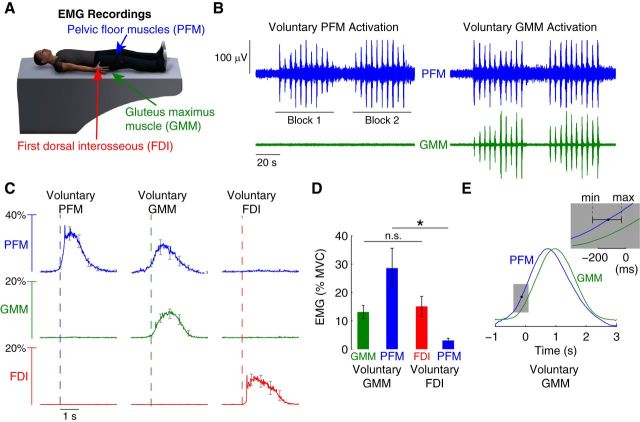
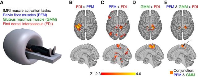
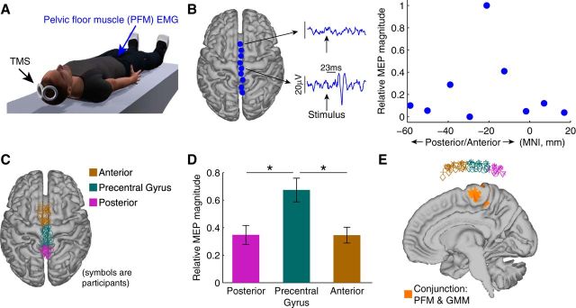
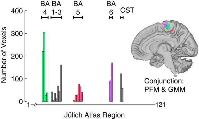
Human pelvic floor muscles have been shown to operate synergistically with a wide variety of muscles, which has been suggested to be an important contributor to continence and pelvic stability during functional tasks. However, the neural mechanism of pelvic floor muscle synergies remains unknown. Here, we test the hypothesis that activation in motor cortical regions associated with pelvic floor activation are part of the neural substrate for such synergies. We first use electromyographic recordings to extend previous findings and demonstrate that pelvic floor muscles activate synergistically during voluntary activation of gluteal muscles, but not during voluntary activation of finger muscles. We then show, using functional magnetic resonance imaging (fMRI), that a region of the medial wall of the precentral gyrus consistently activates during both voluntary pelvic floor muscle activation and voluntary gluteal activation, but not during voluntary finger activation. We finally confirm, using transcranial magnetic stimulation, that the fMRI-identified medial wall region is likely to generate pelvic floor muscle activation. Thus, muscle synergies of the human male pelvic floor appear to involve activation of motor cortical areas associated with pelvic floor control.

Keywords: EMG; TMS; fMRI; motor cortex; pelvic floor; supplementary motor area.


Figures

Figure 1.
Figure 1.
EMG evidence of pelvic floor muscle synergies. A, EMG signals from the PFM (blue), GMM (green), and the FDI (red) were recorded during separate trials that focused on the voluntary activation of each of these muscle groups. B, Example EMG recordings from the PFM and GMM muscles in a single participant during repeated voluntary PFM activation and separate voluntary GMM activation. Participants performed two blocks of 10 activations, each activation lasting 2 s. We observed PFM muscle activation during voluntary GMM muscle activation, but no GMM muscle activation during voluntary PFM muscle activation. C, Group data demonstrating the consistent finding of synergistic activation of the PFM muscles during voluntary GMM muscle activation but not during voluntary FDI muscle activation. Moreover, we did not find evidence of FDI or GMM muscle activation during voluntary PFM muscle activation. Curves show the average EMG transient triggered by the onset of the primary voluntary muscle of the task, averaged across participants (error bars indicate SEM across participants). D, Statistical analysis of group data shows that PFM activity is significantly greater (*p < 0.01) during voluntary GMM activation compared with PFM activity during voluntary FDI activation. The activity in the primary muscles of the tasks (GMM and FDI) was not significantly different (p = 0.40, n.s.; error bars indicate SEM across participants). E, Analysis of the normalized EMG transients for the PFM and GMM muscles during voluntary GMM muscle activation revealed that activation of PFM muscles led GMM muscle activation by an average of 128 ms across participants (minimum of 30.5 ms and maximum of 239.5 ms; p = 0.001).
Figure 2.
Figure 2.
fMRI evidence of overlapping activity during voluntary PFM and voluntary GMM activation. A, fMRI data were collected while participants performed three separate runs identical to the EMG tasks: separate repeated voluntary activation of PFM (run 1), GMM (run 2), and FDI (run 3). B, Contrast of voluntary FDI activation greater than voluntary PFM activation produced significant brain activation in left sensorimotor cortex. C, Contrast of voluntary PFM activation greater than voluntary FDI activation produced significant activation in the medial wall of the precentral gyrus. D, Contrast of voluntary GMM activation greater than voluntary FDI activation produced significant activation in the medial wall of the precentral gyrus E, Anterior medial wall of the precentral gyrus exhibited significant brain activation for both PFM activation and GMM activation compared with FDI activation.
Figure 3.
Figure 3.
TMS evidence that region of brain activation overlap between voluntary PFM activation and voluntary GMM activation is associated with the activity in PFM muscles. A, We collected MEP from the PFM muscles generated by TMS along the midline of the participant's brain. B, Single participant data showing an MEP in the PFM muscles generated by stimulating over precentral gyrus at a latency of 23 ms. Stimulating at points not over the precentral gyrus did not generate significant MEPs. The MEP in the PFM peaked at an MNI coordinate at approximately −20 mm. C, Locations of applied stimulation across all participants confined over the midline and divided (binned) into posterior, middle (precentral gyrus), and anterior bins. D, ANOVA analysis revealed that bin location had a significant main effect on MEP magnitude (*p = 0.003). MEPs corresponding to the middle bin were significantly greater than either the posterior bin (p = 0.001) or anterior bin (p = 0.016). Error bars indicate SEM across participants. E, Stimulation points classified as precentral gyrus were above the fMRI-identified region of activation common to both voluntary PFM activation and voluntary GMM activation.
Figure 4.
Figure 4.
Evidence of motor cortical representation in the overlapping brain activation during voluntary PFM and voluntary GMM activation. We computed the number of voxels in the overlap that were most likely to belong to each of the 121 regions in the Jülich Histological Atlas within FSL. The range of atlas regions included in each BA are labeled. CST, Corticospinal tract.

References

    1. Aagaard P, Simonsen EB, Andersen JL, Magnusson SP, Bojsen-Møller F, Dyhre-Poulsen P. Antagonist muscle coactivation during isokinetic knee extension. Scand J Med Sci Sports. 2000;10:58–67. doi: 10.1034/j.1600-0838.2000.010002058.x. - DOI - PubMed
    1. Aruin AS. The organization of anticipatory postural adjustments. J Automatic Control. 2002;12:31–37. doi: 10.2298/JAC0201031A. - DOI
    1. Ashton-Miller JA, DeLancey JO. Functional anatomy of the female pelvic floor. Ann N Y Acad Sci. 2007;1101:266–296. doi: 10.1196/annals.1389.034. - DOI - PubMed
    1. Ball T, Schreiber A, Feige B, Wagner M, Lücking CH, Kristeva-Feige R. The role of higher-order motor areas in voluntary movement as revealed by high-resolution EEG and fMRI. Neuroimage. 1999;10:682–694. doi: 10.1006/nimg.1999.0507. - DOI - PubMed
    1. Bharucha A. Pelvic floor: anatomy and function. Neurogastroenterol Motility. 2006;18:507–519. doi: 10.1111/j.1365-2982.2006.00803.x. - DOI - PubMed

Publication types

LinkOut - more resources