. 2015 Jan;87(1):37-48.
doi: 10.1002/cyto.a.22575. Epub 2014 Oct 8.

Cryopreservation of MHC multimers: Recommendations for quality assurance in detection of antigen specific T cells

Affiliations
Free PMC article

Cryopreservation of MHC multimers: Recommendations for quality assurance in detection of antigen specific T cells

Sine Reker Hadrup et al. Cytometry A. 2015 Jan.
Free PMC article

Abstract

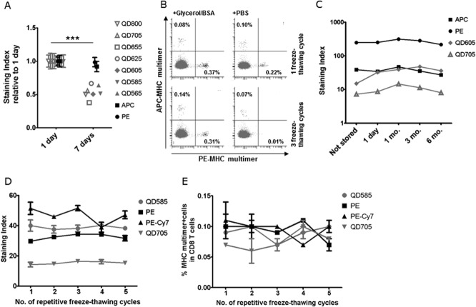
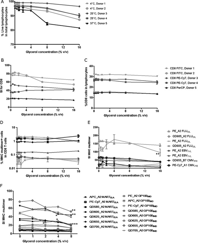
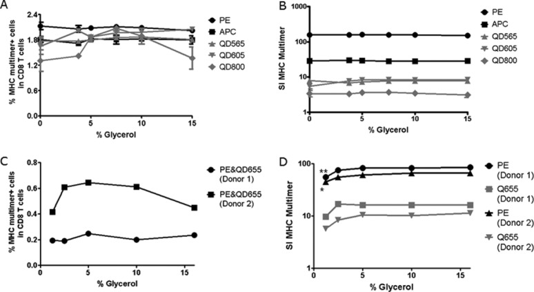
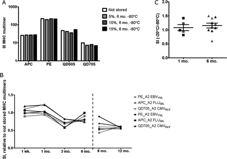
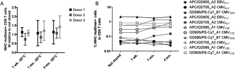
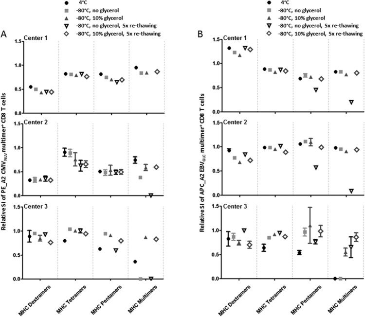
Fluorescence-labeled peptide-MHC class I multimers serve as ideal tools for the detection of antigen-specific T cells by flow cytometry, enabling functional and phenotypical characterization of specific T cells at the single cell level. While this technique offers a number of unique advantages, MHC multimer reagents can be difficult to handle in terms of stability and quality assurance. The stability of a given fluorescence-labeled MHC multimer complex depends on both the stability of the peptide-MHC complex itself and the stability of the fluorochrome. Consequently, stability is difficult to predict and long-term storage is generally not recommended. We investigated here the possibility of cryopreserving MHC multimers, both in-house produced and commercially available, using a wide range of peptide-MHC class I multimers comprising virus and cancer-associated epitopes of different affinities presented by various HLA-class I molecules. Cryopreservation of MHC multimers was feasible for at least 6 months, when they were dissolved in buffer containing 5-16% glycerol (v/v) and 0.5% serum albumin (w/v). The addition of cryoprotectants was tolerated across three different T-cell staining protocols for all fluorescence labels tested (PE, APC, PE-Cy7 and Quantum dots). We propose cryopreservation as an easily implementable method for stable storage of MHC multimers and recommend the use of cryopreservation in long-term immunomonitoring projects, thereby eliminating the variability introduced by different batches and inconsistent stability.

Keywords: MHC multimer; cryopreservation; cryoprotectant; glycerol in T cell staining; quality assurance; recommendations for MHC multimer storage.


Figures

Figure 1
Figure 1
MHC multimer staining is affected by storage conditions. (A) MHC multimers HLA-A2 CMVNLV coupled to Streptavidin QD800, 705, 655, 625, 605, 585, 565 (in gray), APC, or PE (in black) were stored at 4°C for either 1 day or 7 days without glycerol. The effect of storage time is depicted as the average staining index for the multimer specific T-cell population in two different donors, calculated relative to the average staining index at day 1. Error bars indicate range between donors. Performed at Center 2 (n = 1), with additional dataFigure S2 (n = 2). (B) Dot plots of HLA-A1 CMVVTE-PE and CMVYSE-APC multimer stainings of healthy donor PBMC after one (top row) or three (bottom row) freeze-thawing cycles with (left plots) or without (right plots) addition of 10% glycerol/0.5% BSA. Performed at Center 3 (n = 3). (C) MHC multimers: HLA-A2 EBVGLC-APC, -PE, -QD605, or -QD705 were produced and tested immediately (not stored) or frozen with 10% glycerol/0.5% BSA and tested after 1 day, 1 month, 3 months, and 6 months, respectively. Performed at Center 2 (n = 1). (D and E) The effect of repetitive freeze-thawing cycles on D) the staining index and E) the percentage of MHC multimer+ CD8 T cells when staining PBMC from one donor with: HLA-A1 FLUVSD -QD585 or -PE-Cy7 and HLA-A1 CMVYSE -PE or -QD705. MHC multimers were frozen with 10% glycerol/0.5% BSA and subjected to 1–5 freeze-thawing cycles at one-day intervals. All data points are the average of duplicates, error bars indicate range (not always visible). Performed at Center 3 (n = 1).
Figure 2
Figure 2
Effect of glycerol on cell viability and T-cell staining. The effect of increasing concentrations of glycerol was analyzed by addition of 0, 0.5, 1, 2, 4, 8, 16% glycerol to the first step of the MHC multimer staining reaction. MHC multimers were freshly prepared and contained no glycerol themselves. Three independent experiments including a total number of five different donors are shown: Donors 1 and 2 by Center 2 (light gray) (n = 2), Donors 3 and 4 by Center 1 (dark gray) (n = 2), Donor 5 by Center 3 (black) (n = 2). All data points are the average of duplicates, error bars indicate range (often not visible). (A) Cell viability under different staining conditions. The percentage of living lymphocytes (dead cell dye negative) in total lymphocytes (defined by FSC/SSC) is depicted in relation to increasing glycerol concentrations. Three different incubation temperatures were used based on the different staining protocols of each center. (B) Staining indexes for the CD8 staining by use of different antibodies, CD8-PE-Cy7, FITC, and PerCP and (C) Frequency of CD8 T cells in living lymphocytes for the same CD8 antibodies. (D and E) MHC multimer staining results expressed as percentages of specific T cells and staining indexes for eight different virus-specific T-cell populations. (F) MHC multimer staining of TIL specific for HLA-A2 MART-1ELA and GP100IMD with multimers labeled with PE, APC, PE-Cy7, QD585, 605, 625, 655, 705, after addition of increasing concentration of glycerol. The staining index of the MHC multimer positive population is depicted. Performed at Center 3 (n = 1).
Figure 3
Figure 3
Concentration of glycerol for cryopreservation. The concentration of glycerol required for cryopreservation of MHC multimers was tested in two independent experiments (A, B) (Center 2) and (C, D) (Center 3). The x-axis of each plot shows the % glycerol used for storage, x = 0 represents the not stored sample. (A, B) HLA-A2 EBVCLG MHC multimers were generated with five different fluorescence labels, cryopreserved with 3.75, 5, 7.5, 10, or 15% glycerol for 7 days, and compared to not frozen MHC multimers in terms of (A) percentage of MHC multimer+ and (B) staining index for the MHC multimer+ CD8 T cell population in one donor. Each dot is the average of duplicates and error bars indicate range (often not visible). Residual glycerol during staining varied between 0.5 and 2.2% for this experiment. (C, D) HLA-A2 CMVNLV MHC multimers were generated with PE- and QD655-fluorescence labels (dual color coded). MHC multimers were cryopreserved with 1.25, 2.5, 5, 10, and 16% glycerol for 7 days, then (C) percentage of stained CD8 T cells and (D) staining index for the MHC multimer+ population were determined in two donors. Single values of one experiment are plotted. Residual glycerol during staining varied between 0.1 and 1.6% in this experiment.
Figure 4
Figure 4
Long-term storage of cryopreserved MHC multimers. Results obtained in three independent tests performed are shown. (A) HLA-A2 EBVGLC MHC multimers with different fluorescent labels (APC, PE, QD605, QD705) were cryopreserved with 0.5% BSA and 5, 10, or 15% glycerol for 6 months. The staining index of the MHC multimer population is depicted compared to a not stored control prepared each time 1 day prior to staining (performed by Center 2, one donor, n = 1). (B) Long-term stability of three different HLA-A2 multimers (PE-EBVYVL, APC-FLUGIL, QD705 CMVNLV) was assessed after 1 week up to 13 months storage for two different donors (indicated by gray or black symbols). The staining index of the MHC multimer population is depicted relative to a not stored control of the same batch of monomers tested directly after preparation, before cryopreservation (left plot, 1 week-6 months) or compared to a freshly-prepared batch of multimers for the additional time points, 9 and 12 months (right plot), (performed by Center 1, n = 1). (C) SI after storage at −20°C vs. −80°C, for MHC multimers refolded with virus-derived epitopes: A2 CMVNLV, A2 EBVFLY, B7 CMVTPR labeled with PE or QD605, used for staining PBMC (black symbols) or MHC multimers with cancer-associated epitopes A2 MARTELA (modified) and A2 MARTEAA (wt) used for staining TILs (gray symbols) (performed by Center 3, n = 1).
Figure 5
Figure 5
Cryopreservation of MHC multimers for large panels of combinatorial-encoded stainings. We generated a panel of MHC multimers refolded with 27 virus-derived T-cell epitopes and determined the frequency of specific T-cell populations in three different healthy donors (black, dark gray, and light gray). Only responses of >0.002% of CD8 T cells and a minimum of 10 events are included in the graphs. (A) SI of the MHC multimer positive populations when stained using MHC multimers cryopreserved at −20°C for either 1 week, 1 month, or 4 months relative to the SI obtained using not stored MHC multimers. The data represent the average relative SI of the following populations: Donor 1: APC_A2 EBVFLY, QD605_A2 EBVFLY, APC_A2 CMVNLV, QD705_A2 CMVNLV, QD585_B7 CMVTPR, PE-Cy7_B7 CMVTPR; Donor 2: APC_A2 EBVFLY, QD605_A2 EBVFLY, APC_A2 CMVNLV, QD705_A2 CMVNLV, APC_A1 CMVYSE, QD585_A1 CMVYSE, QD605_A1 CMVVTE, PE-Cy7_A1 CMVVTE, QD585_B7 CMVTPR, PE-Cy7_B7 CMVTPR; Donor 3: APC_A2 CMVNLV, QD705_A2 CMVNLV, APC_A1 CMVYSE, QD585_A1 CMVYSE, QD605_A1 CMVVTE, PE-Cy7_A1 CMVVTE. Error bars represent SD. (B) Frequency of MHC multimer+ T cells, when stained using not stored, 1 week, 1 month, or 4 month cryopreserved MHC multimers at −20°C, prepared from the same batch (performed by Center 3, n = 1).
Figure 6
Figure 6
Testing of different storage conditions for commercially available and in-house generated MHC-multimers. We measured the SI of the MHC multimer+ T cell populations after staining using three different commercial MHC multimers: MHC dextramers (Immudex), MHC tetramers (TCMetrix), and MHC pentamers (ProImmune), in parallel with in-house produced MHC multimers from the three different centers. MHC multimers PE_A2 CMVNLV and APC A2_EBVGLC were included and tested shortly after purchase and following 10 days storage under the indicated conditions. (A) The SI of PE_A2 CMVNLV positive T cells at day 10 calculated relative to the SI at day 1, respectively shown for the different storage conditions, separately for each of the three centers. (B) The SI of APC A2_EBVGLC positive T cells at day 10 calculated relative to the SI at day 1, respectively shown for the different storage conditions, separately for each of the three centers. Performed as three independent, but parallel experiments at Centers 1–3. Relative SI = 0 means that no visible MHC multimer+ cells were detected. Each dot is the average of duplicates except staining with the MHC dextramers for Center 1, and error bars indicate range (often not visible).

References

    1. Altman JD, Moss PA, Goulder PJ, Barouch DH, McHeyzer-Williams MG, Bell JI, McMichael AJ, Davis MM. Phenotypic analysis of antigen-specific T lymphocytes. Science. 1996;274:94–96. - PubMed
    1. Davis MM, Altman JD, Newell EW. Interrogating the repertoire: broadening the scope of peptide-MHC multimer analysis. Nat Rev Immunol. 2011;11:551–558. - PMC - PubMed
    1. Appay V, Dunbar PR, Callan M, Klenerman P, Gillespie GM, Papagno L, Ogg GS, King A, Lechner F, Spina CA, et al. Memory CD8+ T cells vary in differentiation phenotype in different persistent virus infections. Nat Med. 2002;8:379–385. - PubMed
    1. Tan LC, Gudgeon N, Annels NE, Hansasuta P, O'Callaghan CA, Rowland-Jones S, McMichael AJ, Rickinson AB, Callan MF. A re-evaluation of the frequency of CD8+ T cells specific for EBV in healthy virus carriers. J Immunol. 1999;162:1827–1835. - PubMed
    1. Khan N, Cobbold M, Cummerson J, Moss PAH. Persistent viral infection in humans can drive high frequency low-affinity T-cell expansions. Immunology. 2010;131:537–548. - PMC - PubMed

Publication types

MeSH terms

LinkOut - more resources