. 2014 Dec 1;74(23):6845-55.
doi: 10.1158/0008-5472.CAN-14-0836. Epub 2014 Oct 8.

Inhibition of adaptive immunity by IL9 can be disrupted to achieve rapid T-cell sensitization and rejection of progressive tumor challenges

Affiliations

Inhibition of adaptive immunity by IL9 can be disrupted to achieve rapid T-cell sensitization and rejection of progressive tumor challenges

Dominique B Hoelzinger et al. Cancer Res. .

Abstract

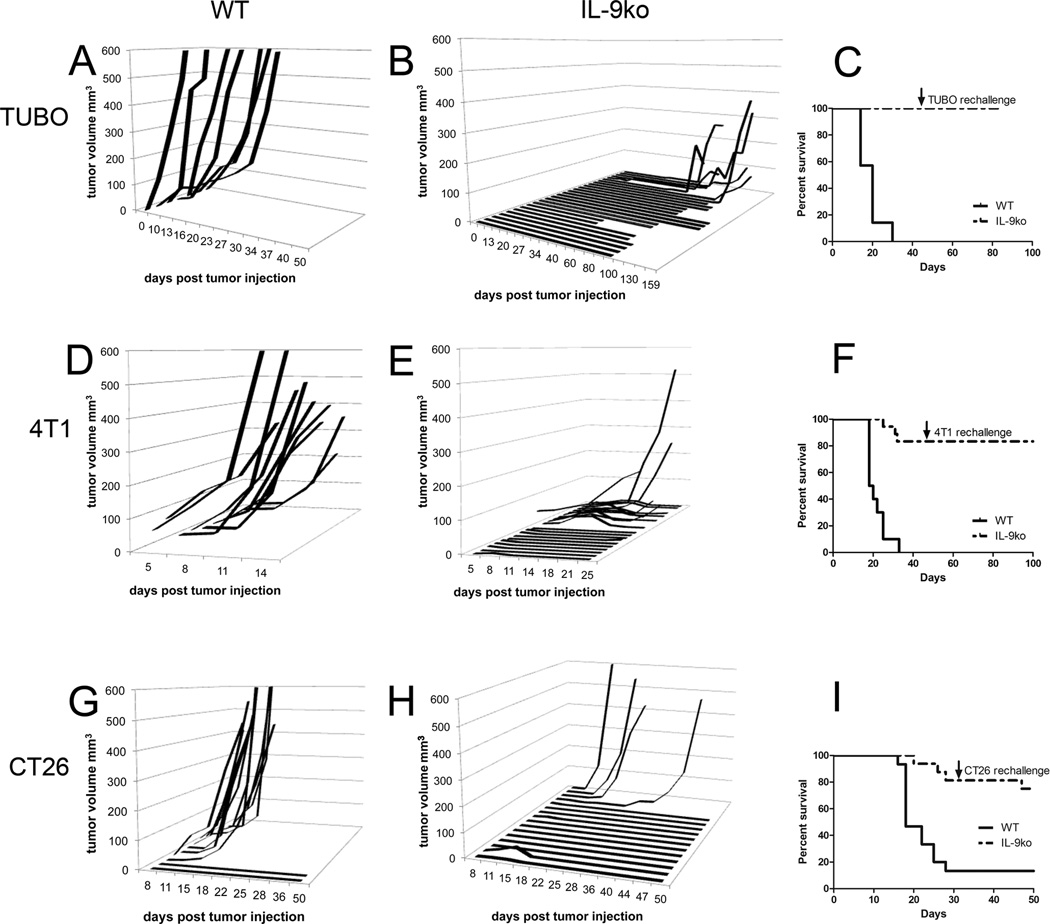
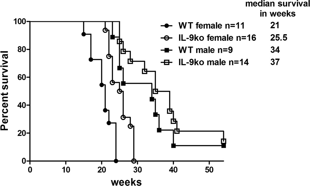
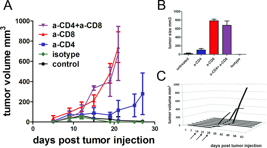
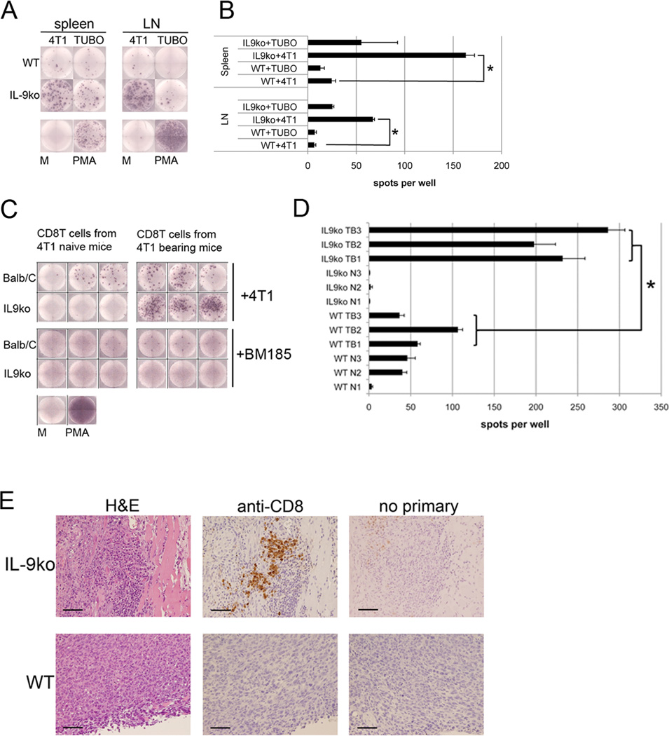
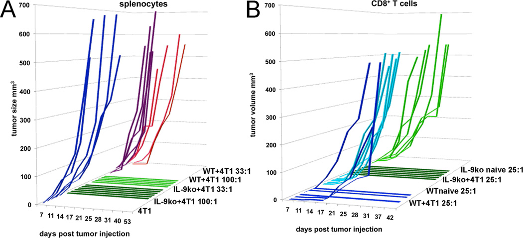
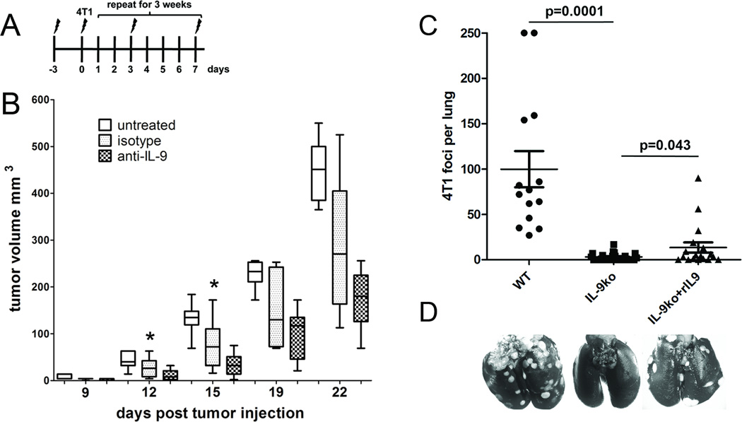
The tolerogenic cytokine IL9 promotes T regulatory cell function and allergic airway inflammation, but it has not been extensively studied in cancer. In this report, we used IL9-deficient mice to investigate the effects of IL9 in multiple models of breast and colon cancer development. Eliminating endogenous IL9 enabled sensitization of host T cells to tumors, leading to their early rejection without the requirement of vaccines or immunomodulatory therapies. Notably, IL9-deficient mice acquired immunologic memory, which actively protected from residual disease and tumor rechallenge, an effect linked to activation of CD8(+) T cells. Depletion of either CD8(+) or CD4(+) T cells abolished the benefits of IL9 loss to tumor control. Adoptive transfer experiments showed that T cells from tumor-rejecting IL9-deficient mice retained their effector competency in wild-type animals. Moreover, neutralizing IL9 antibody phenocopied the effects of IL9 gene deletion by slowing tumor progression in wild-type animals. Our results show the ability of IL9 to function as an inhibitor of adaptive immunity that prevents the formation of immunologic memory to a growing tumor, highlighting the potential for IL9 neutralization as a unique tool for cancer immunotherapy.


Conflict of interest statement

Conflict of interest: The authors declare that they have no conflict of interest.

Figures

Figure 1
Figure 1. TUBO and 4T1 mammary carcinomas are rejected in IL-9ko mice
Growth of 1×10⁶ TUBO cells implanted s.c. in the flank of (A) WT and (B) IL-9ko mice. (C) Survival plot of TUBO bearing mice, showing 100% of IL-9ko mice surviving after day 85 post tumor injection (p<0.0001). Growth of 1×10⁶ 4T1 cells implanted s.c. in the flank of (D) WT and (E) IL-9ko mice. (F) Survival plot of 4T1 bearing mice, showing 75% of IL-9ko mice surviving after day 50 post tumor injection (p<0.0001). Growth of 1×10⁵ CT26 cells implanted s.c. in the flank of (G) WT and (H) IL-9ko mice. (I) Survival plot of CT26 bearing mice, showing 75% of IL-9ko mice surviving after day 50 post tumor injection (p<0.001). Arrows denote the day of rechallenge with 1×10⁶ TUBO or 4T1 cells, or 1×10⁵ CT26 cells (7, 8 and 10 IL-9ko mice respectively). Data for each tumor model are cumulative of a minimum of 2 experiments.
Figure 2
Figure 2. IL-9 deficiency leads to delayed onset of autochthonous mammary tumors in Her2/neu transgenic mice
Survival plot of WT/Her2-neu (WT) and IL-9ko/Her2-neu mice (IL-9ko) showing that IL-9 deficiency lead to increased survival of female Her2/neu transgenic mice (p=0.001).
Figure 3
Figure 3. CD8+ T cells are essential for tumor rejection in IL-9ko mice
(A) Growth of 1×10⁶ 4T1 cells implanted s.c. in the flank of IL9-ko mice treated with neutralizing antibodies against CD4+, CD8+, CD4+and CD8+ T cells or isotype control. There were 6–8 mice per cohort and data shown are cumulative of two separate experiments. (B) Average tumor sizes in each treatment cohort 21 days post tumor injection. (C) Depletion of CD8+ T cells with anti-CD8 antibody in the IL-9ko mice that rejected 4T1 tumors from 2A. Arrows indicate dosage times once weekly for three weeks.
Figure 4
Figure 4. CD8+ T cells are activated in a tumor specific manner and are tumor tropic
(A) ELISpot analysis measuring IFNγ+ spots derived from total splenocytes (spleen) and lymphocytes (LN) from 4T1 bearing WT and IL-9ko mice. (B) Graph showing the average number of IFNγ+ spots from triplicate wells. *asterisk indicates p=0.001. (C) CD8+ T cells derived from naïve or tumor bearing WT and IL-9ko mice co-cultured with either 4T1 or BM185 cells. Wells shown are representative of triplicate determinations from three biological replicates. (D) Graph showing the average numbers of IFNγ+ spots in each well. Biological replicates are denoted as follows: WT naïve (WT N1-3), WT bearing 4T1 (WT TB1-3), IL-9ko naïve (IL9ko N1-3) and IL-9ko bearing 4T1 (IL9ko TB1-3). The *asterisk represents the significance of the difference in number of spots when comparing CD8+ T cells from tumor bearing WT versus IL-9ko mice (p=0.007). Representative data of duplicate experiments with three mice in each condition. (E) Immunohistochemical evaluation of CD8+ T cells present in 4T1 tumor derived from WT (lower panel) and IL-9ko (upper panel) mice. Each series is comprised of sequential slides to show morphology (H&E), anti-CD8 staining, and a negative control. Images are representative of three tumors from three individual mice in each strain. Bar represents 100 µm. M=media, PMA= Phorbol 12-myristate 13-acetate.
Figure 5
Figure 5. Splenocytes, or CD8+ T cells from IL-9ko mice that rejected 4T1 tumors, also impede 4T1 growth in WT mice
(A) Splenocytes derived from tumor bearing WT (WT+4T1) or IL-9ko (IL-9ko+4T1) mice were mixed with 4T1 cells and implanted s.c. in the flank of WT mice in the given ratios (100:1 and 33:1), holding the number of 4T1 cells constant at 2.5×10⁴. Each line represents tumor growth in one mouse, and the cohorts are colored as follows: 1) 4T1 only, blue 2) IL-9ko+4T1 100:1, dark green 3) IL-9ko+4T1 33:1, light green 4) WT+4T1 100:1 purple and 5) WT+4T1 33:1, red. Shown is one of two determinations. (B) Enriched CD8+ T cells derived from either naïve or 4T1 tumor bearing, WT or IL-9ko mice were co-injected s.c. into WT mice in a ratio of 25:1. Each cohort contains duplicate mice injected with 4T1 and cells from three individual donor mice. Each line represents tumor growth in one mouse, and the cohort colors define the CD8+ T cell donor sub-sets as follows: 1) WT+4T1, dark blue 2) WT naïve, light blue 3) IL-9ko+4T1, dark green and 4) IL-9ko naïve, light green.
Figure 6
Figure 6. Anti-IL-9 treatment results in slowed tumor growth in WT mice, whereas addition of recombinant IL-9 increases experimental metastases in IL-9ko mice
(A) Representation of the treatment schedule to measure the effect of IL-9 depletion on tumor growth. Symbols mark the days of anti-IL-9 injections, twice a week for 3 weeks (3×). 2.5×10⁴ 4T1 cells were injected on day 0. (B) Box-whisker plot of tumor growth in 3 cohorts of mice: untreated (white boxes, n=7), isotype control antibody (dotted boxes, n=8) and anti-IL-9 antibody (checkered boxes, n=11). Each box contains a line representing the median, and is bounded by the upper and lower quartiles. Minimum and maximum values are shown as whiskers. The bar frames the period of time (days 0 to 15) during which there is a significant difference in growth between the isotype control and anti-IL-9 treated cohorts (*asterisk, p=0.03). Data are cumulative of two independent experiments. (C) Experimental 4T1 lung metastases in WT and IL-9ko mice. Number of lung metastases per mouse are shown, cross bar indicates the median number of metastases and bounding bars represent upper and lower quartiles. (D) representative images of 4T1 foci in lungs from each treatment group.

References

    1. Stassen M, Schmitt E, Bopp T. From interleukin-9 to T helper 9 cells. Ann N Y Acad Sci. 1247:56–68. - PubMed
    1. Nowak EC, Noelle RJ. Interleukin-9 as a T helper type 17 cytokine. Immunology. 131:169–173. - PMC - PubMed
    1. Visekruna A, Ritter J, Scholz T, Campos L, Guralnik A, Poncette L, et al. Tc9 cells, a new subset of CD8(+) T cells, support Th2-mediated airway inflammation. Eur J Immunol. 43:606–618. - PubMed
    1. Goswami R, Kaplan MH. A brief history of IL-9. J Immunol. 186:3283–3288. - PMC - PubMed
    1. Jabeen R, Kaplan MH. The symphony of the ninth: the development and function of Th9 cells. Curr Opin Immunol. 24:303–307. - PMC - PubMed

Publication types