. 2015 Jan 1;113(1):203-17.
doi: 10.1152/jn.00405.2014. Epub 2014 Oct 8.

Nicotinic and muscarinic acetylcholine receptors shape ganglion cell response properties

Affiliations

Nicotinic and muscarinic acetylcholine receptors shape ganglion cell response properties

Christianne E Strang et al. J Neurophysiol. .

Abstract

The purpose of this study was to evaluate the expression patterns of nicotinic and muscarinic ACh receptors (nAChRs and mAChRs, respectively) in relation to one another and to understand their effects on rabbit retinal ganglion cell response properties. Double-label immunohistochemistry revealed labeled inner-retinal cell bodies and complex patterns of nAChR and mAChR expression in the inner plexiform layer. Specifically, the expression patterns of m1, m4, and m5 muscarinic receptors overlapped with those of non-α7 and α7 nicotinic receptors in presumptive amacrine and ganglion cells. There was no apparent overlap in the expression patterns of m2 muscarinic receptors with α7 nicotinic receptors or of m3 with non-α7 nicotinic receptors. Patch-clamp recordings demonstrated cell type-specific effects of nicotinic and muscarinic receptor blockade. Muscarinic receptor blockade enhanced the center responses of brisk-sustained/G4 On and G4 Off ganglion cells, whereas nicotinic receptor blockade suppressed the center responses of G4 On-cells near the visual streak but enhanced the center responses of nonstreak G4 On-cells. Blockade of muscarinic or nicotinic receptors suppressed the center responses of brisk-sustained Off-cells and the center light responses of subsets of brisk-transient/G11 On- and Off-cells. Only nicotinic blockade affected the center responses of G10 On-cells and G5 Off-cells. These data indicate that physiologically and morphologically identified ganglion cell types have specific patterns of AChR expression. The cholinergic receptor signatures of these cells may have implications for understanding visual defects in disease states that result from decreased ACh availability.

Keywords: cholinergic agonists; cholinergic antagonists; muscarinic acetylcholine receptors; nicotinic acetylcholine receptors; retinal circuitry.


Figures

Fig. 1.
Fig. 1.
Expression patterns of muscarinic ACh receptors (mAChRs) relative to the expression patterns of non-α7 nicotinic AChRs (nAChRs). Confocal image stacks of projections of 5 optical sections (0.5 μm steps) of m1 and m3–m5 mAChRs relative to β2-containing nAChRs. Colocalization was assessed from single confocal optical sections (0.5 μm steps) and masked onto overlay images. Column 1: many ganglion cells (arrows) and subsets of amacrine cells were immunoreactive for both m1 mAChRs and β2-containing nAChRs (arrowheads). Whereas most m1-positive amacrine cells also displayed β2-containing nAChR immunoreactivity (IR), not all amacrine cells that were positive for β2-containing nAChRs were immunoreactive for m1 receptors (notched arrowheads). Whereas both m1 and β2-containing nAChR IR was broadly distributed throughout the inner plexiform layer (IPL), the limited colocalization in the IPL was consistent with the somatic-labeling patterns. Muscarinic m1 IR was present in the outer plexiform layer (OPL), possibly indicating m1 expression by bipolar cells or horizontal cells. Column 2: there was no apparent double-labeling of m3 mAChRs and β2-containing nAChRs. Arrows and notched arrowheads indicate ganglion and amacrine cell somas that displayed β2-containing nAChR IR, whereas small arrowheads indicate bipolar cells that displayed m3 mAChR IR. m3 IR processes were localized to the OPL and to innermost and outermost sublamina (triangles) of the IPL, whereas β2-containing nAChR IR was broadly distributed throughout the IPL. Column 3: m4 muscarinic IR and β2-containing nAChR IR were colocalized almost completely in cell bodies in the ganglion cell layer (GCL; arrows) but were only colocalized to subsets of cell bodies in the inner nuclear layer (INL; arrowheads), whereas other m4-immunoreactive amacrine cell somata did not display β2-containing nAChR IR (notched arrowheads). Double-labeled processes were evident throughout the IPL. Column 4: ganglion cells in the GCL (arrows) and subsets of amacrine cells in the INL (arrowheads) were double labeled for m5 muscarinic and β2-containing nAChRs, and double-labeled processes were clearly visible throughout the IPL. Additional ganglion (triangles) and amacrine cells (notched arrowheads) were immunoreactive for β2-containing nicotinic but not m5 mAChRs. Bipolar cells (small arrowheads) were immunoreactive for m5 mAChRs but not β2-containing nAChRs. Original scale bar, 50 μm. Bottom: boxed areas in detail; original scale bar, 10 μm.
Fig. 2.
Fig. 2.
Expression patterns of mAChRs relative to the expression of α7 nAChRs. Confocal image stacks of projections of 5 optical sections (0.5 μm steps) of m1, m3, and m5 mAChRs relative to α7 nAChRs. Colocalization was assessed from single confocal optical sections and masked onto overlay images. Column 1, A and B: subsets of amacrine cells and ganglion cells (arrowheads) and subsets of bipolar cells (small arrowheads) were immunoreactive for both m1 mAChRs and α7 nAChRs. Other bipolar cells (arrows), amacrine cells (notched arrowheads), and ganglion cells (triangles) were m1 immunoreactive but not α7 immunoreactive. Muscarinic m1 and nicotinic α7 receptor IR were also colocalized in a band in the On sublamina of the IPL. Colocalization of IR throughout the rest of the IPL was limited. Column 2: ganglion cells and subsets of amacrine cells (triangles) were immunoreactive for the α7 nAChR subunit, whereas other amacrine cells were immunoreactive for m2 mAChRs (notched arrowheads). However, there was no apparent colocalization between m2 IR and α7 IR in cell bodies. m2 labeling in cell somas was very dim (notched arrowheads), even though IR was broadly distributed throughout the IPL, with intense labeling in a broad center band. There were double-labeled processes throughout the IPL, with strong masking at the boundary of GCL in the On sublamina of the IPL. Column 3: whereas a small number of bipolar cells were immunoreactive for both α7 nAChR subunits and m3 mAChRs (small arrowheads), the majority of immunoreactive bipolar cells was immunoreactive either for m3 mAChRs (small arrows) or α7 nAChRs (notched arrowheads), although there was a narrow band of colocalization in the OPL. Likewise, immunoreactive cells in the inner INL and GCL, presumptive amacrine and ganglion cells, displayed either α7 or m3 IR but not both. m3 IR was distributed primarily in 2 bands in the inner and outer IPL and sparsely colocalized with α7 IR. Column 4: cells in the GCL were m4 immunoreactive and α7 immunoreactive (arrowheads), whereas ganglion cells (triangles) and amacrine cells (notched arrowheads) were immunoreactive only to antibodies against α7 nAChR subunits. Dendritic stratification of α7-immunoreactive ganglion cells appeared to be in the On sublamina of the IPL, as colocalization through the rest of the IPL was minimal. Column 5: there was no apparent colocalization of m5 IR and α7 IR in ganglion cells (triangles) or amacrine cells (notched arrowheads), but bipolar cells with small cell bodies appeared to be immunoreactive for both m5 mAChR and α7 nAChR subunits (small arrowheads). Double-labeled processes were sparsely distributed throughout the IPL. Original scale bar, 50 μm. Bottom: boxed areas in detail; original scale bar, 10 μm.
Fig. 3.
Fig. 3.
Triple-label studies of mAChR IR, α bungarotoxin (αBgt) labeling, and choline acetyltransferase (ChAT) IR in the IPL. Confocal images of single optical sections of m1–m5 mAChR and αBgt labeling at the level of the labeled ChAT band (green) in the inner IPL. Density and expression patterns of muscarinic receptor subtypes m1–m5 (row 1, blue) varied, whereas the αBgt labeling (row 2, red) and ChAT labeling (row 3, green) were more uniform across experiments. Pseudocolored masks of colocalized pixels for each pair of fluorophores were obtained from single optical sections. The 3 pairwise masks were then merged into a single map of pixels that contained fluorescence from either 2 or 3 fluorophores (row 4). Colocalization of αBgt and mAChR labeling within a single pixel was pseudocolored magenta, mAChR and ChAT labeling was pseudocolored cyan, αBgt and ChAT labeling was pseudocolored yellow, and all 3 colors in a single pixel appear white. m1 IR was punctate with regions of larger puncta that did not colocalize with αBgt or ChAT (arrowheads). In a smaller area, m1 IR colocalized with αBgt labeling but rarely with ChAT IR (notched arrowheads). Colocalization of all 3 markers was minimal (arrows). Although m2 IR was closely apposed to αBgt-labeled puncta (notched arrowheads), m2 had limited colocalization with αBgt labeling or ChAT IR (arrowheads). m3-immunoreactive puncta at the level of the ChAT bands were frequently colocalized with both αBgt labeling and ChAT IR (arrows), although there were regions of m3-immunoreactive puncta without ChAT IR but with (notched arrowheads) and without (arrowheads) αBgt. m4 IR was almost always colocalized with αBgt labeling at this level of the retina (notched arrowheads) but rarely with ChAT IR (arrows). m5 IR was punctate with regions of larger puncta that did not colocalize with αBgt or ChAT. In a smaller area, m5 IR colocalized with αBgt labeling but not with ChAT IR (notched arrowheads). Colocalization of all 3 markers was minimal (arrows). Original scale bar, 20 μm. Optical depth is indicated by a double-headed arrow in vertical reconstructions of Z-stack (row 5) and lower magnification (row 6), with boxed areas demarking regions of interest; original scale bar, 50 μm.
Fig. 4.
Fig. 4.
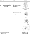
Sustained On-cells responded to the light onset with sustained inward currents that lasted for at least 500 ms and included cells with morphologies consistent with brisk-sustained On and G4 cells. Peak inward currents (pA) at light onset were measured to identify transient components of the light responses, whereas area under the curve (AUC; average nA·1 s) was calculated to measure sustained components of the light responses. The average changes in peak currents and AUC of sustained On-cells with light responses that were significantly affected by pharmacological blockade are shown by cell type. ATR, atropine; HEX, hexamethonium bromide; MLA, methyllycaconitine.
Fig. 5.
Fig. 5.
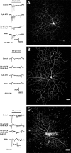
Muscarinic and nicotinic blockade affects sustained and transient On-cell light responses. High-contrast spot stimuli were used to test for interactions of nAChR and mAChR activation in the modulation of ganglion cell light responses in whole-cell, voltage-clamp mode. The application of 3 μM ATR (A, row 2) significantly increased the sustained inward currents by 1.2 nA·1 s (50%) and the number of spikes from 3 to 22 during light stimulation of this brisk-sustained/G4 On-cell (image at right; original scale bar, 50 μm). Blockade of nAChRs with 100 nM MLA and 100 μM HEX (A, row 3) significantly suppressed the peak transient component of the inward currents at light onset by 9 pA (50%). nAChR blockade slightly enhanced the component of the inward currents (115 pA·1 s; 5%) and enhanced the firing (11 spikes) compared with control. Light responses recovered partially after wash (A, row 4). Nicotinic and muscarinic blockade differentially affected the center and surround responses of a subset of transient On center ganglion cells with G11 morphology (B and C). (B) For the cell, blockade of nAChRs with 100 nM MLA and 100 μM HEX (row 2) significantly reduced peak inward currents at light onset from 18 to 3 pA and significantly reduced the peak outward currents at light offset (AUC) from 19 to 10 pA. (C) For the cell, nicotinic blockade (row 2) significantly reduced peak inward currents at light onset from 38 to 16 pA and significantly increased inward currents at light offset from 7 to 12 pA. Blockade of mAChRs with 3 μM ATR also significantly reduced (B, row 3; 8 pA reduction) or eliminated (C, row 3) light-evoked peak inward currents of both cells and revealed inward currents at light offset, i.e., antagonistic surround responses (B, row 3). Full cholinergic blockade, during coapplication of 100 nM MLA and 100 μM HEX with 3 μM ATR, further enhanced inward currents at light offset that were revealed by nicotinic blockade alone, resulting in the complete reversal of the center On response to an Off surround response (C, row 3). Images at right; original scale bars, 50 μm.
Fig. 6.
Fig. 6.
Transient On-cells were defined by inward currents at light onset that lasted <500 ms. The 16 transient On-cells included cells with morphologies consistent with nonlinear brisk-transient, G11 ganglion cells, and cells with morphologies consistent with the G2 or G10 subtypes. The average changes in peak currents of transient On-cells with light responses that were significantly affected by pharmacological blockade are shown by cell type.
Fig. 7.
Fig. 7.
Sustained Off-cells were identified by responses of longer than 500 ms to light decrements. The morphologies were consistent with either brisk-sustained Off-cells or the G4 Off subtype. The average changes in peak currents and AUC of sustained Off-cells with light responses that were significantly affected by pharmacological blockade are shown by cell type.
Fig. 8.
Fig. 8.
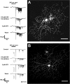
nAChRs and mAChRs contributed to the sustained component of the light-evoked responses of sustained Off-cells. This sustained Off-cell (image at right; original scale bar, 50 μm) responded to light offset with an initial transient and a high rate of sustained spiking (A, row 1). Blockade of nicotinic receptors with 100 nM MLA and 100 μM HEX significantly decreased the initial inward currents (reduction of 67 pA; 49%) and the AUC (reduction of 9.0 pA·1 s; 32%). The number of spikes was decreased from 98 to 36 (A, row 2). Blockade of muscarinic receptors also significantly decreased the peak inward currents by 87 pA (64%), the AUC by 5.6 nA·1 s (36%) control, and the firing at light offset by 38 spikes compared with control. The light responses recovered partially after wash. For the transient Off-cell (B), nicotinic and muscarinic blockade affected the transient and delayed-sustained components of light-evoked responses. Under control conditions, 200 μm light spots evoked transient inward currents at light onset, followed by spikes with a delay of >500 ms (B, row 1). Gray spikes show the transient components of the Off responses in greater detail. Bath application of HEX/MLA significantly decreased the transient Off responses by 106 pA (46%; gray spikes, row 2), whereas 3 μM ATR significantly enhanced the transient responses by 21 pA (9%; gray spikes, row 3). Both 100 nM MLA/100 μM HEX and 3 μM ATR enhanced the duration of the delayed response and increased the firing rate by 38 and 27 spikes, respectively. Image at right; original scale bar, 50 μm.
Fig. 9.
Fig. 9.
Transient Off-cells were defined by transient (<500 ms) inward currents in response to the offset of optimally sized light stimuli. Cells were identified according to previously published morphological criteria (Amthor et al. 1989; Rockhill et al. 2002). Each of the 3 transient Off-cells included in this study had different morphologies and displayed different responses to mAChR blockade. The average changes in peak currents of transient Off-cells with light responses that were significantly affected by pharmacological blockade are shown for each cell.

Similar articles

Cited by

References

    1. Albuquerque EX, Alkondon M, Pereira EF, Castro NG, Schrattenholz A, Barbosa CT, Bonfante-Cabarcas R, Aracava Y, Eisenberg HM, Maelicke A. Properties of neuronal nicotinic acetylcholine receptors: pharmacological characterization and modulation of synaptic function. J Pharmacol Exp Ther 280: 1117–1136, 1997. - PubMed
    1. Alhomida AS, Al-Rajhi AA, Kamal MA, Al-Jafari AA. Kinetic analysis of the toxicological effect of tacrine (Cognex) on human retinal acetylcholinesterase activity. Toxicology 147: 33–39, 2000. - PubMed
    1. Alkondon M, Pereira EF, Cortes WS, Maelicke A, Albuquerque EX. Choline is a selective agonist of alpha7 nicotinic acetylcholine receptors in the rat brain neurons. Eur J Neurosci 9: 2734–2742, 1997. - PubMed
    1. Alkondon M, Pereira EF, Eisenberg HM, Albuquerque EX. Choline and selective antagonists identify two subtypes of nicotinic acetylcholine receptors that modulate GABA release from CA1 interneurons in rat hippocampal slices. J Neurosci 19: 2693–2705, 1999. - PMC - PubMed
    1. Alkondon M, Pereira EF, Wonnacott S, Albuquerque EX. Blockade of nicotinic currents in hippocampal neurons defines methyllycaconitine as a potent and specific receptor antagonist. Mol Pharmacol 41: 802–808, 1992. - PubMed

Publication types

LinkOut - more resources