. 2014 Oct 9;10(10):e1004423.
doi: 10.1371/journal.ppat.1004423. eCollection 2014 Oct.

mRNA Structural constraints on EBNA1 synthesis impact on in vivo antigen presentation and early priming of CD8+ T cells

Affiliations

mRNA Structural constraints on EBNA1 synthesis impact on in vivo antigen presentation and early priming of CD8+ T cells

Judy T Tellam et al. PLoS Pathog. .

Abstract

Recent studies have shown that virally encoded mRNA sequences of genome maintenance proteins from herpesviruses contain clusters of unusual structural elements, G-quadruplexes, which modulate viral protein synthesis. Destabilization of these G-quadruplexes can override the inhibitory effect on self-synthesis of these proteins. Here we show that the purine-rich repetitive mRNA sequence of Epstein-Barr virus encoded nuclear antigen 1 (EBNA1) comprising G-quadruplex structures, limits both the presentation of MHC class I-restricted CD8(+) T cell epitopes by CD11c(+) dendritic cells in draining lymph nodes and early priming of antigen-specific CD8(+) T-cells. Destabilization of the G-quadruplex structures through codon-modification significantly enhanced in vivo antigen presentation and activation of virus-specific T cells. Ex vivo imaging of draining lymph nodes by confocal microscopy revealed enhanced antigen-specific T-cell trafficking and APC-CD8(+) T-cell interactions in mice primed with viral vectors encoding a codon-modified EBNA1 protein. More importantly, these antigen-specific T cells displayed enhanced expression of the T-box transcription factor and superior polyfunctionality consistent with the qualitative impact of translation efficiency. These results provide an important insight into how viruses exploit mRNA structure to down regulate synthesis of their viral maintenance proteins and delay priming of antigen-specific T cells, thereby establishing a successful latent infection in vivo. Furthermore, targeting EBNA1 mRNA rather than protein by small molecules or antisense oligonucleotides will enhance EBNA1 synthesis and the early priming of effector T cells, to establish a more rapid immune response and prevent persistent infection.


Conflict of interest statement

The authors have declared that no competing interests exist.

Figures

Figure 1
Figure 1. Schematic description of EBNA1 expression constructs comprising different GAr mRNA sequences whilst encoding identical protein sequences.
Plasmids expressing EBNA1 were generated in either pcDNA3 for in vitro translation studies (A–B) or in Ad5-adenoviral vectors (C–F) for immunological studies. (A–B) EBNA1 encoding 400 nucleotides of either native GAr (E1-GArN) or codon-modified GAr (E1-GArM) respectively; (C–F) EBNA1 encoding 400 nucleotides of either native GAr (Ad-E1-GArN) or 400 nucleotides of codon-modified GAr (Ad-E1-GArM); full-length EBNA1 (Ad-E1) or EBNA1 with a deleted GAr (Ad-E1-ΔGA) were fused in-frame to a C-terminal Green Fluorescent Protein (GFP) to generate (Ad-E1-GArN-GFP); (Ad-E1-GArM-GFP); (Ad-E1-GFP) or (Ad-E1-ΔGA-GFP) respectively. The Ad-EBNA1-GFP expression constructs (C–F) included insertion of a H2Kb-restricted ovalbumin CTL epitope, SIINFEKL, fused to the EBNA1 C-terminal sequence, allowing analysis of endogenous processing of EBNA1 using a B3Z T-cell hybridoma in the immunological assays as outlined in the Materials and Methods. (G) In vitro translation assay of pcDNA3 expression constructs encoding EBNA1 comprising either a native GAr (E1-GArN) or a codon-modified GAr (E1-GArM). Band intensities from the IVT assays were quantified by densitometric analysis using ImageJ64 software. Mean ± SD shown (n = 4).
Figure 2
Figure 2. Ex vivo antigen presentation by DCs is influenced by mRNA translation efficiency.
(A) Flow chart illustrating the experimental procedure. Female C57BL/6 mice were intramuscularly immunized with Ad-E1-GArN-GFP or Ad-E1-GArM-GFP. DLNs were pooled from 8 mice on day 2 following infection. Pan-DCs enriched from DLNs were incubated with B3Z cells at varying effector to target (E∶T) ratios to assess antigen presentation as described in the Materials and Methods. (B) Representative FACS plots showing β-galactosidase activity in B3Z hybridoma incubated with DCs from mice immunized with Ad-E1-GArN-GFP or Ad-E1-GArM-GFP at an E∶T ratio of 1∶2. (C) Overall activation of B3Z cells at varying B3Z cells/DCs ratios from mice immunized with Ad-E1-GArN-GFP or Ad-E1-GArM-GFP.
Figure 3
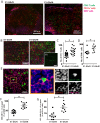
Figure 3. Antigen translation efficiency regulates Ag-specific T cell trafficking and DC-T cell interactions.
Female C57BL/6 mice were adoptively transferred with CFSE+CD8+ OT-1 cells, followed by intramuscular immunization with adenoviral expression vectors Ad-E1-GArN-GFP or Ad-E1-GArM-GFP. DLNs were collected 2 days post-infection for frozen section. These were stained for nucleus, CD11c and Kb-SIINFEKL complex. (A) Representative merged images (×40) show the transferred CFSE+CD8+ OT-1 cells, CD11c-expressing cells and SIINFEKL epitope presenting cells. (B) Representative merged images at high magnification (×100) show the transferred CFSE+CD8+ OT-1 cells, CD11c-expressing cells and SIINFEKL epitope presenting cells. (C) Comparative analysis of the number of CFSE+CD8+ OT-1 cells in DLNs following infection with Ad-E1-GArN-GFP or Ad-E1-GArM-GFP. (D) Comparative analysis of the number of and SIINFEKL epitope presenting cells in DLNs following infection with Ad-E1-GArN-GFP or Ad-E1-GArM-GFP. (E) Representative images show the interactions between CD11c cells and SIINFEKL epitope presenting cells; CFSE+CD8+ OT-1 cells and SIINFEKL epitope presenting cells. (F) Comparative analysis of the interaction between CD11c cells with H-2Kb-SIIN+ cells. (G) Comparative analysis of the interaction between of H-2Kb-SIIN+ CD11c cells with CD8+ OT1 effector cells. (*p<0.05, Mann-Whitney).
Figure 4
Figure 4. T cell proliferation and activation are influenced by differentially translated EBNA1 mRNAs.
(A) Representative FACS plot shows the proliferation of transferred CD8+ OT-1 cells in murine DLNs 3 days post-infection with Ad-E1-GArN-GFP or Ad-E1-GArM-GFP. The numbers in each box represent the percentage of CFSE labeled CD8+ OT-1 cells in murine DLNs. Each box represents one round of cell division. FSC: forward scatter. (B–C) Data represent the mean ± S.E.M. of the percentage of OT-1 CD8+ T cells in each division on days 2 and 3 post-infection with Ad-E1-GArN-GFP or Ad-E1-GArM-GFP at two different viral dosages (1×10⁶ pfu/mouse) or (1×10⁸ pfu/mouse). (D) Expression of CD25, CD122 and CD27 on days 1, 2 and 3 by transferred CD8+ OT-1 cells following infection with Ad-E1-GArN-GFP or Ad-E1-GArM-GFP (1×10⁶ pfu/mouse). Each vertical bar represents the mean ± SD (n = 5).
Figure 5
Figure 5. T cell activation is influenced by differentially translated EBNA1 mRNAs.
Expression of the T cell activation markers CD44, CD62L and CD69 in the DLNs of mice infected with Ad-E1-GArN-GFP or Ad-E1-GArM-GFP (1×10⁶ pfu/mouse or 1×10⁸ pfu/mouse) on days 1, 2 and 3. Each vertical bar represents the mean ± S.E.M. (n = 5).
Figure 6
Figure 6. Differential translation of EBNA1 mRNAs impacts on the expression of transcriptional factors and antigen specific functions in Ag-specific T cells from DLNs.
Female C57BL/6 mice were adoptively transferred with CFSE+CD8+ OT-1 cells, followed by intramuscular immunization with Ad-E1-GArN-GFP or Ad-E1-GArM-GFP virus (106 pfu/mouse) 2 hours following transfer. Mice were sacrificed on days 1, 2 or 3 post-infection. Expression of T-bet and Eomes in transferred CD8+ OT-1 cells was evaluated. (A) Representative FACS plots show the expression of T-bet and Eomes in CFSE+CD8+ OT-1 cells. (B) Overall results of the expression of T-bet and Eomes in CFSE+CD8+ OT-1 cells at different time points post-infection with Ad-E1-GArN-GFP or Ad-E1-GArM-GFP. (C) DLN cells were prepared from mice on day 3 post-infection with Ad-E1-GArN-GFP or Ad-E1-GArM-GFP and the expression of IFN-γ, TNF-α and translocation of CD107α by transferred CD8+ OT-1 cells was evaluated using a standard 6 hour ICS assay. (D–E) Female C57BL/6 mice were adoptively transferred with CFSE-labeled CD8+ OT-1 cells (1×10⁶ cells/mouse) and immunized intramuscularly with Ad-E1-GFP or Ad-E1-ΔGA-GFP virus (1×10⁶ pfu/mouse) 2 hours following transfer with DLNs harvested on day 3 post-infection to assess the activation and functions of antigen-specific CD8+ T cells. (D) The expression of T cell activation markers CD44, CD62L and CD69 on CFSE+CD8+ OT-1 cells (E) Expression of IFN-γ, TNF-α and translocation of CD107α by transferred CD8+ OT-1 cells. Mean ± SD shown (n = 5).
Figure 7
Figure 7. The differential translation of EBNA1 mRNAs impacts on the activation and functional programming of antigen-specific T cells.
Female C57BL/6 mice were immunized intramuscularly with Ad-E1-GArN-GFP or Ad-E1-GArM-GFP virus (A–B). Mice were sacrificed on day 7 post-infection and spleen cells harvested to evaluate the activation and functions of antigen-specific CD8+ T cells. (A) Expression of T cell activation markers CD44, CD62L and CD69. (B) Expression of IFN-γ, TNF-α and translocation of CD107α by Ag-specific CD8+ OT-1 cells. Mean ± SD shown (n = 5).
Figure 8
Figure 8. Differentially translated EBNA1 mRNAs have a minimal effect on Antigen-specific memory responses.
Female C57BL/6 mice were immunized intramuscularly with Ad-E1-GArN-GFP or Ad-E1-GArM-GFP virus. Spleen cells were collected on day 14 (top panel) or day 28 (lower panel) post-infection. (A) Expression of T cell activation markers of CD44, CD62L and CD69 on SIINFEKL-specific CD8+ T cells. (B) Expression of transcriptional factors Eomes and T-bet in SIINFEKL-specific CD8+ T cells. (C) The expression of IFN-γ by SIINFEKL-specific CD8+ T cells was evaluated using a standard 6- hour ICS assay. Mean ± SD shown (n = 5).

Similar articles

Cited by

References

    1. Doherty PC, Christensen JP, Belz GT, Stevenson PG, Sangster MY (2001) Dissecting the host response to a gamma-herpesvirus. Philosophical transactions of the Royal Society of London Series B, Biological sciences 356: 581–593. - PMC - PubMed
    1. Tellam J, Connolly G, Green KJ, Miles JJ, Moss DJ, et al. (2004) Endogenous presentation of CD8+ T cell epitopes from Epstein-Barr virus-encoded nuclear antigen 1. J Exp Med 199: 1421–1431. - PMC - PubMed
    1. Voo KS, Fu T, Wang HY, Tellam J, Heslop HE, et al. (2004) Evidence for the presentation of major histocompatibility complex class I-restricted Epstein-Barr virus nuclear antigen 1 peptides to CD8+ T lymphocytes. J Exp Med 199: 459–470. - PMC - PubMed
    1. Yewdell JW (2005) Serendipity strikes twice: the discovery and rediscovery of defective ribosomal products (DRiPS). Cell Mol Biol (Noisy-le-grand) 51: 635–641. - PubMed
    1. Yewdell JW, Anton LC, Bennink JR (1996) Defective ribosomal products (DRiPs): a major source of antigenic peptides for MHC class I molecules? J Immunol 157: 1823–1826. - PubMed

Publication types

MeSH terms