. 2014 Oct 9;10(10):e1004440.
doi: 10.1371/journal.ppat.1004440. eCollection 2014 Oct.

Vaginal challenge with an SIV-based dual reporter system reveals that infection can occur throughout the upper and lower female reproductive tract

Affiliations

Vaginal challenge with an SIV-based dual reporter system reveals that infection can occur throughout the upper and lower female reproductive tract

Daniel J Stieh et al. PLoS Pathog. .

Abstract

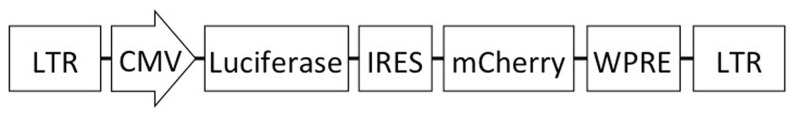
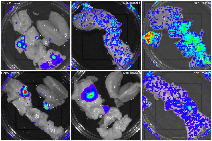
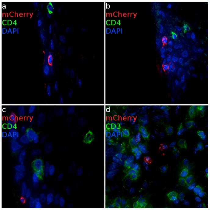
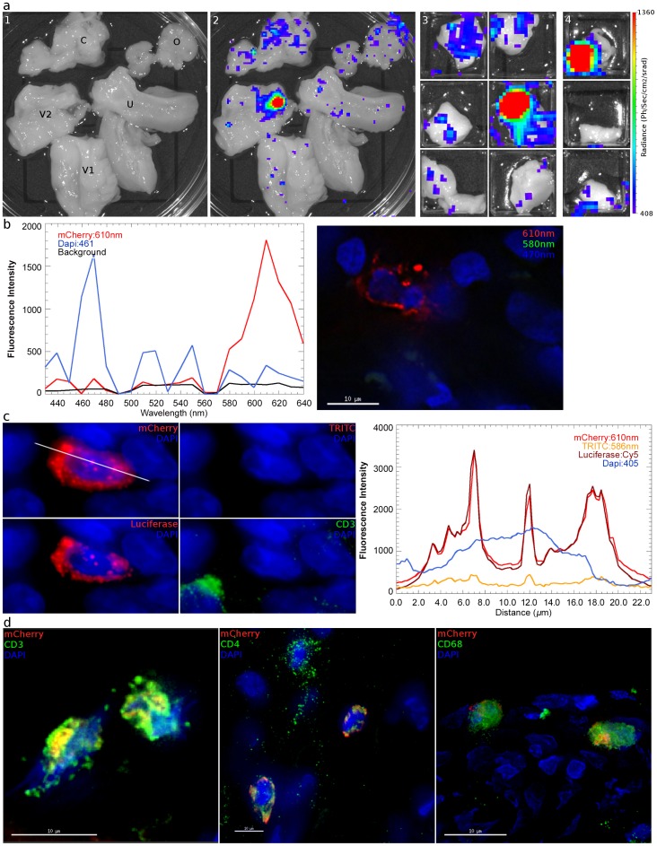
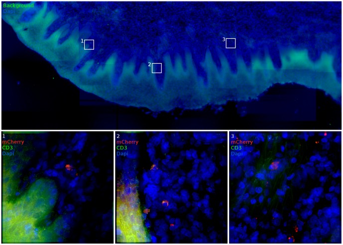
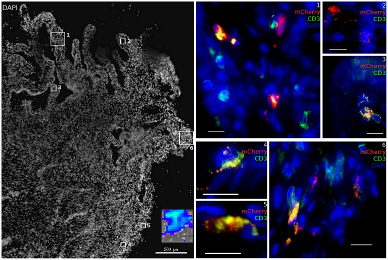
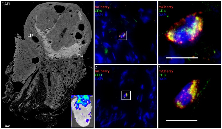
The majority of new HIV infections occur in women as a result of heterosexual intercourse, overcoming multiple innate barriers to infection within the mucosa. However, the avenues through which infection is established, and the nature of bottlenecks to transmission, have been the source of considerable investigation and contention. Using a high dose of a single round non-replicating SIV-based vector containing a novel dual reporter system, we determined the sites of infection by the inoculum using the rhesus macaque vaginal transmission model. Here we show that the entire female reproductive tract (FRT), including the vagina, ecto- and endocervix, along with ovaries and local draining lymph nodes can contain transduced cells only 48 hours after inoculation. The distribution of infection shows that virions quickly disseminate after exposure and can access target cells throughout the FRT, with an apparent preference for infection in squamous vaginal and ectocervical mucosa. JRFL enveloped virions infect diverse CD4 expressing cell types, with T cells resident throughout the FRT representing the primary target. These findings establish a new perspective that the entire FRT is susceptible and virus can reach as far as the ovary and local draining lymph nodes. Based on these findings, it is essential that protective mechanisms for prevention of HIV acquisition must be present at protective levels throughout the entire FRT to provide complete protection.


Conflict of interest statement

The authors have declared that no competing interests exist.

Figures

Figure 1
Figure 1. Schematic of the lentiviral vector used in this study.
The vector contains an effLuc expression cassette, expression of which is driven by the CMV promoter. An internal ribosomal entry site (IRES) and a mCherry cassette are downstream of luciferase. This vector contains the SIV promoter in the 5′ long-terminal repeat (LTR) for efficient virus production in the context of a Tat-free packaging system, and self-inactivating mutations in the 3′ LTR.
Figure 2
Figure 2. Luciferase reporter expression throughout the female reproductive tract in VSV-G pseudotyped vector inoculated Rhesus Macaques.
6 animals (4 pretreated with Depo-Provera) were inoculated with VSV-G pseudotyped dual-reporter vectors and sacrificed 48 hours later. Exposure to luciferin reveals reporter expression is present throughout the reproductive tract of Depo-Provera treated and normally cycling monkeys and that expression pattern varies between animals.
Figure 3
Figure 3. Infected cells from ectocervical and vaginal tissue of VSV-G enveloped reporter inoculated Rhesus Macaques.
(a, b) Virus primarily infects epithelial cells within the ectocervix. CD4 expressing cells cluster proximal to mCherry expressing cells but are not infected. (Animal code: FM27) (c) Epithelial and (d) sub-epithelial cells in the vagina are also susceptible to infection. Where infection is found, there are abundant numbers of T cells are present. (Animal code: FM28) mCherry signal is shown in red. CD3 or CD4 is shown in green. Nuclei (DAPI) are shown in blue. Scale bars, 30 µm.
Figure 4
Figure 4. Identification and phenotyping of infected cells from female reproductive tract. a,
48 hours after inoculation with JRFL pseudotyped LICh vector, the reproductive tract is separated into large sections (panel 1; V1: labia and lower vagina; V2: upper vagina; C: cervix; U: uterus; O: ovary), soaked in d-luciferin to identify luciferase expressing regions with in vivo imaging systems, and dissected into sequentially smaller pieces (panels 2–4) to isolate strongly luminescent regions measuring 2×2 mm2. (animal code: EH99) b, Left panel; The spectral emission profile of a putative infected cell from the tissue identified in a is measured on laser scanning confocal microscope with resonant scanner. Emission profile matches the defined profile of mCherry. Right panel; Pseudo-colored reconstructed image on the right shows the localization of mCherry (red), DAPI (blue) and background signals (green). c, Vaginal tissue is stained for firefly luciferase expression. The combination of mCherry+ (red), TRITC−/low (red), luciferase+ (red) fluorescence is criteria for confirming infection. The right panel shows relative fluorescence intensity of mCherry, TRITC, luciferase, and nucleus is along the line in mCherry image. (animal code: HP79) d, Phenotyping of infected cells by surface marker staining, shown in green, for CD3 (T Cell), CD4 (HIV-1 primary receptor), or CD68 (tissue resident macrophages) shows all conventionally defined HIV susceptible cell types are transduced within the vaginal tissue shown in a. (animal code: EH99).
Figure 5
Figure 5. Vaginal tissue with intact epithelium is the most common target for target cell transduction.
48 hours post JRFL pseudotyped vector administration, the FRT was removed and luminescent foci identified by IVIS analysis (inset). In vaginal domains without biopsy, the stratified epithelium is intact. In these portions of the vaginal vault, infected cells can still be found. mCherry expression (red), CD3 staining (green) and nuclei label (blue) are used to show transduced cells. (animal code: GK26).
Figure 6
Figure 6. Diverse cell types are infected within the cervix.
Within the cervix, both endocervical (Region 1–3) and ectocervical (Region 4–6) domains in the area of the transformation zone contain many resident T cells which are susceptible to infection with JRFL pseudotyped LICh vector. Non-T cells are also infected. Inset shows luciferase expression within dissected tissue before freezing. Scale bars measure 10 µm unless otherwise specified. mCherry expression (red), CD3 staining (green) and nuclei label (blue) are used to show transduced cells. (animal code: CT82).
Figure 7
Figure 7. The ovary is susceptible to infection.
After inoculation with JRFL pseudotyped LICh vector, tissues demonstrating luminescent signal (inset) is stained for luciferase and CD3 or CD4. Inset shows luciferase expression within tissue before freezing. Transduced cells within each region are shown as numbered close-up. Infected cells are found within the cortical zone of the ovary. Infected cells are not found within developing ova. The cell in region 1 and 3 expresses HIV-1 receptor CD4. Staining the adjacent tissue section identifies the transduced cell in region 2 and 4 as being CD3+. (animal code: GK26).
Figure 8
Figure 8. Nested PCR of macaque tissues.
Distribution of LICh reporter DNA-positive tissues in the female reproductive tract after inoculation with JRFL pseudotyped LICh vector. Results are based on the number of mCherry positive reactions, with at least 24 reactions per experiment. Gel shown is representative of three independent DNA extractions and amplifications.

References

    1. Joint United Nations Programme on HIV/AIDS. (2010) Global report: UNAIDS report on the global AIDS epidemic. Geneva, Switzerland: Joint United Nations Programme on HIV/AIDS. pp. v.
    1. Haynes BF, McElrath MJ (2013) Progress in HIV-1 vaccine development. Curr Opin HIV AIDS 8: 326–332. - PMC - PubMed
    1. Shattock RJ, Moore JP (2003) Inhibiting sexual transmission of HIV-1 infection. Nat Rev Microbiol 1: 25–34. - PubMed
    1. Haase AT (2011) Early events in sexual transmission of HIV and SIV and opportunities for interventions. Annu Rev Med 62: 127–139. - PubMed
    1. Keele BF, Estes JD (2011) Barriers to mucosal transmission of immunodeficiency viruses. Blood 118: 839–846. - PMC - PubMed

Publication types