. 2014 Oct 9;10(10):e1004636.
doi: 10.1371/journal.pgen.1004636. eCollection 2014 Oct.

Developmentally-regulated excision of the SPβ prophage reconstitutes a gene required for spore envelope maturation in Bacillus subtilis

Affiliations

Developmentally-regulated excision of the SPβ prophage reconstitutes a gene required for spore envelope maturation in Bacillus subtilis

Kimihiro Abe et al. PLoS Genet. .

Abstract

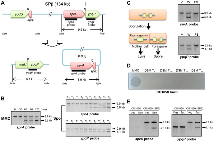
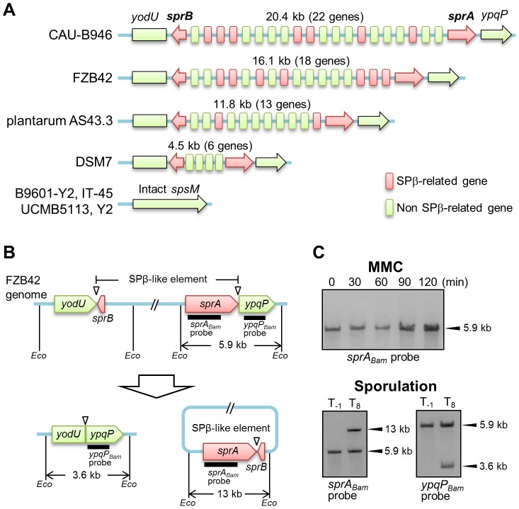
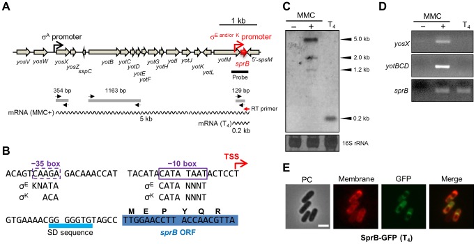
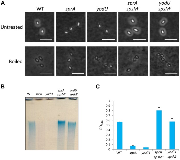
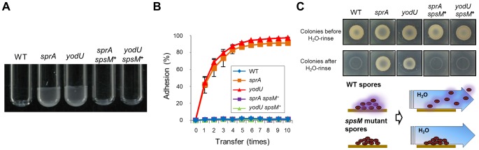
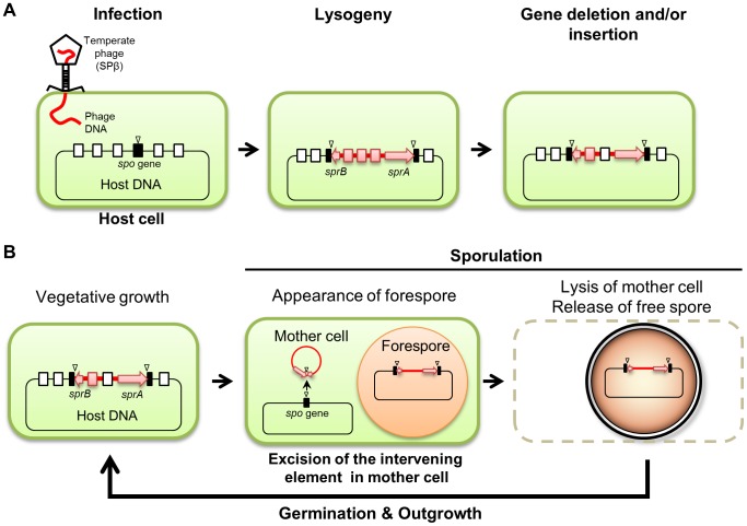
Temperate phages infect bacteria by injecting their DNA into bacterial cells, where it becomes incorporated into the host genome as a prophage. In the genome of Bacillus subtilis 168, an active prophage, SPβ, is inserted into a polysaccharide synthesis gene, spsM. Here, we show that a rearrangement occurs during sporulation to reconstitute a functional composite spsM gene by precise excision of SPβ from the chromosome. SPβ excision requires a putative site-specific recombinase, SprA, and an accessory protein, SprB. A minimized SPβ, where all the SPβ genes were deleted, except sprA and sprB, retained the SPβ excision activity during sporulation, demonstrating that sprA and sprB are necessary and sufficient for the excision. While expression of sprA was observed during vegetative growth, sprB was induced during sporulation and upon mitomycin C treatment, which triggers the phage lytic cycle. We also demonstrated that overexpression of sprB (but not of sprA) resulted in SPβ prophage excision without triggering the lytic cycle. These results suggest that sprB is the factor that controls the timing of phage excision. Furthermore, we provide evidence that spsM is essential for the addition of polysaccharides to the spore envelope. The presence of polysaccharides on the spore surface renders the spore hydrophilic in water. This property may be beneficial in allowing spores to disperse in natural environments via water flow. A similar rearrangement occurs in Bacillus amyloliquefaciens FZB42, where a SPβ-like element is excised during sporulation to reconstitute a polysaccharide synthesis gene, suggesting that this type of gene rearrangement is common in spore-forming bacteria because it can be spread by phage infection.


Conflict of interest statement

The authors have declared that no competing interests exist.

Figures

Figure 1
Figure 1. DNA rearrangement at the spsM locus.
(A) Diagram showing SPβ excision in Bacillus subtilis 168. The thick lines indicate the location of the digoxigenin (DIG)-labeled probes used for Southern blotting. Nde indicates NdeI sites. Triangles point to the attachment sites for SPβ. (B) SPβ excision upon mitomycin C (MMC) treatment and during sporulation. Left panel shows induction of SPβ excision by MMC treatment. B. subtilis 168 cells were grown in LB medium. Vegetative cells in the early log phase (OD600 = 0.25) were treated with 0.5 µg/ml MMC. Time 0 indicates the time point immediately after MMC addition. Right panels show SPβ excision (top panel) and spsM reconstitution (bottom panel) during sporulation. B. subtilis 168 cells were grown in DSM, and samples were taken at the indicated times (in h) after the onset of sporulation (T0). The DNA samples were digested with NdeI and subjected to Southern blotting. (C) Mother cell-specific SPβ excision. Chromosomal DNA from the vegetative cells (V) at T−1, whole sporangia (W) at T8, and the forespores (FS) at T8 were isolated, digested with NdeI, and subjected to Southern blotting. (D) Lytic activity of SPβ phages. SPβ phage lysate, which was prepared by treating the B. subtilis 168 vegetative cells with MMC, was spotted on the plate (MMC). The DSM culture of B. subtilis 168 at T6, T12, T24, and T48 was centrifuged and the supernatant was filtrated with 0.44 µm Millex filter (Millipore). The filtrate was spotted on the lawn of a SPβ sensitive strain CU1050 (DSM T24 and DSM T48). (E) Horizontal transfer of spsM rearrangement system. A new SPβ-lysogen, CU1050 (SPβ) was obtained by infecting CU1050 cells with the SPβ phage lysate. The CU1050 and CU1050 (SPβ) cells were induced to sporulate on DSM-agar plates at 37°C for 3 (Vegetative cells, Veg) and 12 hours (Sporulating cells, Spo). Chromosomal DNA of the CU1050 and CU1050 (SPβ) cells was subjected to Southern blotting.
Figure 2
Figure 2. DNA rearrangement of spsM in B. amyloliquefaciens.
(A) Schematic representation of the gene organization of SPβ-like elements at the spsM locus of B. amyloliquefaciens strains. Eight B. amyloliquefaciens strains with genome sequences deposited in KEGG are shown here as representative examples. The yodU and ypqP ORFs are located at the left and right ends, respectively. The red arrows indicate sprA and sprB, which are required for SPβ excision. The size (kb) of the element and number of genes in the element are shown above the diagram. The red and green boxes indicate SPβ-related and non-SPβ-related genes, respectively. The conserved SPβ genes in B. amyloliquefaciens strains are listed in Table S1. (B) Diagram of SPβ-like element excision in B. amyloliquefaciens FZB42. The thick lines indicate the DIG-labeled probes used for Southern blotting. Eco indicates EcoRV sites. Triangles point to the attachment sites for SPβ. (C) DNA rearrangement of spsM in B. amyloliquefaciens FZB42. B. amyloliquefaciens FZB42 cells were cultured at 37°C in DSM medium. Chromosomal DNA samples from the cells in the vegetative (T−1) and sporulation phases (T8) were digested with EcoRV and subjected to Southern blotting. The sprABam and ypqPBam probes were specific to B. amyloliquefaciens sprA and ypqP, respectively.
Figure 3
Figure 3. Expression of spsM, sprA, and sprB in response to mitomycin C treatment and during sporulation.
(A) β-galactosidase activity of B. subtilis strains carrying lacZ reporter constructs during sporulation. The B. subtilis strains, YODUd (yodU–lacZ), SPRAd (sprA–lacZ), and BsINDB (sprB–lacZ), were sporulated at 37°C in liquid DSM. Aliquots were collected at various time points during sporulation, and the β-galactosidase activity (Miller units, MU) was determined using ortho-nitrophenyl-β-galactoside (ONPG) as a substrate. To compare the expression pattern of yodU (5′-spsM) to that of cotG, a previously-known σK-dependent sporulation gene , the β-galactosidase activity of cotG–lacZ (COTGd) is shown on the left panel (gray line, right axis) along yodU–lacZ (purple line, left axis). SPβ excision and spsM rearrangement occurred at T3 and later time points (blue-shaded areas). The background activity was subtracted from the values. Error bars indicate ± standard deviations based on three independent experiments. (B) β-galactosidase activity of B. subtilis vegetative cells carrying the lacZ reporter construct fused transcriptionally to the promoters of spsM, sprA, and sprB in response to MMC treatment. The B. subtilis strains, YODUd (yodU–lacZ), SPRAd (sprA–lacZ), and BsINDB (sprB–lacZ), were cultured in liquid LB medium. MMC was added to a final concentration of 0.5 µg/ml when the cells reached an OD600 of 0.5. The culture was sampled at 0, 20, 40, 60, 80, 100, and 120 min after the addition of MMC. SPβ excision occurred at 60 min and later (blue-shaded areas). Error bars indicate ± standard deviations based on three independent experiments.
Figure 4
Figure 4. SPβ genes required for prophage excision.
(A) Chromosomal DNA from the vegetative (T−1) and the sporulating cells (T8) of strain 168 (WT), SPRAd (sprA), and SPRBd (sprB) were digested with NdeI and subjected to Southern blotting. The genetic maps of SPRAd and SPRBd were shown in Figure S2. (B) The schematic shows construct of the SPmini strain. Thick lines indicate sprA and ypqP probes for Southern blotting. Bgl denotes BglII restriction sites. (C) Southern blotting. Chromosomal DNA was isolated from vegetative (left panels, T−1) and sporulating cells (left panels, T8) in the DSM culture and from the SPmini vegetative cells (OD600 = 0.25) grown in LB with or without MMC treatment (0.5 µg/ml) at 37°C for 60 min (right panels). DNA was digested with BglII and subjected to Southern blotting using the sprA and the ypqP probes.
Figure 5
Figure 5. Mother cell-specific expression of sprB during sporulation.
(A) Genetic organization of the sprB region. The black and red promoter symbols indicate the promoter upstream of yosX and the mother cell-specific promoter directly upstream of sprB, respectively. The thick black line indicates the sprB probe for Northern blotting. The wavy lines indicate the sprB transcripts with their respective lengths (kb). The red and black arrows indicate the sprB-specific primer for reverse transcription (RT primer) and the yosX-, yotBCD-, and sprB-specific primers for the PCR reactions, respectively. The gray lines show the products of RT followed by PCR amplification. (B) Nucleotide sequence of the sprB promoter region. The transcriptional start site (TSS) of sprB is indicated by the red arrow. Boxes indicate −35 and −10 elements of the sprB promoter. The consensus sequences for σE and σK binding are shown below (K = G or T; N = A, T, G, or T). (C) Northern blotting. Total RNA was isolated from B. subtilis 168 vegetative cells treated with (+) or without (−) 0.5 µg/ml MMC at 37°C for 60 min and from sporulating cells 4 hours after onset of the sporulation (T4). The RNA samples were subjected to Northern blotting using the sprB probe. The bottom panel shows methylene blue-stained 16S rRNA as a loading control. (D) RT-PCR. The sprB cDNA was synthesized using the sprB-specific primer (Figure 5A, the red arrow RT primer) and total RNA from the B. subtilis 168 vegetative cells treated with (+) or without (−) MMC and from sporulating cells (T4). Internal regions of the cDNA were amplified with the yosX-, yotBCD-, and sprB-specific primer sets. The PCR product was analyzed by 2% agarose gel electrophoresis. (E) Compartmentalization of SprB–GFP expression. BsSPRBG, carrying the sprBgfp fusion gene under the control of the mother cell specific sprB promoter, was cultured at 37°C in liquid DSM containing FM4-64 (0.25 µg/ml) and kanamycin (10 µg/ml). Sporulating cells at T4 were observed using fluorescence microscopy. PC, phase contrast; Membrane, cell membranes stained with FM4-64; GFP, GFP fluorescence; Merge, merged images of Membrane and GFP. Scale bar, 2 µm.
Figure 6
Figure 6. Analysis of B. subtilis spore surface components.
(A) Negative staining with Indian ink of the B. subtilis wild-type and mutants. The purified spores from strain 168 (WT), SPRAd (sprA), YODUd (yodU), SPRAc (sprA spsM +), and YODUc (yodU spsM +) were negatively stained with Indian ink and observed using phase-contrast microscopy. Untreated, native spores; boiled, heat-treated spores at 98°C 10 min in SDS buffer. Scale bars, 4 µm. (B) Electrophoresis of B. subtilis spore surface extracts. Spore surface extracts from strain 168 (WT), SPRAd (sprA), YODUd (yodU), SPRAc (sprA spsM +), and YODUc (yodU spsM +) were loaded onto a 5% native polyacrylamide gel. The gel was stained with Stains-All after electrophoresis. (C) Quantification of the polysaccharides in spore surface extracts. The spore surface polysaccharides from B. subtilis spores were ethanol-precipitated. The precipitants were dissolved in water and reacted with Stains-All. The amounts of polysaccharides were determined by measuring the OD640 according to the method described by Hammerschmidt et al. . Error bars indicate ± standard deviations based on three independent experiments.
Figure 7
Figure 7. Spore properties.
(A) Adhesion of the mutant and wild-type spores to glass tubes. The spores purified from strain 168 (WT), SPRAd (sprA), YODUd (yodU), SPRAc (sprA spsM +), and YODUc (yodU spsM +) were resuspended in water and the final OD600 was adjusted to 15. Each 30 µl of spore resuspension was added to a Pyrex tube (13×100 mm, Corning) and vortexed gently for 30 s. After removing the spore resuspensions, the glass tubes were briefly dried and images were acquired. (B) Adhesion of the mutant and wild-type spores to polypropylene tubes. Adhesion (%) was determined by 10 successive binding reactions of the spores to the tubes. Error bars indicate ± standard deviations based on three independent experiments. (C) The polysaccharide layer facilitates spore dispersal through water flow. Overnight cultures of B. subtilis cells grown in LB medium were spotted onto DSM-agar plates. The plates were incubated at 37°C for 1 week. Each colony was confirmed as containing>95% free spores using phase-contrast microscopy. The images show the spore colonies on the DSM plates before (upper panels) and after rinsing with 1 ml of DDW (lower panels). The wild-type spores on the plates were dispersed by water, whereas the mutant spores stuck to the plates.
Figure 8
Figure 8. Model of the phage-mediated DNA rearrangement.
(A) A model of the sporulation-specific phage-mediated gene rearrangement, based on the cases of SPβ in B. subtilis and B. amyloliquefaciens. (B) Maintenance of the intervening element in the host genome. Sporulation gene (spo gene), black box; attachment sites, triangle; intervening element, red line; sprA and sprB, red arrow; phage-related genes, red box; host genes, open box.

References

    1. Tonegawa S (1983) Somatic generation of antibody diversity. Nature 302: 575–581 10.1038/302575a0 - DOI - PubMed
    1. Golden JW, Robinson SJ, Haselkorn R (1985) Rearrangement of nitrogen fixation genes during heterocyst differentiation in the cyanobacterium Anabaena. Nature 314: 419–423 10.1038/314419a0 - DOI - PubMed
    1. Golden JW, Mulligan ME, Haselkorn R (1987) Different recombination site specificity of two developmentally regulated genome rearrangements. Nature 327: 526–529 10.1038/327526a0 - DOI - PubMed
    1. Carrasco CD, Buettner JA, Golden JW (1995) Programmed DNA rearrangement of a cyanobacterial hupL gene in heterocysts. Proc Natl Acad Sci USA 92: 791–795 10.1073/pnas.92.3.791 - DOI - PMC - PubMed
    1. Ramaswamy KS, Carrasco CD, Fatma T, Golden JW (1997) Cell-type specificity of the Anabaena fdxN-element rearrangement requires xisH and xisI. Mol Microbiol 23: 1241–1249 10.1046/j.1365-2958.1997.3081671.x - DOI - PubMed

Publication types

MeSH terms