. 2014 Oct 9;10(10):e1004709.
doi: 10.1371/journal.pgen.1004709. eCollection 2014 Oct.

The formation of endoderm-derived taste sensory organs requires a Pax9-dependent expansion of embryonic taste bud progenitor cells

Affiliations

The formation of endoderm-derived taste sensory organs requires a Pax9-dependent expansion of embryonic taste bud progenitor cells

Ralf Kist et al. PLoS Genet. .

Abstract

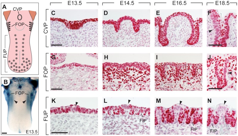
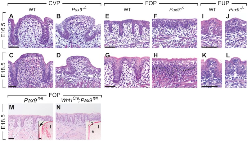
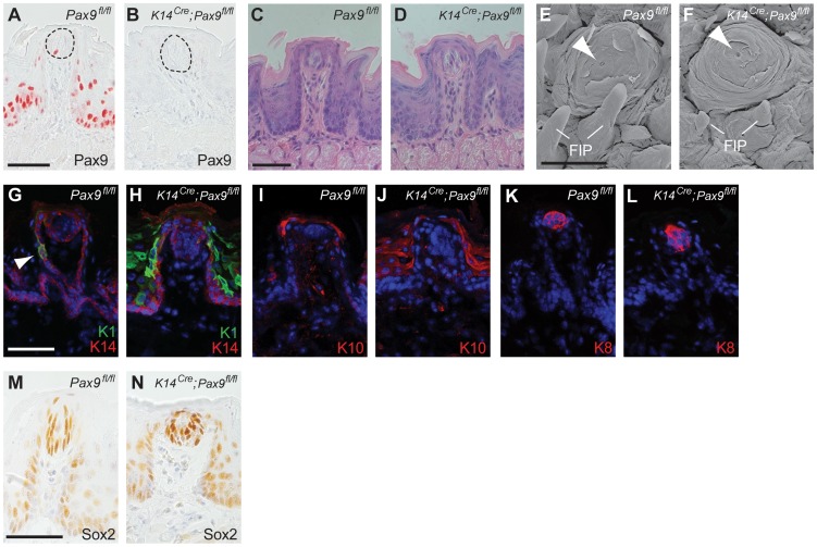
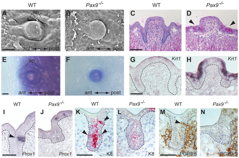
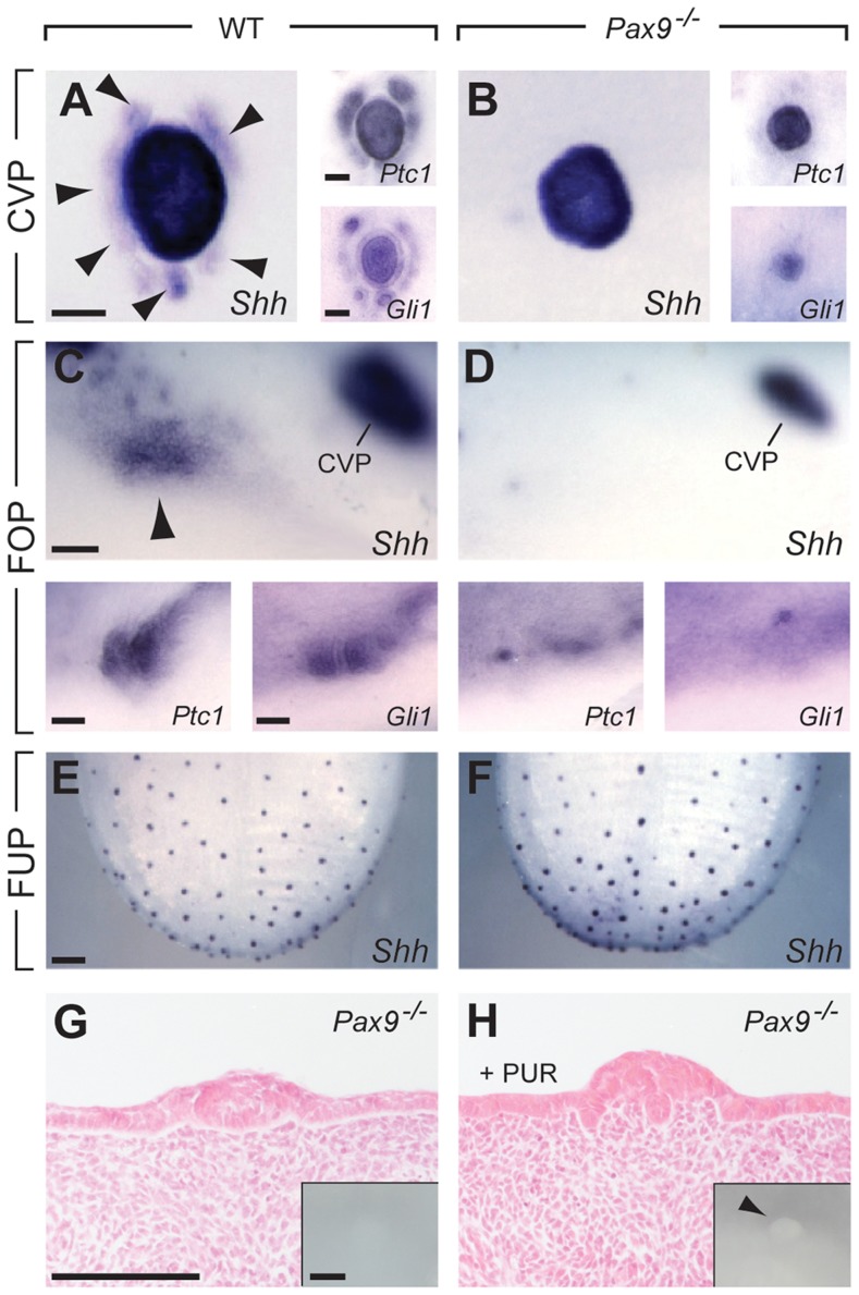
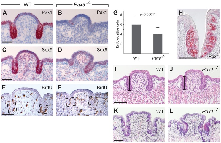
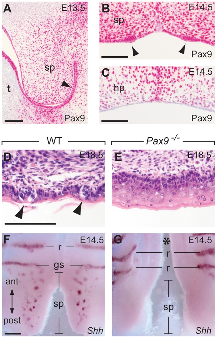
In mammals, taste buds develop in different regions of the oral cavity. Small epithelial protrusions form fungiform papillae on the ectoderm-derived dorsum of the tongue and contain one or few taste buds, while taste buds in the soft palate develop without distinct papilla structures. In contrast, the endoderm-derived circumvallate and foliate papillae located at the back of the tongue contain a large number of taste buds. These taste buds cluster in deep epithelial trenches, which are generated by intercalating a period of epithelial growth between initial placode formation and conversion of epithelial cells into sensory cells. How epithelial trench formation is genetically regulated during development is largely unknown. Here we show that Pax9 acts upstream of Pax1 and Sox9 in the expanding taste progenitor field of the mouse circumvallate papilla. While a reduced number of taste buds develop in a growth-retarded circumvallate papilla of Pax1 mutant mice, its development arrests completely in Pax9-deficient mice. In addition, the Pax9 mutant circumvallate papilla trenches lack expression of K8 and Prox1 in the taste bud progenitor cells, and gradually differentiate into an epidermal-like epithelium. We also demonstrate that taste placodes of the soft palate develop through a Pax9-dependent induction. Unexpectedly, Pax9 is dispensable for patterning, morphogenesis and maintenance of taste buds that develop in ectoderm-derived fungiform papillae. Collectively, our data reveal an endoderm-specific developmental program for the formation of taste buds and their associated papilla structures. In this pathway, Pax9 is essential to generate a pool of taste bud progenitors and to maintain their competence towards prosensory cell fate induction.


Conflict of interest statement

The authors have declared that no competing interests exist.

Figures

Figure 1
Figure 1. Expression patterns of Pax9 in different taste papillae of the embryonic mouse tongue.
(A) Drawing showing the localization of the circumvallate papilla (CVP), foliate papillae (FOP), and fungiform papillae (FUP) in the mouse tongue. (B) Whole mount X-Gal staining of a Pax9+/LacZ mouse tongue at embryonic day 13.5 (E13.5). Note that expression is also seen in the mesenchyme adjacent to the developing FOP (arrowheads) and that the color reaction was stopped before epithelial staining began to obscure the mesenchymal expression domain. (C–N) Pax9 immunostaining of taste papillae during development on cross sections (C–F; K–N) and horizontal sections of the tongue (G–J). (C–F) Pax9 is expressed in the epithelium during CVP morphogenesis and is down-regulated in some regions of the trenches at E18.5 (arrowhead in F). (G–J) In addition to the epithelium, Pax9 is also expressed in the mesenchyme during FOP development, while reduced Pax9 levels were observed in the trenches at E18.5 (arrowhead in J). (K–N) In the anterior part of the tongue Pax9 is expressed in the FUP epithelium and in filiform papillae (FIP). Note that the expression is very weak or absent in the taste placodes (arrowheads). Scale bars: 200 µm in B; 50 µm in other panels.
Figure 2
Figure 2. Arrest of CVP and FOP development in Pax9-deficient mouse embryos.
(A,C) In wild type (WT) embryos, the invaginating CVP epithelium forms deep trenches. (B,D) Rudimentary CVP trenches form in Pax9−/− embryos at E16.5 (B) but these trenches fail to invaginate (D). (E,G) A series of invaginations develop in the FOP of wild type embryos. (F,H) FOP trenches are absent in Pax9 mutants. (I–L) FUP development on the dorsal tongue. The FUP of wild type embryos (I,K) and Pax9−/− embryos (J,L) are morphologically indistinguishable. (M,N) FOP development in Pax9fl/fl embryos. (M) Without Cre expression, FOP development at E14.5 is normal and Pax9 expression is detectable in both epithelium and mesenchyme of the tongue (t), as well as in the adjacent lower jaw mesenchyme (arrow; inset shows a coronal section of the posterior region of the tongue). (N) Wnt1Cre-mediated inactivation of Pax9fl/fl did not disrupt the formation of epithelial invaginations. Note that Pax9-positive cells are not detectable in the tongue mesenchyme (asterisk in inset) or in the mesenchyme of the non-elevated secondary palate (p). Scale bars: 50 µm.
Figure 3
Figure 3. FUP maintenance and FUP taste bud renewal do not require Pax9 functions.
All analyses were carried out using 3–5 months old mice. (A,B) Pax9 immunostaining of FUPs. In Pax9fl/fl mice (A), Pax9 expression is detected in the FUP epithelium and in isolated taste bud cells (area of taste bud is indicated by dotted line). (B) No Pax9-positive cells are detectable in the FUP after K14Cre-mediated recombination of Pax9fl/fl. (C,D) Histological sections of FUP. Pax9fl/fl FUP (C) and K14Cre;Pax9fl/fl FUP (D) are morphologically indistinguishable. (E,F) Scanning electron microscopy images of FUP. The FUP of both Pax9fl/fl (E) and K14Cre;Pax9fl/fl (F) form taste pores (arrowhead), whereas the non-sensory FIP of the mutants (F) are hypoplastic. (G–L) Indirect immunofluorescent detection of keratins. Nuclei were stained with DAPI (blue). (G) In Pax9fl/fl mice, K14 is expressed in basal cells of the epithelium and K1 expression was seen in isolated epithelial cells of the FUP epithelium (arrowhead). (H) While K14 expression was not affected in the FUP of K14Cre;Pax9fl/fl mice, the number K1 expressing cells was strongly increased. (I,J) K10 expression is mainly restricted to the apical end of the FUP in Pax9fl/fl mice (I) whereas its expression is more extended in K14Cre;Pax9fl/fl mice (J). (K,L) K8 expression marks taste bud cells in both genotypes. (M,N) Immunohistochemical staining showing that Sox2 is expressed in mature taste buds of both Pax9fl/fl (M) and K14Cre;Pax9fl/fl (N) mice. Scale bars: 50 µm in A,C,G,M; 500 µm in E.
Figure 4
Figure 4. Differentiation defects and lack of proneural induction in the Pax9-deficient CVP trench epithelium.
(A–N) Analyses of mouse embryos at E18.5. Anterior (ant) to posterior (post) orientation is indicated where appropriate. (A,B) SEM images of the CVP showing that both central dome and accessory papillae (ap) are well developed and separated by trenches in the wild type (A) but not in the Pax9−/− embryo (B). (C,D) PAS staining indicates intensively increased concentration of mucopolysaccharides in the mutant CVP trenches (arrowheads in (D). (E,F) Whole-mount barrier assay revealing that the CVP and the posterior tongue epithelium is permeable to toluidine blue in the wild type embryo (E), while a premature barrier has formed in the epithelium surrounding the mutant CVP (F). (G,H) Krt1 RNA in situ hybridization showing that Krt1 expression is strongly up-regulated in the Pax9−/− CVP (H). Dashed lines indicate the margin of the trench epithelium. (I,J) In situ hybridization of Prox1. Groups of epithelial trench cells express the proneural marker Prox1 in the wild type (I) but not in the Pax9−/− embryo (J). (K,L) Immunostaining of K8. Similar to Prox1, K8 is locally expressed in the wild type CVP (arrowheads in K). In contrast, only weak expression of K8 was detectable in the mutant CVP (L). (M,N) Immunostaining of PGP9.5. In the wild type CVP (M), nerve fibers contact the CVP trench epithelium (arrowhead; this section is directly adjacent to that shown in (K)), while nerve endings fail to invade the CVP trench epithelium of the Pax9−/− embryo (N). Scale bars: 100 µm in A,C,E; 50 µm in G,I,K,M.
Figure 5
Figure 5. Absence of Pax9 causes an endoderm-specific disruption of the Shh pathway in taste papillae.
(A–F) Whole mount in situ hybridization of Shh, its receptor (Ptc1) and the downstream effector transcription factor (Gli1) at E14.5. (A,B) In the wild type CVP (A), Shh is expressed in the central dome as well as in a ring of accessory papillae (arrowheads). Ptc1 and Gli are expressed in a similar pattern. In the absence of Pax9, Shh, Ptc1 and Gli1 are only expressed in the central dome of the CVP (B). (C,D) In wild type embryos (C), patches of Shh, Ptc1 and Gli1 expression are detectable in the region of the developing FOP, whereas these expression patterns are missing (Shh) or are greatly reduced (Ptc1, Gli1) in Pax9−/− embryos (D). (E,F) Shh expression in FUP placodes is similar in wild type (E) and Pax9-deficient (F) embryos. (G, H) Histological sections of Pax9-deficient, cultured embryonic tongues. (G) In control medium the Pax9−/− CVP of cultured tongues is small and is not visible externally (inset). (H) In the presence of purmorphamine (PUR) the number of epithelial cells is increased in the dome of the CVP. Note the absence of trenches. Inset shows enlarged, protruded CVP dome (arrowhead) of the cultured tongue. Scale bars: 100 µm in A,C,G: 200 µm in E.
Figure 6
Figure 6. Pax1 and Sox9 are Pax9 targets in the proliferating compartment of the CVP trenches.
(A–F) Immunohistochemical staining on sections of the CVP at E15.5. (A,B) Pax1 is strongly expressed in the tips of epithelial trenches and in periderm cells covering the central dome of the wild type CVP (A), but not in the Pax9-deficient CVP (B). (C,D) Similarly, Sox9 expression is strongest in the epithelial trenches (C) and is barely detectable in the Pax9 mutant CVP (D). (E,F) BrdU-positive cells were counted in defined areas (boxed) of the CVP trenches from three wild type (n = 29 sections) and three Pax9 mutant CVPs (n = 28 sections). (G) The number of proliferating cells in the Pax9-deficient CVP is significantly reduced. Error bars illustrate standard deviation. (H) Pax1 immunostaining of one CVP trench in a 3 months old wild type mouse. (I,J) Morphology of the CVP at E18.5. The lengths of the CVP trenches (indicated by bars) were measured and shown to be reduced in the absence of Pax1 (for summary of measurements see Table S1). (K,L) Morphology of the CVP at postnatal day 16. In Pax1 mutants (n = 3) the trenches are growth-retarded and contain fewer taste buds. Scale bars: 50 µm in A,C,E; 100 µm in H,I,K.
Figure 7
Figure 7. Pax9 is essential for taste placode formation in the soft palate.
(A–C) Pax9 immunostaining of the secondary palate. (A) At E13.5, Pax9 expression is found in the mesenchyme as well as in those epithelial cells (arrowhead) of the soft palate (sp) facing the oral cavity after palatal shelf elevation. (B) At E14.5 the palatal shelves have elevated and Pax9 expression is seen in epithelial placodes (arrowheads) of the soft palate. (C) Pax9 is not expressed in the epithelium of the hard palate (hp). (D,E) Histological staining revealed taste bud precursors in wild type (D), but not in the Pax9-deficient (E) epithelium of the soft palate at E18.5. (F,G) Whole-mount Shh in situ hybridization at E14.5. In the wild type soft palate (F), Shh expression marks the taste placodes of the soft palate as well as the “Geschmacksstreifen” (gs). Note that palatal rugae (r) also express Shh at this stage. (G) Shh expression is not detectable in the soft palate of Pax9 mutants, which also form a cleft secondary palate (asterisk). Scale bars: 100 µm in A–D; 200 µm in F.

Similar articles

Cited by

References

    1. Iwasaki S (2002) Evolution of the structure and function of the vertebrate tongue. J Anat 201: 1–13. - PMC - PubMed
    1. Northcutt RG (2004) Taste buds: development and evolution. Brain Behav Evol 64: 198–206. - PubMed
    1. Kapsimali M, Barlow LA (2013) Developing a sense of taste. Semin Cell Dev Biol 24: 200–209. - PMC - PubMed
    1. Iwatsuki K, Liu HX, Grónder A, Singer MA, Lane TF, et al. (2007) Wnt signaling interacts with Shh to regulate taste papilla development. Proc Natl Acad Sci U S A 104: 2253–2258. - PMC - PubMed
    1. Liu F, Thirumangalathu S, Gallant NM, Yang SH, Stoick-Cooper CL, et al. (2007) Wnt-beta-catenin signaling initiates taste papilla development. Nat Genet 39: 106–112. - PubMed

Publication types

MeSH terms

LinkOut - more resources