. 2014 Oct 9;5(10):e1455.
doi: 10.1038/cddis.2014.417.

c-Rel is a critical mediator of NF-κB-dependent TRAIL resistance of pancreatic cancer cells

Affiliations

c-Rel is a critical mediator of NF-κB-dependent TRAIL resistance of pancreatic cancer cells

C Geismann et al. Cell Death Dis. .

Abstract

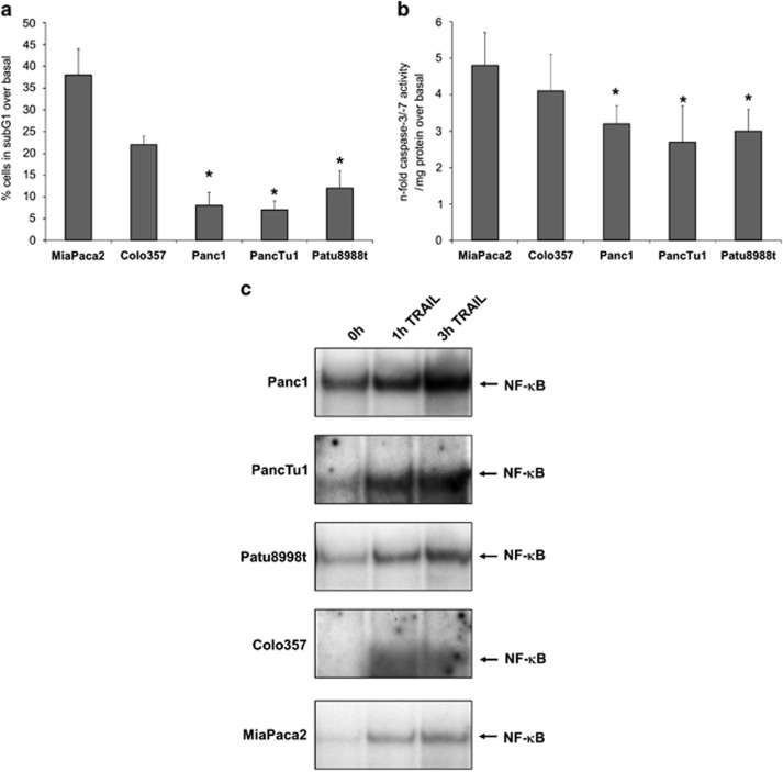
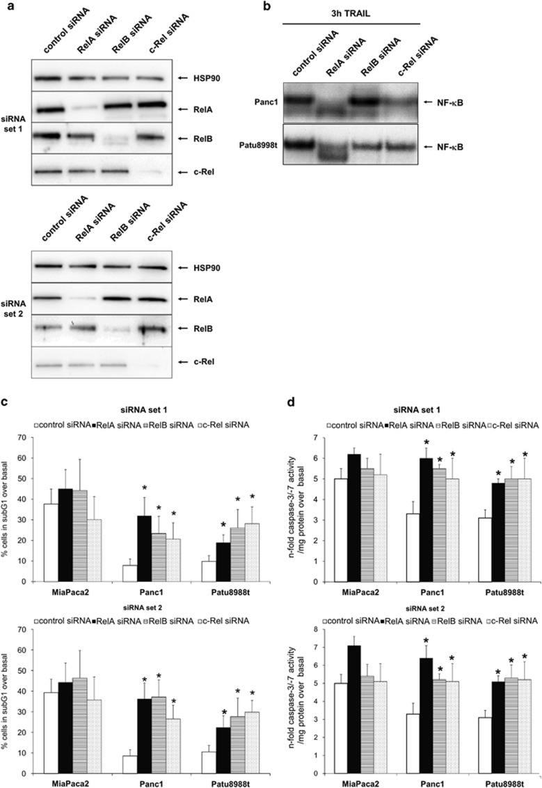
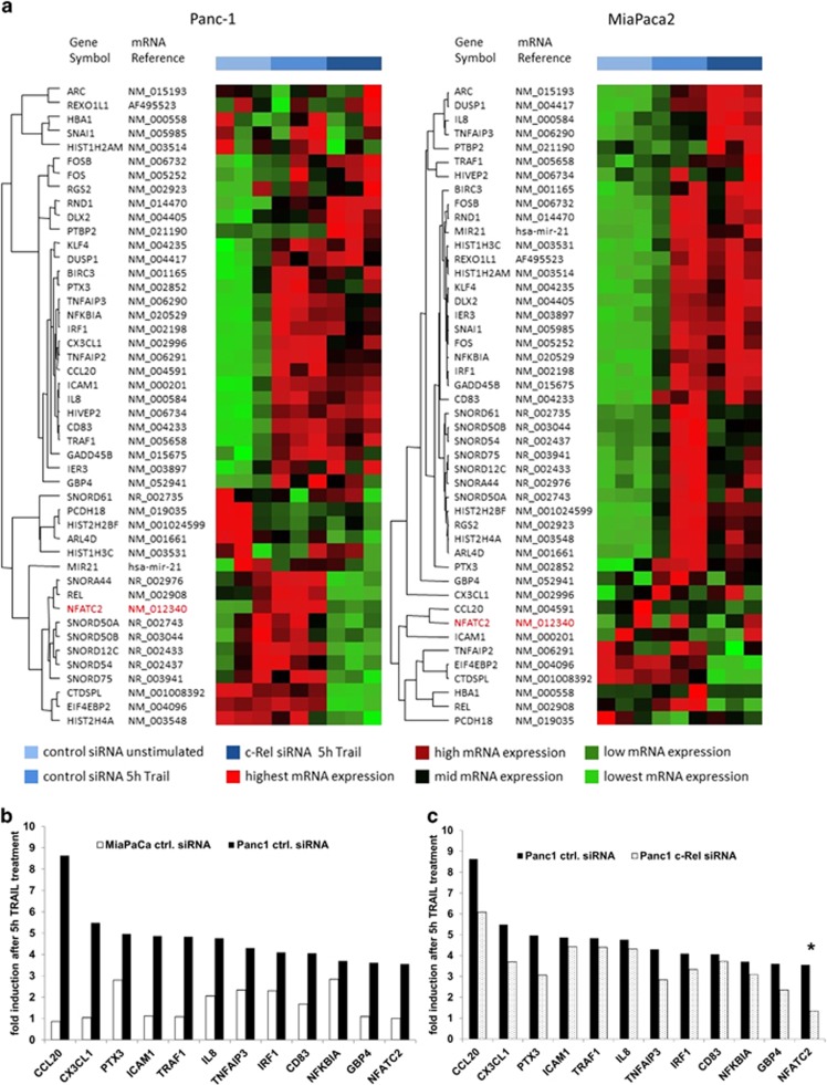
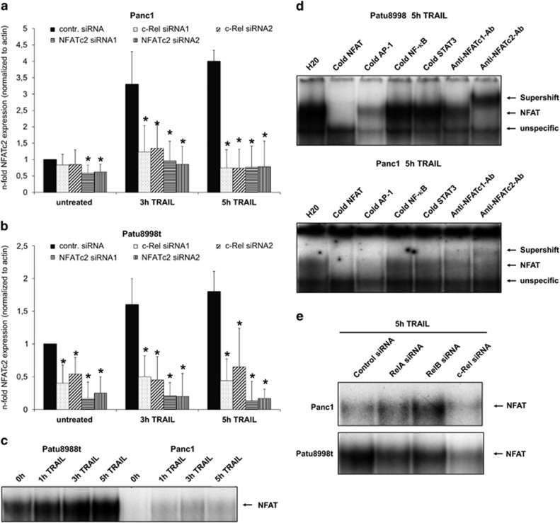
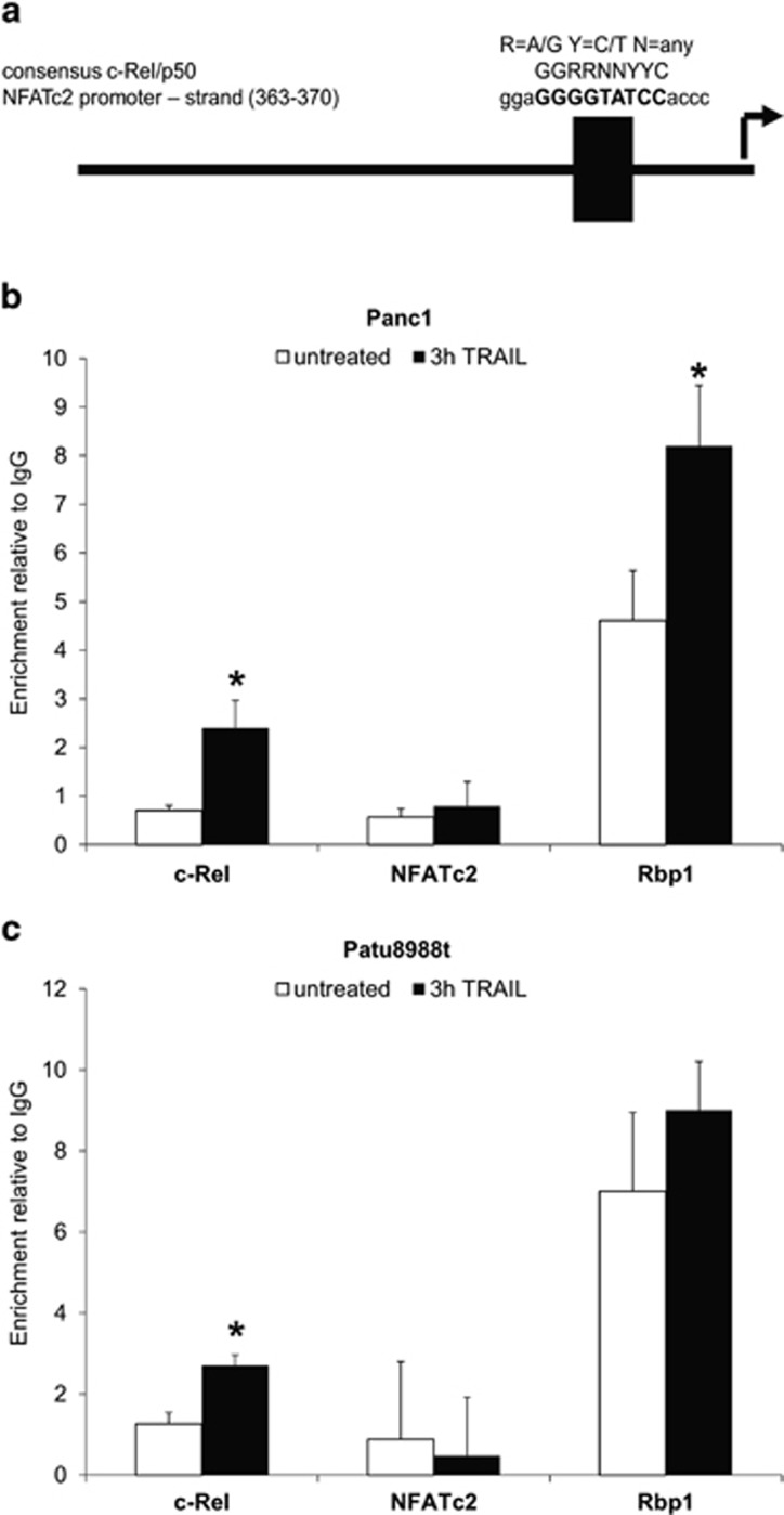
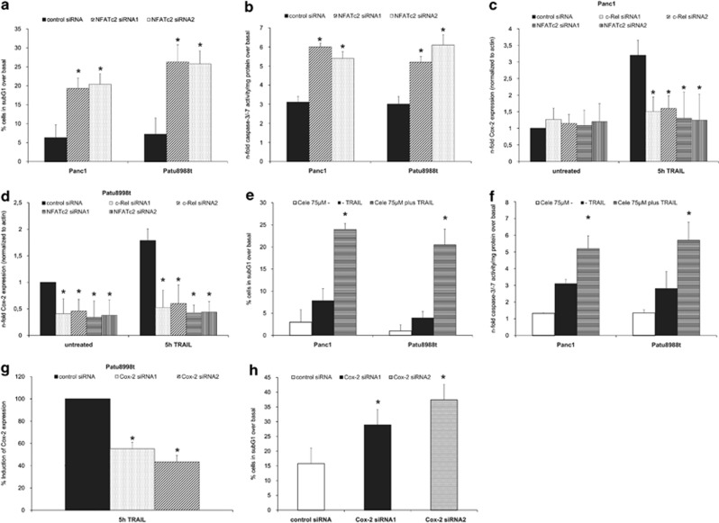
Pancreatic ductal adenocarcinoma (PDAC) represents one of the deadliest malignancies with an overall life expectancy of 6 months despite current therapies. NF-κB signalling has been shown to be critical for this profound cell-autonomous resistance against chemotherapeutic drugs and death receptor-induced apoptosis, but little is known about the role of the c-Rel subunit in solid cancer and PDAC apoptosis control. In the present study, by analysis of genome-wide patterns of c-Rel-dependent gene expression, we were able to establish c-Rel as a critical regulator of tumour necrosis factor-related apoptosis-inducing ligand (TRAIL)-induced apoptosis in PDAC. TRAIL-resistant cells exhibited a strong TRAIL-inducible NF-κB activity, whereas TRAIL-sensitive cells displayed only a small increase in NF-κB-binding activity. Transfection with siRNA against c-Rel sensitized the TRAIL-resistant cells in a manner comparable to siRNA targeting the p65/RelA subunit. Gel-shift analysis revealed that c-Rel is part of the TRAIL-inducible NF-κB complex in PDAC. Array analysis identified NFATc2 as a c-Rel target gene among the 12 strongest TRAIL-inducible genes in apoptosis-resistant cells. In line, siRNA targeting c-Rel strongly reduced TRAIL-induced NFATc2 activity in TRAIL-resistant PDAC cells. Furthermore, siRNA targeting NFATc2 sensitized these PDAC cells against TRAIL-induced apoptosis. Finally, TRAIL-induced expression of COX-2 was diminished through siRNA targeting c-Rel or NFATc2 and pharmacologic inhibition of COX-2 with celecoxib or siRNA targeting COX-2, enhanced TRAIL apoptosis. In conclusion, we were able to delineate a novel c-Rel-, NFATc2- and COX-2-dependent antiapoptotic signalling pathway in PDAC with broad clinical implications for pharmaceutical intervention strategies.


Figures

Figure 1
Figure 1
TRAIL sensitivity correlates with NF-κB activity in PDAC cell lines. (a and b) Cells were treated with 10 ng/ml TRAIL for 24  and 5 h, respectively. Apoptosis was determined by analysing sub-G1 content (a) or caspase-3/-7 activity (b). Data are presented as % of sub-G1 content over basal (untreated cells) or expressed as n-fold caspase-3/-7 activity normalized to the cellular protein content and represent the mean value±S.D. from six independent experiments. *P-values <0.05. (c) Cells were left untreated or were treated with 10 ng/ml TRAIL for the indicated time periods and submitted to EMSA for measurement of NF-κB-binding activity. All samples was loaded on the same gel to compare the binding activity. Only for the presentation each cell line is presented as a single row but shows the same amount of total nuclear protein and the same exposition time. A representative of four independent experiments is shown
Figure 2
Figure 2
All Rel subunits are involved in TRAIL-induced NF-κB and apoptosis. (a) Whole-cell extracts of Panc1 cells transfected with the indicated siRNA for 48 h were submitted to western blotting for the indicated Rel subunit and Hsp90 as the loading control. A representative of four independent experiments with two different sets of siRNA is shown. (b) Cells were transfected with the indicated siRNA (set1) for 48 h, treated with 10 ng/ml TRAIL for 3 h and nuclear extracts were submitted to EMSA for measurement of NF-κB-binding activity. A representative of four independent experiments is shown. (c and d) Cells were transfected with with two different sets of siRNA (as indicated) for 48 h and then treated with 10 ng/ml TRAIL for 24 and 5 h, respectively. Apoptosis was determined by analysing sub-G1 content (c) or caspase-3/-7 activity (d). Data are presented as % of sub-G1 content over basal (untreated cells) or expressed as n-fold caspase-3/-7 activity normalized to the cellular protein content and represent the mean value±S.D. from six independent experiments. *P-values <0.05
Figure 3
Figure 3
NFATc2 is a c-Rel target gene in TRAIL-resistant Panc1 cells. (a) Hierarchical clustering of top 50 differentially expressed genes in Panc1 cells and MiaPaca2 cells. Transcripts were selected for being differentially expressed in either of the cell lines and associated with characterized genes. Transcripts are displayed in rows and samples are listed in columns, whereas the row dendogram represents the similarity between the individual transcripts. Transcription levels and experimental groups are colour coded. Hierarchical clustering was performed using the unweight pair group method with arithmetic mean as a cluster algorithm, and similarities between transcripts are based on their Spearman rank correlation. For better readability, all transcripts were z-score normalized before cluster analysis. (b) Transcript levels were normalized to unstimulated cells and the 12 genes with the highest fold induction in the resistant Panc1 cells transfected with the control siRNA (black columns) are presented in comparison with the sensitive MiaPaca2 cell line transfected with the control siRNA (white columns). Data represent the mean of three independent experiments. (c) Transcript levels were normalized to unstimulated cells and the effect of the transfection of resistant Panc1 cells with the c-Rel siRNA in comparison with the resistant Panc1 cells transfected with the control siRNA are presented. Data represent the mean of three rindependent experiments. *P-values <0.05 for the effect of the c-Rel siRNA on TRAIL-mediated gene induction
Figure 4
Figure 4
TRAIL-induced NFATc2 expression and DNA binding are c-Rel dependent. (a and b) Panc1 (a) and Patu8998t (b) cells were transfected with the indicated siRNA for 48 h. Then cells were left untreated or treated with 10 ng/ml TRAIL for 3 or 5 h. Total RNA was submitted to reverse transcription and real-time PCR detecting NFATc2. For normalization, beta-actin was analysed as control. Data are expressed as normalized mRNA level and represent the mean values±S.D. from four independent experiments performed in duplicates. *P-values <0.05. (c) Patu8998t (left part) and Panc1 (right part) cells were left untreated or were treated with 10 ng/ml TRAIL for the indicated time periods and submitted to EMSA for measurement of NFAT-binding activity. A representative of four independent experiments is shown. (d) Nuclear extracts from Panc1 (upper part) or Patu8998t cells (lower part) treated for 5 h with 10 ng/ml TRAIL were submitted to EMSA with the indicated antibodies and was performed using the indicated oligonucleotides. A representative of four independent experiments is shown. (e) Panc1 (upper part) and Patu8998t (lower part) cells were transfected with the indicated siRNA (set1) for 48 h, and then treated with 10 ng/ml TRAIL for 5 h and submitted to EMSA for measurement of NFAT-binding activity. A representative of four independent experiments is shown
Figure 5
Figure 5
Characterization of a functional c-Rel binding site in the NFATc2 promoter. (a) Schematic representation of the putative c-Rel binding site (MatInspector query). For comparison, the c-Rel/p50 consensus site GGRRNNYYC (where R is for A/G, Y for C/T and N for any base) is shown. The putative binding motive in the NFATc2 promoter is highlighted in bold. (b and c) Panc1 (b) or Patu8998t cells (c) were left untreated (withe columns) or treated with 10 ng/ml TRAIL for 3 h (black columns) and qChIP were conducted with IgG, anti-c-Rel, anti-NFATc2 and anti-Rbp1 antibodies. Data are expressed as enrichment relative to IgG levels and represent the mean values±S.D. from six independent experiments performed in duplicates. *P-values <0.05
Figure 6
Figure 6
NFATc2 and COX-2 are downstream mediators of c-Rel in TRAIL resistance. (a and b) Cells were transfected with control or NFATc2 siRNA for 48 h and treated with 10 ng/ml TRAIL for 24 and 5 h, respectively. Apoptosis was determined by analysing sub-G1 content (a) or caspase-3/-7 activity (b). Data are presented as % of sub-G1 content over basal activity (untreated cells) or expressed as n-fold caspase-3/-7-activity normalized to the cellular protein content and represent the mean value±S.D. from six independent experiments. (c and d) Panc1 (c) and Patu8998t (d) cells were transfected with the indicated siRNA for 48 h. Then, cells were left untreated or treated with 10 ng/ml TRAIL for 5 h. Total RNA was submitted to reverse transcription and real-time PCR detecting COX-2. For normalization, beta-actin was analysed as control. Data are expressed as normalized mRNA level and represent the mean values±S.D. from four independent experiments performed in duplicates. *P-values <0.05. (e and f) Cells were pretreated with 75 μM celecoxib (Cele) for 1/2 h and then treated with 10 ng/ml TRAIL for 24 and 5 h, respectively. Apoptosis was determined by analysing sub-G1 content (e) or caspase-3/-7 activity (f). Data are presented as % of sub-G1 content over basal activity (untreated cells) or expressed as n-fold caspase-3/-7 activity normalized to the cellular protein content and represent the mean value±S.D. from six independent experiments. *P-values <0.05. (g) Patu8998t cells were transfected with control or COX-2 siRNA for 48 h. Then, cells were left untreated or treated with 10 ng/ml TRAIL for 5 h. Total RNA was submitted to reverse transcription and real-time PCR detecting COX-2. For normalization, beta-actin was analysed as control. Data are expressed % induction of COX-2 mRNA with control siRNA-transfected cells set as 100% and represent the mean values±S.D. from four independent experiments performed in duplicates. *P-values <0.05. (h) Cells were transfected with control or COX-2 siRNA for 48 h and treated with 10 ng/ml TRAIL for 24 and 5 h, respectively. Apoptosis was determined by analysing sub-G1 content. Data are presented as % of sub-G1 content over basal activity (untreated cells) and represent the mean value±S.D. from six independent experiments

Similar articles

Cited by

References

    1. 1Siegel R, Naishadham D, Jemal A. Cancer statistics, 2013. Cancer J Clin 2013; 63: 11–30. - PubMed
    1. 2Conroy T, Desseigne F, Ychou M, Bouche O, Guimbaud R, Becouarn Y et al. FOLFIRINOX versus gemcitabine for metastatic pancreatic cancer. N Engl J Med 2011; 364: 1817–1825. - PubMed
    1. 3Hill R, Rabb M, Madureira PA, Clements D, Gujar SA, Waisman DM et al. Gemcitabine-mediated tumour regression and p53-dependent gene expression: implications for colon and pancreatic cancer therapy. Cell Death Dis 2013; 4: e791. - PMC - PubMed
    1. 4Arlt A, Muerkoster SS, Schafer H. Targeting apoptosis pathways in pancreatic cancer. Cancer Lett 2013; 332: 346–358. - PubMed
    1. 5Wormann SM, Diakopoulos KN, Lesina M, Algul H. The immune network in pancreatic cancer development and progression. Oncogene 2013; 33: 2956–2967. - PubMed

Publication types

MeSH terms