. 2014 Oct 28;8(10):10998-1006.
doi: 10.1021/nn505162u. Epub 2014 Oct 13.

Light-scattering detection below the level of single fluorescent molecules for high-resolution characterization of functional nanoparticles

Affiliations

Light-scattering detection below the level of single fluorescent molecules for high-resolution characterization of functional nanoparticles

Shaobin Zhu et al. ACS Nano. .

Abstract

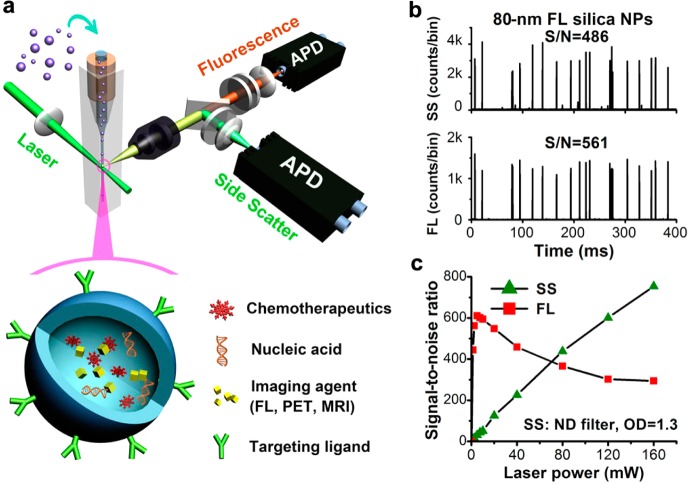
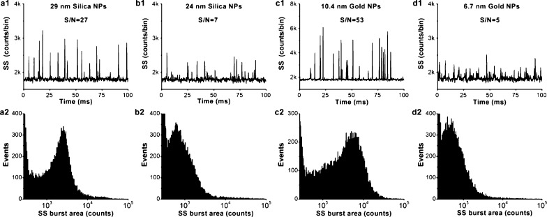
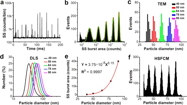
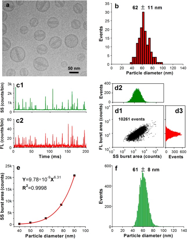
Ultrasensitive detection and characterization of single nanoparticles (<100 nm) is important in nanotechnology and life sciences. Direct measurement of the elastically scattered light from individual nanoparticles represents the simplest and the most direct method for particle detection. However, the sixth-power dependence of scattering intensity on particle size renders very small particles indistinguishable from the background. Adopting strategies for single-molecule fluorescence detection in a sheathed flow, here we report the development of high sensitivity flow cytometry (HSFCM) that achieves real-time light-scattering detection of single silica and gold nanoparticles as small as 24 and 7 nm in diameter, respectively. This unprecedented sensitivity enables high-resolution sizing of single nanoparticles directly based on their scattered intensity. With a resolution comparable to that of TEM and the ease and speed of flow cytometric analysis, HSFCM is particularly suitable for nanoparticle size distribution analysis of polydisperse/heterogeneous/mixed samples. Through concurrent fluorescence detection, simultaneous insights into the size and payload variations of engineered nanoparticles are demonstrated with two forms of clinical nanomedicine. By offering quantitative multiparameter analysis of single nanoparticles in liquid suspensions at a throughput of up to 10 000 particles per minute, HSFCM represents a major advance both in light-scattering detection technology and in nanoparticle characterization.

Keywords: flow cytometry; light scattering; nanomedicine; nanoparticle characterization; single-molecule detection; single-nanoparticle detection; size distribution.


Figures

Figure 1
Figure 1
Laboratory-built HSFCM system and preliminary performance evaluation. (a) Schematic diagram of the laboratory-built apparatus and the structure of a nanomedicine. (b) Representative side-scatter and fluorescence (524/24 band pass) burst traces of 80 nm diameter fluorescent silica nanoparticles (425 MESF AF532) at a laser power of 16 mW, with a focused laser spot of 16 μm. (c) Effect of the laser power on the signal-to-noise ratios of the SS and FL detection of the 80 nm fluorescent silica nanoparticles. The focused laser spot was 6.4 μm, and a neutral density filter (OD = 1.3) was placed in the SS light path to avoid APD saturation.
Figure 2
Figure 2
Light-scattering detection of single particles in the low nanometer range using HSFCM. (a–d) Representative SS burst traces (1) and burst-area distribution histograms (2) for 29 nm diameter silica nanoparticles (a), 14 nm diameter silica nanoparticles (b), 10.4 nm diameter gold nanoparticles (c), and 6.7 nm diameter gold nanoparticles (d). The laser excitation power was 160 mW, the focused laser spot was 6.4 μm, and no ND filter was placed in the SS light path. Each distribution histogram was derived from 1 min of data, and the nanoparticle concentrations were approximately 1–5 × 10⁹/mL.
Figure 3
Figure 3
Comparison of HSFCM and conventional approaches for nanoparticle size distribution analysis. (a) Typical SS burst traces of a mixture of silica nanoparticles of five different sizes detected using the HSFCM. (b) SS burst-area distribution histogram derived from 1 min of data and the fit to a sum of Gaussian peaks. (c) Compiled particle size distribution histograms with a bin width of 2 nm for individual samples of five sizes of nanoparticles measured separately via TEM. (d) Compiled particle size distribution histograms (by particle number) for individual samples of five sizes of nanoparticles measured separately via DLS. (e) Plot of Gaussian-fitted SS burst areas as a function of the particle sizes determined via TEM. (f) Particle size distribution histogram with a bin width of 0.5 nm for the mixture of silica nanoparticles of five different sizes obtained using HSFCM.
Figure 4
Figure 4
Characterization of doxorubicin-encapsulating liposomes. (a) Representative cryo-TEM image of Doxoves. (b) Particle size distribution histogram of Doxoves liposomes obtained from cryo-TEM images. (c) Representative SS and FL burst traces of Doxoves (diluted 4000-fold in 5% glucose) measured using HSFCM. (d) Bivariate dot-plot of the FL versus the SS burst area for Doxoves preparation. (e) Plot of nanoparticle SS burst area as a function of particle size for monodisperse silica nanoparticle standards. (f) Particle size distribution profile of Doxoves liposomes measured using HSFCM and the calibration curve presented in panel e.
Figure 5
Figure 5
Characterization of siRNA nanomedicine using HSFCM. (a) Bivariate dot-plot of the FL burst area versus the SS burst area for a mixture of 80 nm diameter fluorescent nanoparticles with unstained siRNA-loaded LNPs. (b,c) Representative SS and FL burst traces for empty LNPs (b) and siRNA-loaded LNPs (c). (d) Compiled bivariate dot-plot of the FL burst area versus the SS burst area for empty and siRNA-loaded LNPs.

References

    1. Jain R. K.; Stylianopoulos T. Delivering Nanomedicine to Solid Tumors. Nat. Rev. Clin. Oncol. 2010, 7, 653–664. - PMC - PubMed
    1. Abeylath S. C.; Ganta S.; Iyer A. K.; Amiji M. Combinatorial-Designed Multifunctional Polymeric Nanosystems for Tumor-Targeted Therapeutic Delivery. Acc. Chem. Res. 2011, 44, 1009–1017. - PubMed
    1. Bourzac K. Nanotechnology: Carrying Drugs. Nature 2012, 491, S58–S60. - PubMed
    1. Hubbell J. A.; Langer R. Translating Materials Design to the Clinic. Nat. Mater. 2013, 12, 963–966. - PubMed
    1. Chauhan V. P.; Jain R. K. Strategies for Advancing Cancer Nanomedicine. Nat. Mater. 2013, 12, 958–962. - PMC - PubMed

Publication types

Substances