. 2015 Jan 1:104:146-55.
doi: 10.1016/j.neuroimage.2014.09.068. Epub 2014 Oct 6.

Regional quantification of cerebral venous oxygenation from MRI susceptibility during hypercapnia

Affiliations

Regional quantification of cerebral venous oxygenation from MRI susceptibility during hypercapnia

Audrey P Fan et al. Neuroimage. .

Abstract

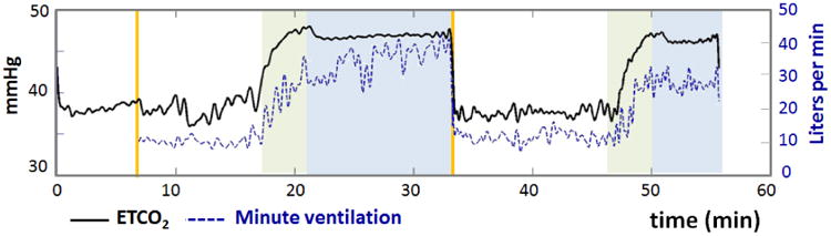
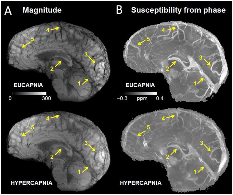
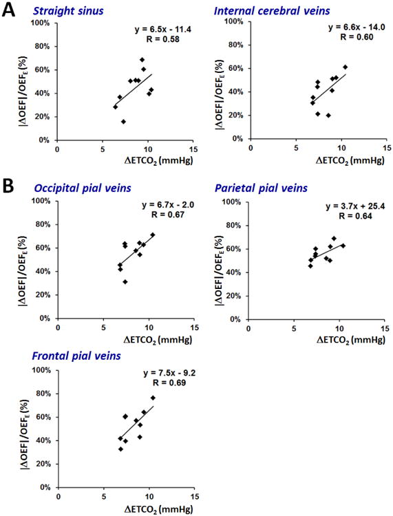
There is an unmet medical need for noninvasive imaging of regional brain oxygenation to manage stroke, tumor, and neurodegenerative diseases. Oxygenation imaging from magnetic susceptibility in MRI is a promising new technique to measure local venous oxygen extraction fraction (OEF) along the cerebral venous vasculature. However, this approach has not been tested in vivo at different levels of oxygenation. The primary goal of this study was to test whether susceptibility imaging of oxygenation can detect OEF changes induced by hypercapnia, via CO2 inhalation, within selected a priori brain regions. Ten healthy subjects were scanned at 3T with a 32-channel head coil. The end-tidal CO2 (ETCO2) was monitored continuously and inspired gases were adjusted to achieve steady-state conditions of eucapnia (41±3mmHg) and hypercapnia (50±4mmHg). Gradient echo phase images and pseudo-continuous arterial spin labeling (pcASL) images were acquired to measure regional OEF and CBF respectively during eucapnia and hypercapnia. By assuming constant cerebral oxygen consumption throughout both gas states, regional CBF values were computed to predict the local change in OEF in each brain region. Hypercapnia induced a relative decrease in OEF of -42.3% in the straight sinus, -39.9% in the internal cerebral veins, and approximately -50% in pial vessels draining each of the occipital, parietal, and frontal cortical areas. Across volunteers, regional changes in OEF correlated with changes in ETCO2. The reductions in regional OEF (via phase images) were significantly correlated (P<0.05) with predicted reductions in OEF derived from CBF data (via pcASL images). These findings suggest that susceptibility imaging is a promising technique for OEF measurements, and may serve as a clinical biomarker for brain conditions with aberrant regional oxygenation.

Keywords: Hypercapnia; Oxygen extraction fraction; Oxygenation imaging; Quantitative susceptibility.


Figures

Figure 1
Figure 1
Physiological time courses of end-tidal CO2 (ETCO2) in mmHg and minute ventilation in L/min for one healthy subject. Green regions indicate transition periods (∼4 minutes) between eucapnia and hypercapnia, and blue regions indicate periods of stable hypercapnia. As expected, ETCO2 increased during hypercapnia and was associated with an increase in minute ventilation.
Figure 2
Figure 2
(a) Minimum intensity projection of gradient echo magnitude images and (b) maximum intensity projections of quantitative susceptibility maps (ppm) over 20-mm corresponding to eucapnia and hypercapnia in one subject. Notice the diminished vessel contrast due to decreased venous blood susceptibility during the hypercapnic condition relative to the eucapnic condition on both magnitude and susceptibility images. Yellow arrows indicate individual vessels of interest including (1) the straight sinus, (2) the internal cerebral veins, (3) occipital pial veins, (4) parietal pial veins, and (5) frontal pial veins.
Figure 3
Figure 3
Regions of interest (ROI) defined from Freesurfer (http://surfer.nmr.mgh.harvard.edu) cortical segmentation for quantification of local cerebral blood flow (CBF) on arterial spin labeling data in one healthy subject. The regions correspond to (1) deep gray matter, (2) thalamus, (3) occipital cortex, (4) parietal cortex, and (5) frontal cortex. The thalami (2) were also included in quantification of regional CBF in the deep gray matter.
Figure 4
Figure 4
Scatter plots across subjects of normalized % change in oxygen extraction fraction (OEF) versus increase in end-tidal CO2 (ETCO2) in mmHg. The plots are generated separately for (a) the straight sinus and internal cerebral veins draining deep gray matter; and (b) cortical pial vessels draining surface gray matter. Linear fits are shown for each graph with slope (% OEF/mmHg) indicating reactivity of vessel OEF to the hypercapnic challenge, and R value to characterize the goodness of fit.
Figure 5
Figure 5
Scatter plots across subjects of measured versus predicted percent change of oxygen extraction fraction (OEF) in (a) deep gray matter, and in (b) superficial cortical regions. Measured OEF values (vertical axis) derive from susceptibility measurements in individual veins; while the OEF predictions (horizontal axis) are determined solely by cerebral blood flow (CBF) values from the arterial spin labeling acquisitions. Linear fits are shown for each graph with slope with R value to characterize the goodness of fit.

References

    1. Aslan S, Xu F, Wang PL, Uh J, Yezhuvath US, van Osch M, Lu H. Estimation of labeling efficiency in pseudocontinuous arterial spin labeling. Magn Reson Med. 2010;63:765–771. - PMC - PubMed
    1. Banzett RB, Garcia RT, Moosavi SH. Simple contrivance “clamps” end-tidal PCO(2) and PO(2) despite rapid changes in ventilation. J Appl Physiol (1985) 2000;88:1597–1600. - PubMed
    1. Banzett RB, Lansing RW, Evans KC, Shea SA. Stimulus-response characteristics of CO2-induced air hunger in normal subjects. Respir Physiol. 1996;103:19–31. - PubMed
    1. Bilgic B, Fan AP, Polimeni JR, Cauley SF, Bianciardi M, Adalsteinsson E, Wald LL, Setsompop K. Fast quantitative susceptibility mapping with L1-regularization and automatic parameter selection. Magn Reson Med 2013 - PMC - PubMed
    1. Bright MG, Donahue MJ, Duyn JH, Jezzard P, Bulte DP. The effect of basal vasodilation on hypercapnic and hypocapnic reactivity measured using magnetic resonance imaging. J Cereb Blood Flow Metab. 2011;31:426–438. - PMC - PubMed

Publication types