. 2014 Nov 10;42(20):12735-45.
doi: 10.1093/nar/gku908. Epub 2014 Oct 9.

Dye label interference with RNA modification reveals 5-fluorouridine as non-covalent inhibitor

Affiliations

Dye label interference with RNA modification reveals 5-fluorouridine as non-covalent inhibitor

Felix Spenkuch et al. Nucleic Acids Res. .

Abstract

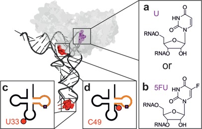
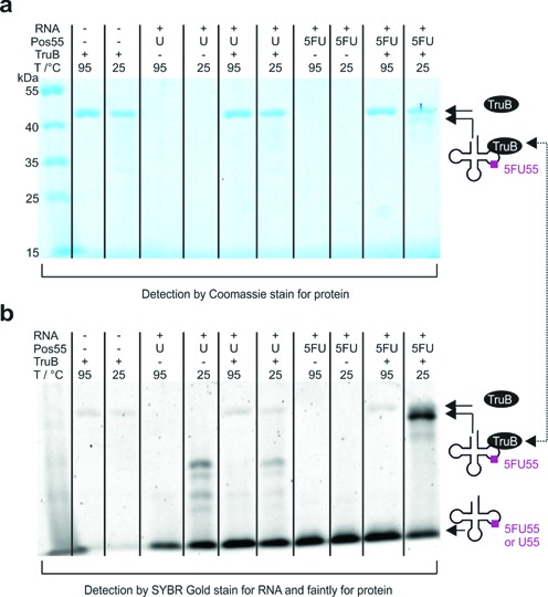
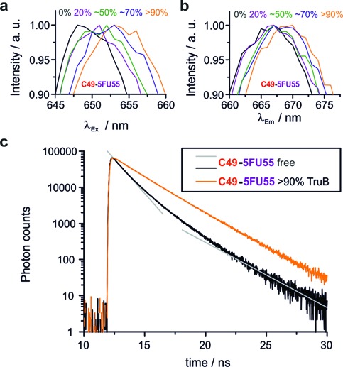
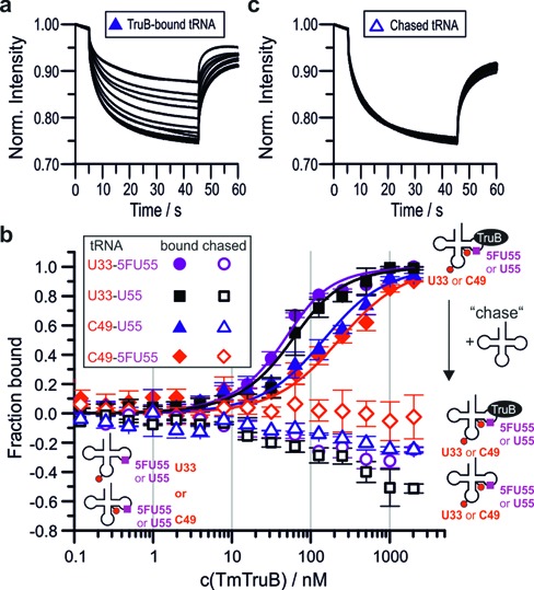
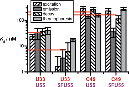
The interest in RNA modification enzymes surges due to their involvement in epigenetic phenomena. Here we present a particularly informative approach to investigate the interaction of dye-labeled RNA with modification enzymes. We investigated pseudouridine (Ψ) synthase TruB interacting with an alleged suicide substrate RNA containing 5-fluorouridine (5FU). A longstanding dogma, stipulating formation of a stable covalent complex was challenged by discrepancies between the time scale of complex formation and enzymatic turnover. Instead of classic mutagenesis, we used differentially positioned fluorescent labels to modulate substrate properties in a range of enzymatic conversion between 6% and 99%. Despite this variegation, formation of SDS-stable complexes occurred instantaneously for all 5FU-substrates. Protein binding was investigated by advanced fluorescence spectroscopy allowing unprecedented simultaneous detection of change in fluorescence lifetime, anisotropy decay, as well as emission and excitation maxima. Determination of Kd values showed that introduction of 5FU into the RNA substrate increased protein affinity by 14× at most. Finally, competition experiments demonstrated reversibility of complex formation for 5FU-RNA. Our results lead us to conclude that the hitherto postulated long-term covalent interaction of TruB with 5FU tRNA is based on the interpretation of artifacts. This is likely true for the entire class of pseudouridine synthases.


Figures

Figure 1.
Figure 1.
Tertiary (above) and secondary (below) structure representations of the yeast tRNAPhe constructs. The substrate position 55 (purple) was either a U55 (a) or a 5FU55 (b). The Cy5-label (red) was either attached at position U33 outside the minimal substrate (orange) (c) or at position C49 inside the minimal substrate (d). The enzyme TruB bound to the tRNA as predicted by a docking model (27) is shown in gray.
Figure 2.
Figure 2.
Analysis of SDS-stable complex formation of TruB with 5FU55- or U55-tRNA, respectively. Following 1 h incubation at 1 μM concentration and 70°C the complex was incubated with 0.5 vol SDS buffer for 5 min either at 25°C or at 95°C. (a) Coomassie stain for protein and (b) SYBR Gold stain for RNA. Note that SYBR Gold stains TruB faintly.
Figure 3.
Figure 3.
(a) Yield of Ψ generation in U55-tRNAs (assessed by LC-MS/MS in duplicate) compared to yield of SDS-stable complex by 5FU55-tRNAs (data quantified from b). (b and c) Kinetics of TruB binding to labeled and unlabeled 5FU55-tRNA analyzed by SDS-PAGE. Samples were incubated at 25°C for the time indicated and 5 min in 0.5 vol. SDS buffer prior to gel loading. Coomassie stain for protein (b), SYBR Gold stain for RNA (c) (excitation 488 nm, emission 670BP30). FRET from SYBR Gold to Cy5 generates the stronger signal of labeled tRNAs.
Figure 4.
Figure 4.
Protein binding (∼% binding is given) causes bathochromic shifts of excitation (a, λEm = 668 nm) and emission spectra (b, λEx = 647 nm). Fluorescence decay curves (c) differ in absence of TruB and with >90% TruB binding. Gray straight lines indicate bi-exponential fast decay (absence of TruB). The bi-exponential slow decay for >90% TruB binding appears to be mono-exponential due to the log-scale of the y-axis.
Figure 5.
Figure 5.
Spectroscopic titration curves of TruB tRNA interaction (ctRNA = 200 nM) for U55-tRNAs (a and b) and 5FU55-tRNAs (c and d), for all spectroscopic parameters with fits to a 1:1 binding model as solid lines. (e) Schematic representation of the chase experiment. (f) Chase experiments for C49-tRNA TruB interaction: The fraction bound is averaged over the values obtained from all spectroscopic parameters without TruB (free), with TruB (>90% binding) and after addition of unlabeled U55-tRNA in excess to the complex.
Figure 6.
Figure 6.
Results of MST experiments. (a) Typical set of thermophoresis curves for protein binding. (b) Titration curves for tmTruB binding (filled symbols) fitted to a one-to-one binding model (solid lines). Empty symbols represent the same samples after addition of unlabeled U55-tRNA in excess (chase). Note that negative values in the chase experiments are presumably caused by the different spectral properties of the bound and the unbound species. Error bars are standard deviations from triplicate measurements. (c) Typical thermophoresis curves in chase experiments by addition of unlabeled U55-tRNA.
Figure 7.
Figure 7.
Overview of determined Kds. Error bars represent standard errors derived from fits to the data using Origin 7 software employing the Levenberg–Marquardt algorithm. In case of thermophoresis the standard error results from a simultaneous fit to all three data sets. Red horizontal drop lines indicate arithmetic means over the different Kd values for each construct.

References

    1. Carell T., Brandmayr C., Hienzsch A., Müller M., Pearson D., Reiter V., Thoma I., Thumbs P., Wagner M. Structure and function of noncanonical nucleobases. Angewandte Chemie. 2012;51:7110–7131. - PubMed
    1. Machnicka M.A., Milanowska K., Osman Oglou O., Purta E., Kurkowska M., Olchowik A., Januszewski W., Kalinowski S., Dunin-Horkawicz S., Rother K.M., et al. MODOMICS: a database of RNA modification pathways–2013 update. Nucleic Acids Res. 2013;41:D262–D267. - PMC - PubMed
    1. Kellner S., Neumann J., Rosenkranz D., Lebedeva S., Ketting R.F., Zischler H., Schneider D., Helm M. Profiling of RNA modifications by multiplexed stable isotope labelling. Chem. Commun. 2014;50:3516–3518. - PubMed
    1. Tollervey J.R., Lunyak V.V. Epigenetics: judge, jury and executioner of stem cell fate. Epigenetics. 2012;7:823–840. - PMC - PubMed
    1. Ramamurthy V., Swann S.L., Paulson J.L., Spedaliere C.J., Mueller E.G. Critical aspartic acid residues in pseudouridine synthases. J. Biol. Chem. 1999;274:22225–22230. - PubMed

Publication types