. 2014 Dec:28:513-23.
doi: 10.1016/j.meegid.2014.09.021. Epub 2014 Oct 6.

Comparative genomic analysis of genogroup 1 (Wa-like) rotaviruses circulating in the USA, 2006-2009

Collaborators, Affiliations

Comparative genomic analysis of genogroup 1 (Wa-like) rotaviruses circulating in the USA, 2006-2009

Sunando Roy et al. Infect Genet Evol. 2014 Dec.

Abstract

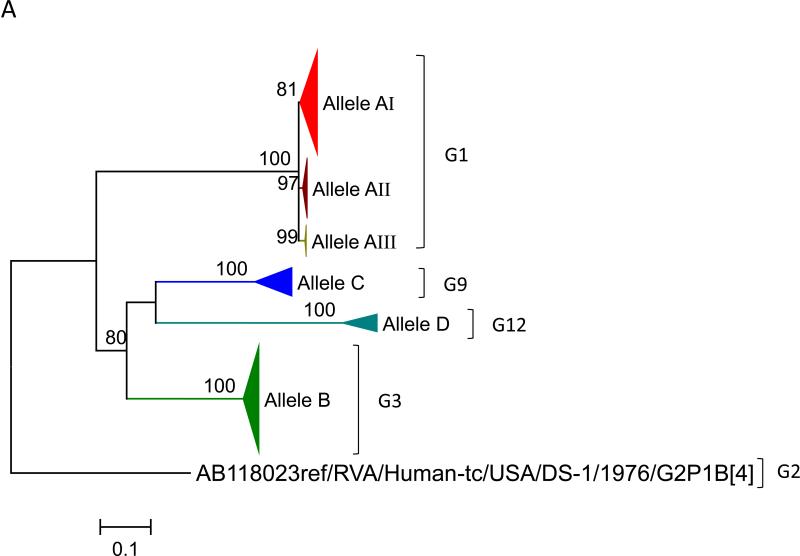
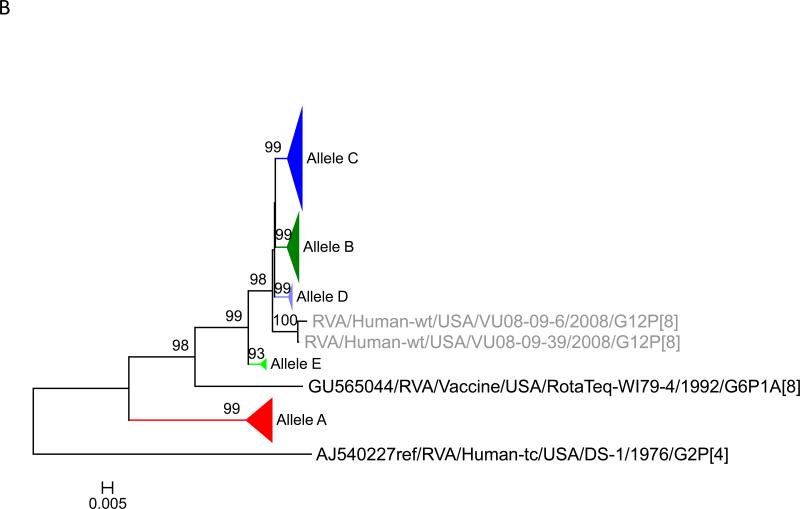
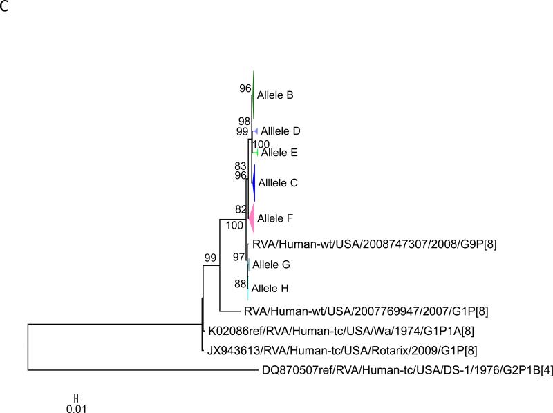
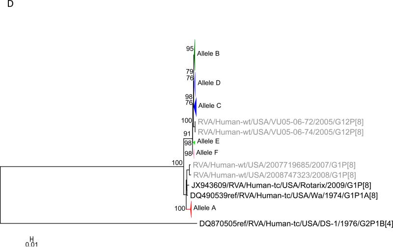
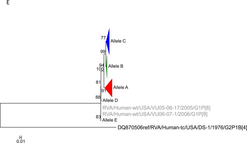
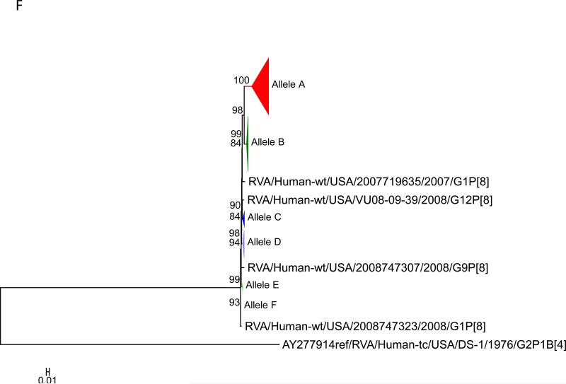
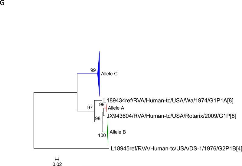
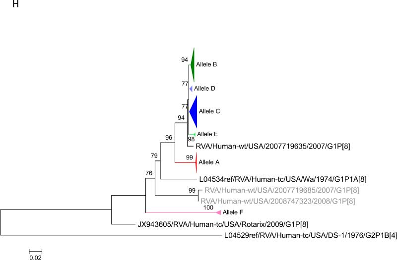
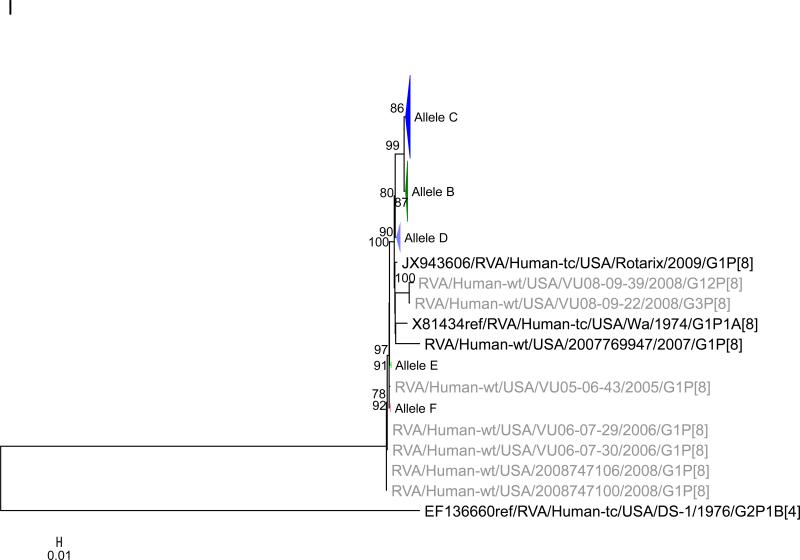
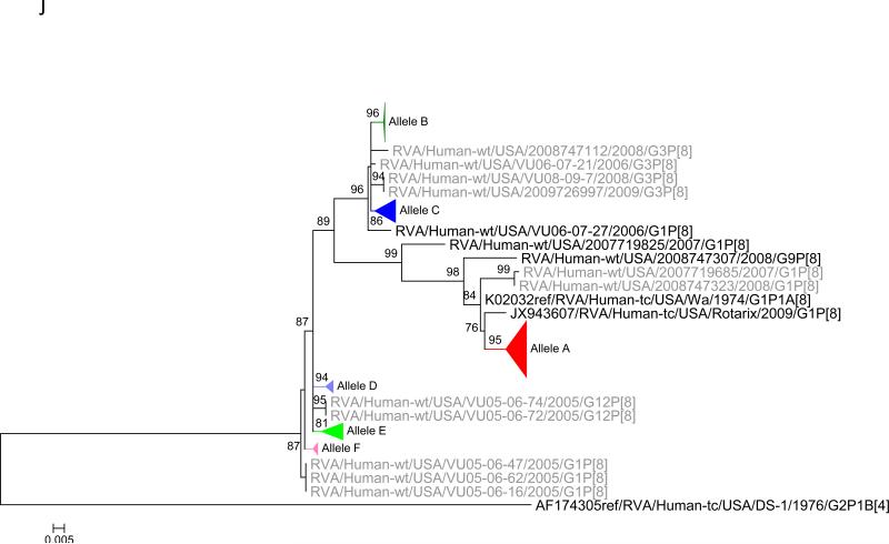
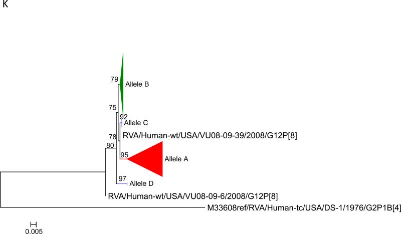
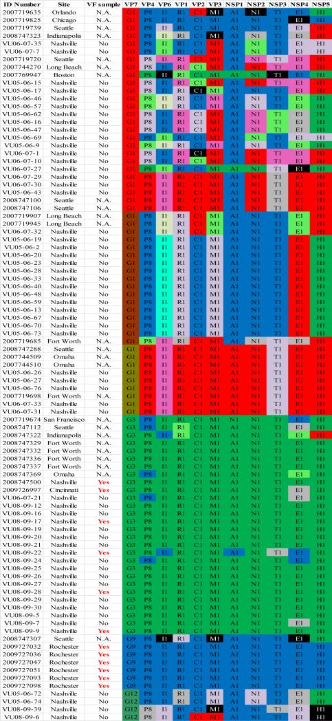
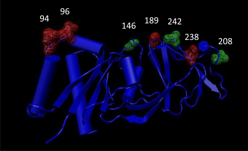
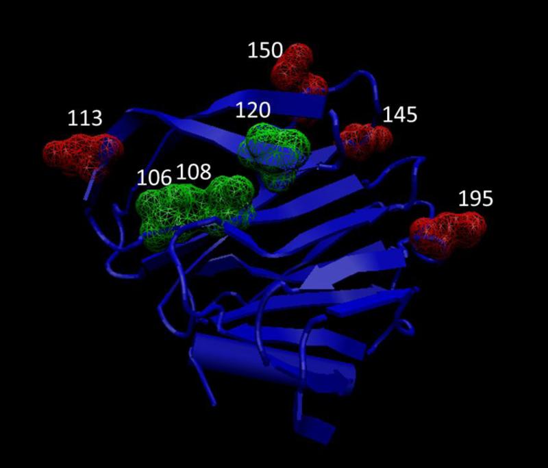
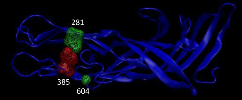
Group A rotaviruses (RVA) are double stranded RNA viruses that are a significant cause of acute pediatric gastroenteritis. Beginning in 2006 and 2008, respectively, two vaccines, Rotarix™ and RotaTeq®, have been approved for use in the USA for prevention of RVA disease. The effects of possible vaccine pressure on currently circulating strains in the USA and their genome constellations are still under investigation. In this study we report 33 complete RVA genomes (ORF regions) collected in multiple cities across USA during 2006-2009, including 8 collected from children with verified receipt of 3 doses of rotavirus vaccine. The strains included 16 G1P[8], 10 G3P[8], and 7 G9P[8]. All 33 strains had a Wa like backbone with the consensus genotype constellation of G(1/3/9)-P[8]-I1-R1-C1-M1-A1-N1-T1-E1-H1. From maximum likelihood based phylogenetic analyses, we identified 3-7 allelic constellations grouped mostly by respective G types, suggesting a possible allelic segregation based on the VP7 gene of RVA, primarily for the G3 and G9 strains. The vaccine failure strains showed similar grouping for all genes in G9 strains and most genes of G3 strains suggesting that these constellations were necessary to evade vaccine-derived immune protection. Substitutions in the antigenic region of VP7 and VP4 genes were also observed for the vaccine failure strains which could possibly explain how these strains escape vaccine induced immune response. This study helps elucidate how RVA strains are currently evolving in the population post vaccine introduction and supports the need for continued RVA surveillance.

Keywords: Allele; Failure; Rotavirus; VP4; VP7; Vaccine.


Figures

Figure 1
Figure 1
Maximum likelihood trees with aLRT values showing branch support for the 11 RVA genes. The different alleles are colored in red, green, blue, purple, lime, pink, teal and aqua for Alleles A, B, C, D, E, F, G and H respectively. Doublets and singletons are shown in grey and black, respectively (Fig. 1A) VP7. The Allele A in further divided into three clusters: red strains that mostly cluster with G1 (Wa) strains and maroon and olive strains that forms a distinct clusters from the reference Wa Strain. (1B) VP4; (1C) VP6; (1D) VP1; (1E) VP2; (1F) VP3; (1G) NSP1; (1H) NSP2; (1I) NSP3; (1J) NSP4; (1K) NSP5. To see the individual strains comprising each colored triangle, consult the supplemental figures (see supplementary material).
Figure 1
Figure 1
Maximum likelihood trees with aLRT values showing branch support for the 11 RVA genes. The different alleles are colored in red, green, blue, purple, lime, pink, teal and aqua for Alleles A, B, C, D, E, F, G and H respectively. Doublets and singletons are shown in grey and black, respectively (Fig. 1A) VP7. The Allele A in further divided into three clusters: red strains that mostly cluster with G1 (Wa) strains and maroon and olive strains that forms a distinct clusters from the reference Wa Strain. (1B) VP4; (1C) VP6; (1D) VP1; (1E) VP2; (1F) VP3; (1G) NSP1; (1H) NSP2; (1I) NSP3; (1J) NSP4; (1K) NSP5. To see the individual strains comprising each colored triangle, consult the supplemental figures (see supplementary material).
Figure 2
Figure 2
Allelic clusters for the 91 RVA strains. Genes from strains that clustered with Allele A (G1) are shown in red, maroon and olive. Allele B (G3) genes are shown in green and Allele C (G9) strains are shown in blue, Independent clusters are shown in purple (Allele D), lime (Allele E), pink (Allele F), teal (Allele G) and aqua (Allele H). Doublet strains are shown in grey and singleton strains in black.
Figure 3
Figure 3
Crystal structure of RVA VP7 protein (3FMG). Substitutions at the antigenic site are marked in green and substitutions at reported neutralization escape mutation sites are marked in red.
Figure 4
Figure 4
Crystal structure of RVA VP8* region (2DWR). Substitutions at the antigenic site are marked in green and substitutions at reported neutralization escape mutation sites are marked in red.
Figure 5
Figure 5
Crystal structure of RVA VP5* region (2B4I). Substitutions at the antigenic site are marked in green and substitutions at reported neutralization escape mutation sites are marked in red.

Similar articles

Cited by

References

    1. Anisimova M, Gascuel O. Approximate likelihood-ratio test for branches: A fast, accurate, and powerful alternative. Systematic biology. 2006;55:539–552. - PubMed
    1. Aoki ST, Settembre EC, Trask SD, Greenberg HB, Harrison SC, Dormitzer PR. Structure of rotavirus outer-layer protein VP7 bound with a neutralizing Fab. Science. 2009;324:1444–1447. - PMC - PubMed
    1. Arista S, Giammanco GM, De Grazia S, Ramirez S, Lo Biundo C, Colomba C, Cascio A, Martella V. Heterogeneity and temporal dynamics of evolution of G1 human rotaviruses in a settled population. Journal of virology. 2006;80:10724–10733. - PMC - PubMed
    1. Armah GE, Sow SO, Breiman RF, Dallas MJ, Tapia MD, Feikin DR, Binka FN, Steele AD, Laserson KF, Ansah NA, Levine MM, Lewis K, Coia ML, Attah-Poku M, Ojwando J, Rivers SB, Victor JC, Nyambane G, Hodgson A, Schodel F, Ciarlet M, Neuzil KM. Efficacy of pentavalent rotavirus vaccine against severe rotavirus gastroenteritis in infants in developing countries in sub-Saharan Africa: a randomised, double-blind, placebo-controlled trial. Lancet. 2010;376:606–614. - PubMed
    1. Banyai K, Laszlo B, Duque J, Steele AD, Nelson EA, Gentsch JR, Parashar UD. Systematic review of regional and temporal trends in global rotavirus strain diversity in the pre rotavirus vaccine era: insights for understanding the impact of rotavirus vaccination programs. Vaccine. 2012;30(Suppl 1):A122–130. - PubMed

Publication types

MeSH terms

Substances