. 2015 Dec;52(3):1119-1134.
doi: 10.1007/s12035-014-8902-7. Epub 2014 Oct 10.

Bryostatin-1 Restores Blood Brain Barrier Integrity following Blast-Induced Traumatic Brain Injury

Affiliations

Bryostatin-1 Restores Blood Brain Barrier Integrity following Blast-Induced Traumatic Brain Injury

Brandon P Lucke-Wold et al. Mol Neurobiol. 2015 Dec.

Abstract

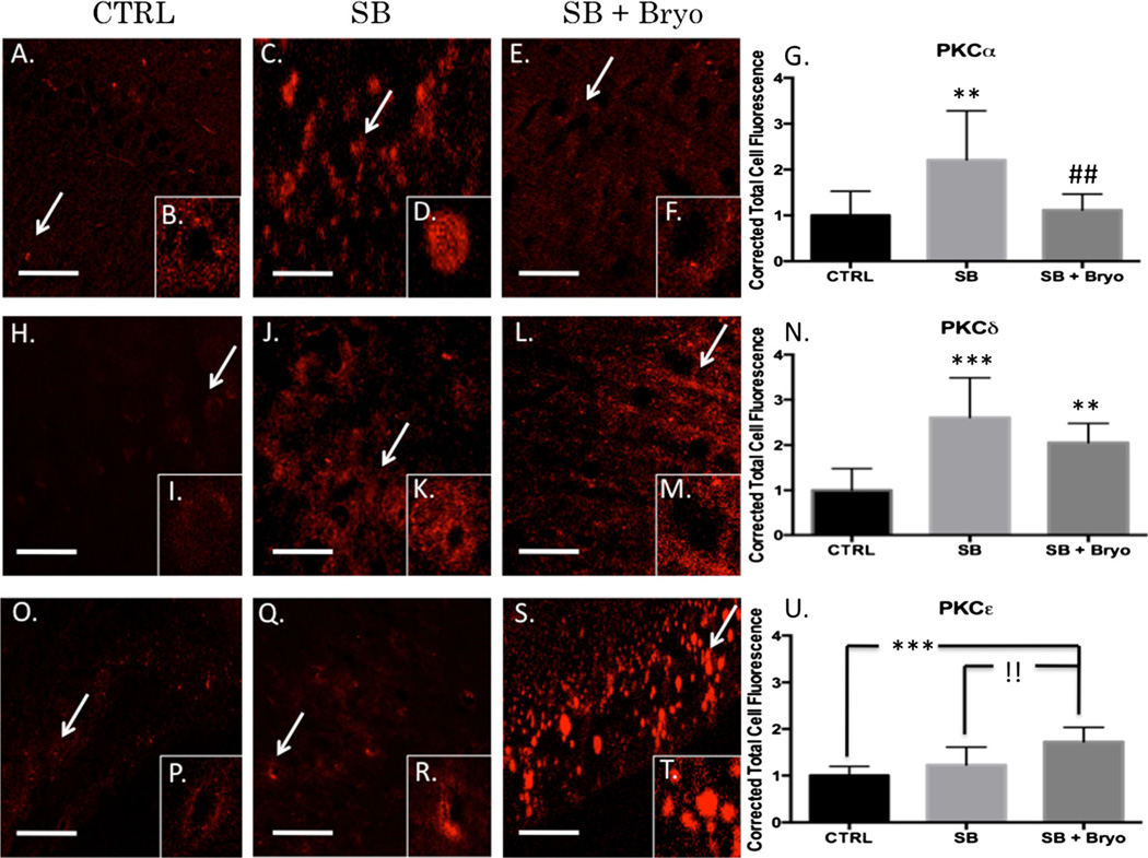
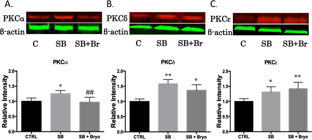
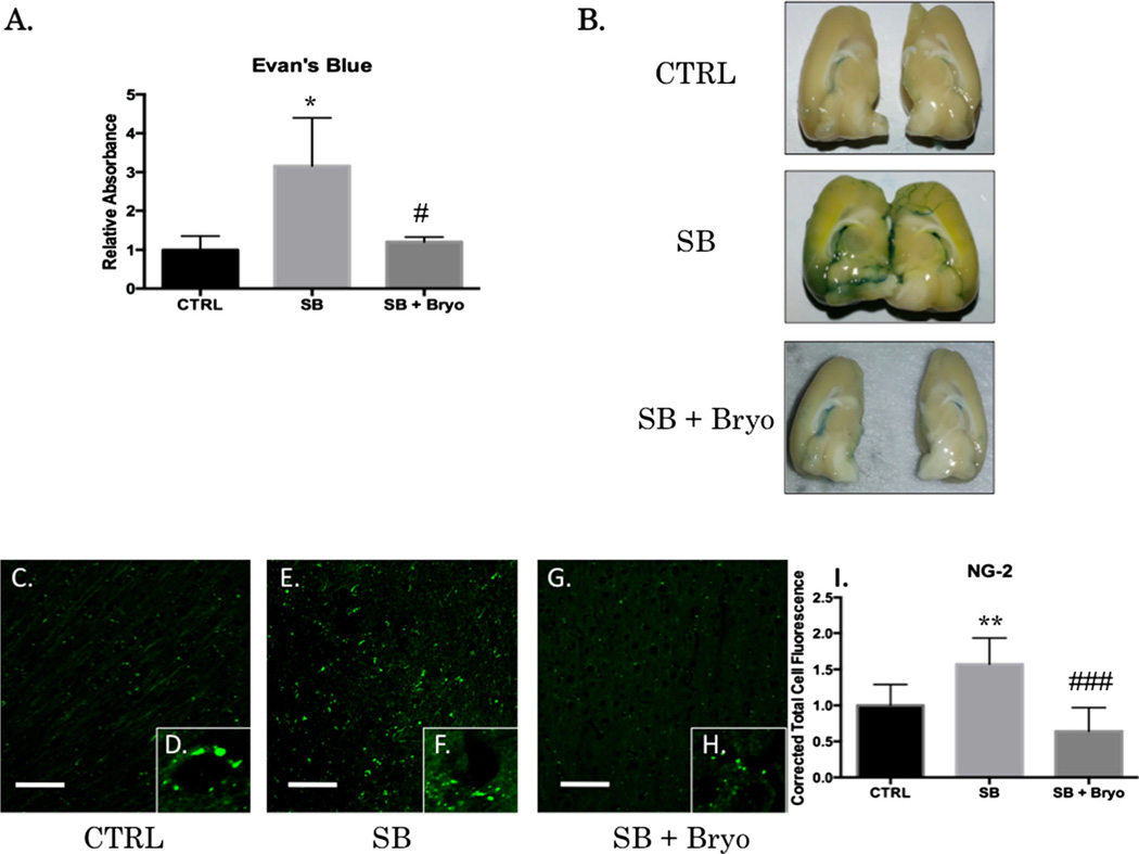
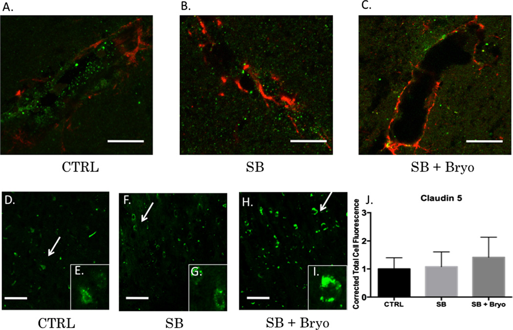
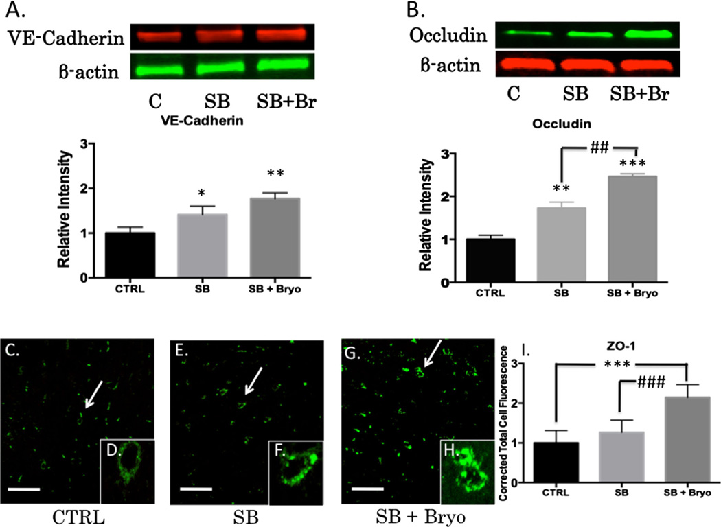
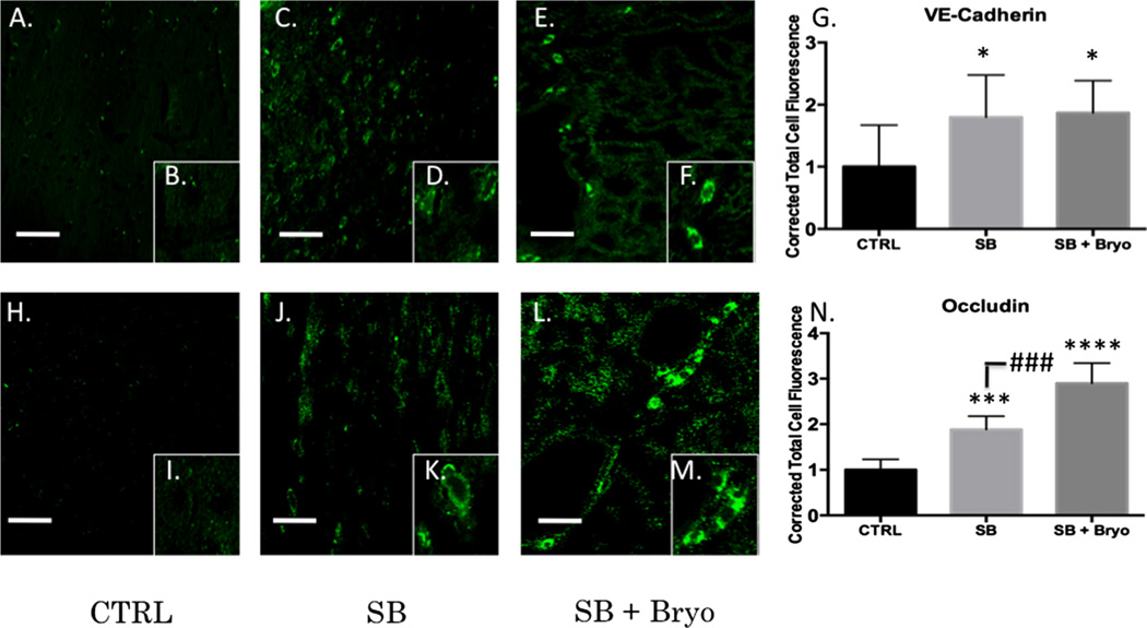
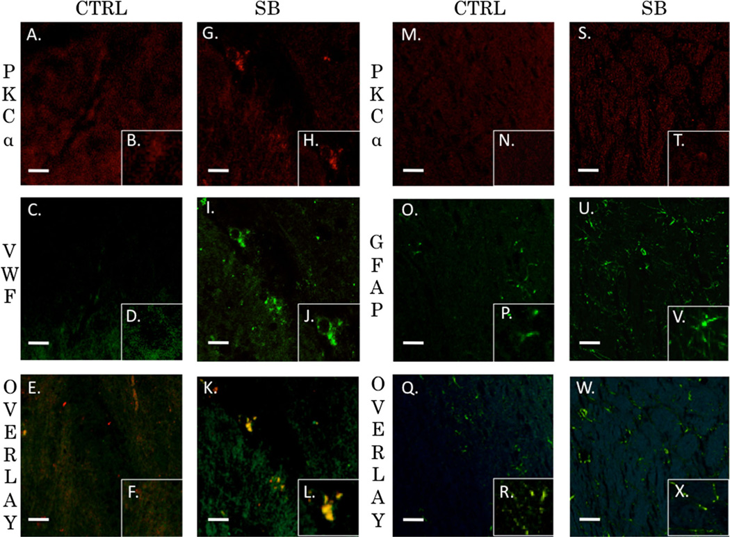
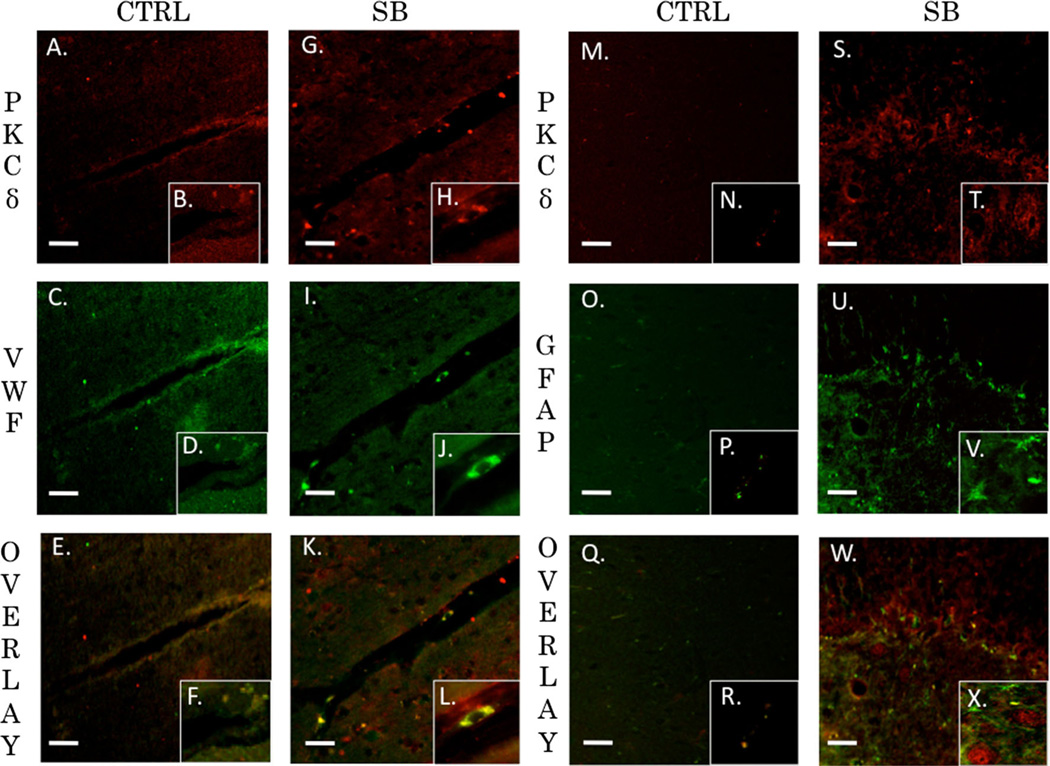
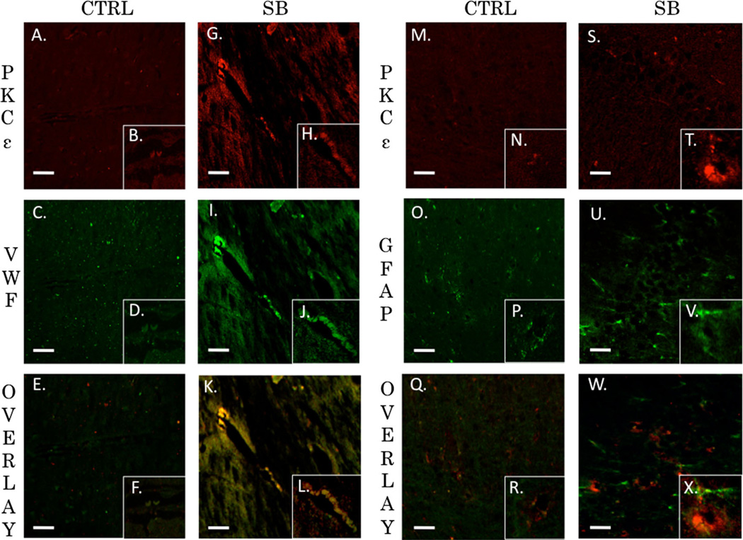
Recent wars in Iraq and Afghanistan have accounted for an estimated 270,000 blast exposures among military personnel. Blast traumatic brain injury (TBI) is the 'signature injury' of modern warfare. Blood brain barrier (BBB) disruption following blast TBI can lead to long-term and diffuse neuroinflammation. In this study, we investigate for the first time the role of bryostatin-1, a specific protein kinase C (PKC) modulator, in ameliorating BBB breakdown. Thirty seven Sprague-Dawley rats were used for this study. We utilized a clinically relevant and validated blast model to expose animals to moderate blast exposure. Groups included: control, single blast exposure, and single blast exposure + bryostatin-1. Bryostatin-1 was administered i.p. 2.5 mg/kg after blast exposure. Evan's blue, immunohistochemistry, and western blot analysis were performed to assess injury. Evan's blue binds to albumin and is a marker for BBB disruption. The single blast exposure caused an increase in permeability compared to control (t = 4.808, p < 0.05), and a reduction back toward control levels when bryostatin-1 was administered (t = 5.113, p < 0.01). Three important PKC isozymes, PKCα, PKCδ, and PKCε, were co-localized primarily with endothelial cells but not astrocytes. Bryostatin-1 administration reduced toxic PKCα levels back toward control levels (t = 4.559, p < 0.01) and increased the neuroprotective isozyme PKCε (t = 6.102, p < 0.01). Bryostatin-1 caused a significant increase in the tight junction proteins VE-cadherin, ZO-1, and occludin through modulation of PKC activity. Bryostatin-1 ultimately decreased BBB breakdown potentially due to modulation of PKC isozymes. Future work will examine the role of bryostatin-1 in preventing chronic neurodegeneration following repetitive neurotrauma.

Keywords: Blood brain barrier; Bryostatin-1; Protein kinase C; Tight junction proteins.


Conflict of interest statement

The authors claim to have no conflicts of interest.

Figures

Fig. 1
Fig. 1
Table showing the experimental techniques used for each time point
Fig. 2
Fig. 2
Bryostatin-1 decreases PKCα and increases PKCε 24 h after blast exposure. Our data show that bryostatin-1 has a profound effect after blast traumatic brain injury using fluorescent IHC. Scale bar=100 µm in left prefrontal cortex. PKCα control (a) with inlay (b) compared to single blast exposure (c) with inlay (d), and single blast exposure + bryostatin-1 (e) with inlay (f) showed significant difference between groups. Post-hoc comparison between control and single blast (**p<0.01) and between single blast and single blast + bryostatin-1 (##p<0.01) as depicted in bar graph (g). PKCγ control (h) with inlay (i) compared to single blast exposure (j) with inlay (k), and single blast exposure + bryostatin-1 (l) with inlay (m) showed significant difference between groups. Post-hoc comparison between control and single blast (***p<0.001) and between control and single blast + bryostatin-1 (**p<0.01) as depicted in bar graph (n). PKCε control (o) with inlay (p) compared to single blast exposure (q) with inlay (r), and single blast exposure + bryostatin-1 (s) with inlay (t) showed a significant difference between groups. Post-hoc comparison between control and single blast + bryostatin-1 (***p<0.001) and between single blast and single blast + bryostatin-1 (!!p<0.01) as depicted in bar graph (u)
Fig. 3
Fig. 3
Bryostatin-1 is a potent PKC modulator. It has been used to regulate PKC activity in a time-specific manner for multiple neural injury models. Protein concentrations were measured in the left prefrontal cortex 24 h after blast expsoure using western blot analysis. A significant difference between groups was observed for PKCα. Post-hoc comparison between control and single blast (*p<0.05), and between single blast and single blast + bryostatin-1 (##p<0.01) (a). A significant difference between groups was observed for PKCδ. Post-hoc comparison between control and single blast (**p<0.01), and control and single blast + bryostatin-1 (*p<0.05) (b). A significant difference between groups was observed for PKCε. Post-hoc comparison between control and single blast (*p<0.05), and between control and single blast + bryostatin-1 (**p<0.01) (c). Bryostatin-1 significantly decreased PKCα levels and increased PKCε levels when administered after blast exposure
Fig. 4
Fig. 4
Bryostatin-1 preserves BBB integrity. Evan’s blue binds to albumin and is a widely used marker for detecting breaches in the BBB. NG-2 is a proteoglycan found in pericytes that will be increased when the BBB is disrupted. Scale bar=100 µm in left prefrontal cortex. A significant difference between groups was observed for EB absorbance in the brain after femoral vein injection post-blast. Post-hoc comparison revealed a significant difference between control and single blast (*p<0.05), and between single blast and single blast + bryostatin-1 (#p<0.05) (a). Gross examination revealed increased EB staining in the left hemisphere following blast exposure that was decreased when bryostatin-1 was given following blast (b). A significant difference between groups was observed for NG-2 IHC fluorescent staining (c–h). Post-hoc comparison revealed a significant difference between control and single blast (**p<0.01), and between single blast and single blast + bryostatin-1 (###p<0.001) (i)
Fig. 5
Fig. 5
Vasculature disruption after blast exposure is independent of claudin-5 regulation. PKCα specifically regulates occludin, ZO-1, and VE-Cadherin, but not claudin-5. Claudin-5 levels may therefore be independent of bryostatin-1 modulation. Scale bar=5 µm in the left prefrontal cortex (a–c). Scale bar=100 µm in the left prefrontal cortex (d–j). VWF (green) was co-localized with GFAP (red) to give a visual representation of cerebral vasculature. Control vasculature in the left prefrontal cortex was visibly intact (a). Twenty-four hours after blast exposure left prefrontal cortex vasculature was visibly disrupted as indicated by the sparse VWF staining (b). Bryostatin-1 preserved vasculature integrity when administered after blast exposure (c). No significant differences were observed between groups for claudin-5 using fluorescent IHC (d–j)
Fig. 6
Fig. 6
Bryostatin-1 significantly increases tight junction proteins. Our data show that bryostatin-1 significantly upregulated tight junction proteins leading to maintenance of BBB integrity following blast TBI. Scale bar=100 µm in left prefrontal cortex. A significant difference was observed between groups using western blot for VE-Cadherin with post hoc comparison showing significance between control and single blast (*p<0.05), and between control and single blast + bryostatin-1 (**p<0.01) 24 h after blast exposure (a). A significant difference was observed between groups using western blot for occludin with post hoc comparison showing significance between control and single blast (**p<0.01), between control and single blast + bryostatin-1 (***p<0.001), and between single blast and single blast + bryostatin-1 (##p<0.01) 24 h after blast exposure (b). A significant difference was observed between groups using fluorescent IHC for ZO-1 with post hoc comparison showing significance between control and single blast + bryostatin-1 (***p<0.001), and between single blast and single blast + bryostatin-1 (###p<0.001) 24 h after blast exposure (c)
Fig. 7
Fig. 7
Tight junction protein expression was increased by bryostatin-1 at vasculature. Tight junction protein return of function is necessary for restoration of BBB integrity. Scale bar=75 µm in left prefrontal cortex. A significant difference was observed between groups using fluorescent IHC for VE-Cadherin with post hoc comparison showing significance between control and single blast (*p<0.05), and between control and single blast + bryostatin-1 (*p<0.05) 24 h after blast exposure (a–g). A significant difference was observed between groups using fluorescent IHC for occludin with post-hoc comparison showing significance between control and single blast (***p<0.001), between control and single blast + bryostatin-1 (****p<0.0001), and between single blast and single blast + bryostatin-1 (###p<0.001) 24 h after blast exposure (h–n)
Fig. 8
Fig. 8
PKCα co-localized with endothelial cells but not astrocytes. Both astrocytes and endothelial cells are critical for maintenance of the BBB. PKCα activity within endothelial cells plays an intimate role in regulating extracellular tight junction proteins. Fluorescnt IHC red staining for PKCα, green staining for VWF (endothelial) or GFAP (astocyte), and yellow is overlay. Scale bar=20 µm in left prefrontal cortex. PKCα (a) with inlay (b) and VWF (c) with inlay (d) have a weak overlay with a Pearson’s coefficient of r=0.395 seen in (e) with inlay (f) for control animals. PKCα (g) with inlay (h) and VWF (i) with inlay (j) have a very strong overlay with an overlap coefficient of r=0.954 seen in (k) with inlay (l) 24 h post blast exposure. PKCα (m) with inlay (n) and GFAP (o) with inlay (p) have a weak overlay with a Pearson’s coefficient of r=0.351 seen in (q) with inlay (r) for control animals. PKCα (s) with inlay (t) and GFAP (u) with inlay (v) have a weak overlay with an overlap coefficient of r=0.379 seen in (w) with inlay (x) 24 h post blast exposure
Fig. 9
Fig. 9
PKCδ co-localized with endothelial cells but not astrocytes. PKCδ plays an important role in mediating vascular tone. Its role in regulation of tight junction proteins is not completely understood. Fluorescent IHC red staining for PKCδ, green staining for VWF (endothelial) or GFAP (astocyte), and yellow is overlay. Scale bar=20 µm in left prefrontal cortex. PKCδ (a) with inlay (b) and VWF (c) with inlay (d) have a moderate overlay with a Pearson’s coefficient of r=0.61 seen in (e) with inlay (f) for control animals. PKCδ (g) with inlay (h) and VWF (i) with inlay (j) have a strong overlay with an overlap coefficient of r=0.88 seen in (k) with inlay (l) 24 h post blast exposure. PKCδ (m) with inlay (n) and GFAP (o) with inlay (p) have a very weak overlay with a Pearson’s coefficient of r=0.029 seen in (q) with inlay (r) for control animals. PKCδ (s) with inlay (t) and GFAP (u) with inlay (v) have a moderate overlay with an overlap coefficient of r=0.449 seen in (w) with inlay (x) 24 h post blast exposure
Fig. 10
Fig. 10
PKCε co-localized with endothelial cells but not astrocytes. PKCε contributes to neuroprotection when increased after brain injury. PKCε has been associated with improved cognitive performance and decreased neurodegeneration. Fluorescent IHC red staining for PKCε, green staining for VWF (endothelial) or GFAP (astocyte), and yellow is overlay. Scale bar=20 µm in left prefrontal cortex. PKCε (a) with inlay (b) and VWF (c) with inlay (d) have a very weak overlay with a Pearson’s coefficient of r=0.004 seen in (e) with inlay (f) for control animals. PKCε (g) with inlay (h) and VWF (i) with inlay (j) have a strong overlay with an overlap coefficient of r=0.978 seen in (k) with inlay (l) 24 h post blast exposure. PKCε (m) with inlay (n) and GFAP (o) with inlay (p) have a weak overlay with a Pearson’s coefficient of r=0.133 seen in (q) with inlay (r) for control animals. PKCε (s) with inlay (t) and GFAP (u) with inlay (v) have a weak overlay with an overlap coefficient of r=0.394 seen in (w) with inlay (x) 24 h post blast exposure

References

    1. Kou Z, Vandevord PJ. Traumatic white matter injury and glial activation: from basic science to clinics. Glia. 2014 - PubMed
    1. Schneider EB, Sur S, Raymont V, Duckworth J, Kowalski RG, Efron DT, Hui X, Selvarajah S, Hambridge HL, Stevens RD. Functional recovery after moderate/severe traumatic brain injury: a role for cognitive reserve? Neurology. 2014;82(18):1636–1642. - PMC - PubMed
    1. Puvenna V, Brennan C, Shaw G, Yang C, Marchi N, Bazarian JJ, Merchant-Borna K, Janigro D. Significance of ubiquitin carboxy-terminal hydrolase l1 elevations in athletes after subconcussive head hits. PLoS One. 2014;9(5):e96296. - PMC - PubMed
    1. Simard JMMDPD, Pampori A, Keledjian K, Tosun C, Schwartzbauer G, Ivanova S, Gerzanich V. Exposure of the thorax to a sublethal blast wave causes a hydrodynamic pulse that leads to perivenular inflammation in the brain. J Neurotrauma. 2014 - PMC - PubMed
    1. Hue CD, Cao S, Dale Bass CR, Meaney DF, Morrison B., 3rd Repeated primary blast injury causes delayed recovery, but not additive disruption, in an in vitro blood–brain barrier model. J Neurotrauma. 2014;31(10):951–960. - PubMed

Publication types

MeSH terms

LinkOut - more resources