Case Reports
. 2014 Oct 9:8:334.
doi: 10.1186/1752-1947-8-334.

The retrieval of unerupted teeth in pedodontics: two case reports

Affiliations
Case Reports

The retrieval of unerupted teeth in pedodontics: two case reports

Simona Tecco et al. J Med Case Rep. .

Abstract

Introduction: The retrieval of unerupted teeth in pedodontics is always significant to preserve the trophism of adjacent tissues, establish the correct space, provide adequate function and maintain good esthetics for the patient. The treatment plan is based on radiographic examinations and measurements, and on an accurate clinical evaluation; it aims to achieve the best treatment possible depending on the complexity of the specific case.In the most difficult clinical cases it is very important to have an early diagnosis, which is essential to plan the treatment and achieve success. In these cases, the pediatrician is in a strategic position to give an early diagnosis through a child's medical history and by counting the child's teeth.

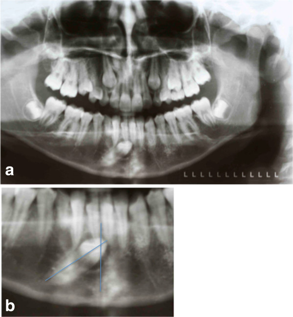
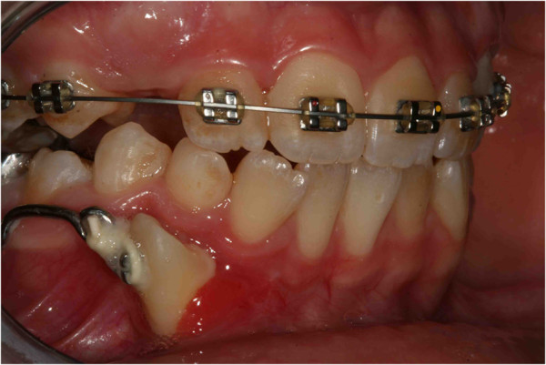
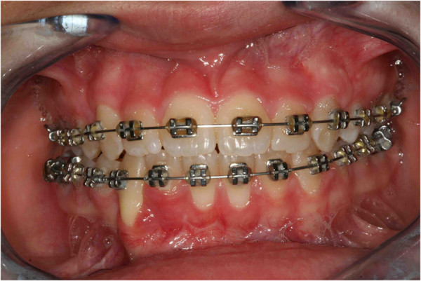
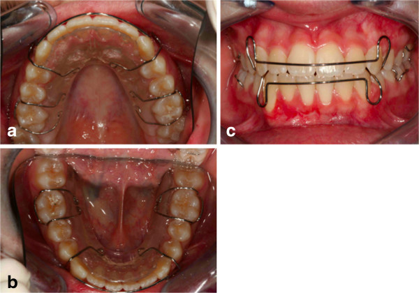
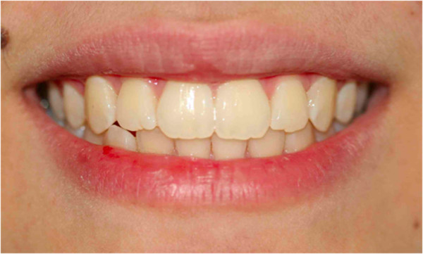
Case presentation: This article presents two different difficult clinical cases of impacted teeth diagnosed during pediatric age, with a radiological analysis, and successfully treated with orthodontic devices designed for these specific cases. Clinical case 1 describes a 13-year-old Italian girl; clinical case 2 describes a 9-year-old Italian girl. The use of these devices achieved the desired treatment goals. The problems associated with impacted teeth and the biomechanical interventions used for these patients are discussed.

Conclusions: An early and careful diagnosis followed by an accurate treatment plan for the individual cases can lead to retrieval of the impacted teeth without affecting other anatomic structures and adjacent teeth. In these cases, the pediatrician is in a strategic position to give an early diagnosis through a child's medical history and by counting the child's teeth.


Figures

Figure 1
Figure 1
Pretreatment records. (a) Pretreatment panoramic radiograph. (b) The unerupted canine is going to migrate across the mandibular midline, and its crown tip is near the apex of the lower right first incisor root.
Figure 2
Figure 2
The occlusal radiograph confirms that the crown of the impacted canine is vestibular.
Figure 3
Figure 3
The canine angulation to the midline is 55°.
Figure 4
Figure 4
Surgical outbreak; a vestibular repositioned, full thickness mucoperiosteal flap is elevated, and the crown of the canine is exposed.
Figure 5
Figure 5
The orthodontic devices: the Fishing-rod. (a) Fishing-rod in occlusal view. (b) Fishing-rod in lateral view. The lever arm of the device allows for a push in the occlusal-distal direction of the crown of the impacted tooth.
Figure 6
Figure 6
Intraoral photographs with fishing-rod; the appliance is used to tie up and drive into the canine’s eruption.
Figure 7
Figure 7
After 5 months, the cusp of the canine was visible in the mouth.
Figure 8
Figure 8
After 8 months; pre-informed brackets and straight archwires are used.
Figure 9
Figure 9
After 12 months. When the canine was present in the oral cavity (a), a bracket was bonded to it and linked directly to the arch by an elastic ligation. To keep the space in the arch for the canine, since it was not aligned, an open coil spring (b) was used.
Figure 10
Figure 10
After 18 months the canine is well positioned in the arch.
Figure 11
Figure 11
After 18 months the patient was advised that she needed an attached gingiva graft on the restored tooth to improve esthetics and the periodontal health compromised by the treatment.
Figure 12
Figure 12
Criss-cross elastic to improve the intercuspidation between the upper right first molar and the lower right first molar.
Figure 13
Figure 13
Retention was established with removable appliances on the upper arch (a) and the lower arch (b), to maintain the obtained result (c). After orthodontic treatment finished and the canine positioned in an acceptable way in the dental arch, the patient was advised that she needed an attached gingiva graft on the restored tooth to improve esthetics and the periodontal health compromised by the treatment.
Figure 14
Figure 14
Post-treatment photograph of the smile.
Figure 15
Figure 15
Post-treatment orthopantomograph.
Figure 16
Figure 16
Post-treatment intraoral photographs: (a) frontal view; (b) upper occlusal view; (c) lower occlusal view. One year after, only a partial recurrence was observed in the position of the upper first right molar, as the patient had not observed the restraint protocol. The periodontal surgery (attached gengiva graft of the lower right canine) has not been performed as requested by the same patient. The need for an attached gingiva graft on the restored tooth remained.
Figure 17
Figure 17
First orthopantomograph, before the patient came to our attention: a general dentist had suggested the extraction of the impacted left lower first molar (in the blue circle), after viewing this orthopantomograph.
Figure 18
Figure 18
Pretreatment intraoral frontal photograph. In correspondence with the missing tooth you see an “empty area” in the occlusion of the patient (indicated by the blue arrows).
Figure 19
Figure 19
Second orthopantomograph prescribed to the patient by us before our intervention to assess the evolution of the clinical case and formulate a new plan of treatment: the situation was even worse; the roots were all sizes and showed closed apexes (red arrows), and the presence of bone above was increased with respect to the first evaluation (blue arrows).
Figure 20
Figure 20
The appliances used. (a) Intraoral frontal photographs. (b) Lingual arch. (c) Upper appliance. The lingual arch shows an extension distal to the deciduous molar, to allow the extrusion of the retained first molar (blue circles).
Figure 21
Figure 21
Surgery (a) and placement of buttons (b). Two buttons were bonded on the molar crown in order to prevent the reopening of the surgical site in case one of the buttons came off accidentally during orthodontic treatment.
Figure 22
Figure 22
Orthopantomograph 3 months after surgery: it confirmed that the tooth was moving, as evidenced by the increase in the distance between the lower edge of the jaw and the roots of the impacted tooth (blue arrows).
Figure 23
Figure 23
Ten months after surgery; when the second molar was erupting, and the first molar was impacting against it (as seen in the orthopenthomograph, a), it was decided to wait for the eruption of the second molar (b) before continuing the treatment. So the lingual arch was eliminated to avoid the mesialization of the second molar.
Figure 24
Figure 24
One year later.
Figure 25
Figure 25
After 6 months. (a) Intraoral photo. (b) Intraoral radiograph. Pre-informed brackets and straight archwires were used; approximately 6 months after the beginning of the fixed treatment, the tooth appeared in the dental arch.
Figure 26
Figure 26
The molar is extruded. (a) Frontal intraoral. (b) Occlusal photo.
Figure 27
Figure 27
Post-treatment occlusal photographs; (a) upper occlusal view; (b) lower occlusal view.
Figure 28
Figure 28
Post-treatment radiograph.
Figure 29
Figure 29
Intraoral photographs 1 year post-treatment. (a) lower occlusal view; (b) the treated teeth; no tooth morbidity is evident. The gums are healthy, and the gingival attachment in the molar region is intact.

References

    1. Almonaitiene R, Balciuniene I, Tutkuviene J. Factors influencing permanent teeth eruption. Part one – general factors. Stomatologija. 2010;12(3):67–72. Review. - PubMed
    1. Albani F, Ballesio I, Campanella V, Marzo G. Pit and fissure sealants: results at five and ten years. Eur J Paediatr Dent. 2005;6(2):61–65. - PubMed
    1. Valletta G, Matarasso S, Mignogna M. Malattie Odontostomatologiche. Piccin: Milano; 2005.
    1. Andreasen JO. In: Textbook and Color Atlas of Tooth Impactions. Andreasen JO, Kolsen Petersen J, Laskin DM, editor. Copenhagen: Munksgaard; 1997. Treatment strategies for eruption disturbances. Chapter 3; pp. 65–91.
    1. Frank CA. Treatment options for impacted teeth. J Am Dent Assoc. 2000;131:623–632. doi: 10.14219/jada.archive.2000.0236. - DOI - PubMed

Publication types

LinkOut - more resources