. 2015 Jan 15;125(3):465-9.
doi: 10.1182/blood-2014-07-588954. Epub 2014 Oct 9.

Progressive maturation toward hematopoietic stem cells in the mouse embryo aorta

Affiliations

Progressive maturation toward hematopoietic stem cells in the mouse embryo aorta

Jean-Charles Boisset et al. Blood. .

Abstract

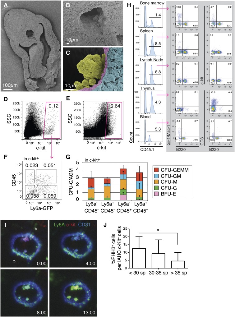
Clusters of cells attached to the endothelium of the main embryonic arteries were first observed a century ago. Present in most vertebrate species, such clusters, or intraaortic hematopoietic clusters (IAHCs), derive from specialized hemogenic endothelial cells and contain the first few hematopoietic stem cells (HSCs) generated during embryonic development. However, some discrepancies remained concerning the spatio-temporal appearance and the numbers of IAHCs and HSCs. Therefore, the exact cell composition and function of IAHCs remain unclear to date. We show here that IAHCs contain pre-HSCs (or HSC precursors) that can mature into HSCs in vivo (as shown by the successful long-term multilineage reconstitution of primary neonates and secondary adult recipients). Such IAHC pre-HSCs could contribute to the HSC pool increase observed at midgestation. The novel insights in pre-HSC to HSC transition represent an important step toward generating transplantable HSCs in vitro that are needed for autologous HSC transplantation therapies.


Figures

Figure 1
Figure 1
Intraaortic hematopoietic clusters at E10 are phenotypically heterogeneous and contain very few progenitors but contain pre-HSCs able of long-term multilineage hematopoietic reconstitution after transplantation in WT neonates. (A-C) Scanning electron microscopy of E10 (28-34 somite pairs, sp) thick embryo slices. Before tissue fixation, the circulating blood was flushed out of the aorta to ascertain that attached IAHCs and not circulating cells were observed. (A) Whole E10 embryo slice. (B) Close-up view of the aorta where IAHCs are visible inside the aorta. (C) Close-up view of an IAHC with sphere shape in the aorta of E10 embryos. IAHC (yellow), endothelium (pink), and sub-aortic mesenchyme (blue) were artificially colored to show the delimitation between the structures. Top, dorsal side; bottom, ventral side. No morphologic heterogeneity was observed in the cells within single IAHCs or between IAHCs. (D-E) Flow cytometry analysis of AGM cells (E10 embryos) stained with c-kit antibody by intraaortic injection before AGM dissociation (D) or on dissociated AGM cells (E). (F) CD45 and Ly6a-GFP expression within c-kit+ AGM cells from E10 Ly6a-GFP embryos. Percentages of viable cells are indicated inside the gates. The absolute numbers of each population per ee are 216 ± 114 (c-kit+Ly6a-GFPCD45), 52 ± 16 (c-kit+Ly6a-GFP+CD45), 37 ± 17 (c-kit+Ly6a-GFPCD45+), and 49 ± 23 (c-kit+Ly6a-GFP+CD45+) (n = 4, mean ± standard deviation [SD]). (G) CFU-C assays for the 4 IAHC cell populations isolated from E10 AGMs (28-34 somite pairs) and sorted based on (F) (n = 3). Bars represent the mean ± SD. The number of CFU-C is indicated per flushed AGM. (H) Analysis of a WT neonate recipient (CD45.2) transplanted with 50 IAHC c-kit+ cells from mid-E10 (33-38 sp) AGM (CD45.1) up to 4 months posttransplantation. FACS analysis shows donor-cell contribution (CD45.1) in bone marrow, spleen, lymph nodes, thymus and blood, represented in the histogram on the left panel (CD45.1, blue; control, black line). Lines indicate the percentages of donor contribution in the whole tissue. Multilineage donor contribution (dot plots on gray background) was analyzed in all organs for myeloid (Gr-1/Mac-1) and B cells (B220) in the bone marrow and spleen for HSCs and progenitor cells (c-kit), and in blood and thymus for T cells (CD3). Percentages of each donor population are indicated per quadrant. The analyzed reconstituted mouse is shown in Table 1 (#). (I) Time-lapse series pictures from supplemental Movie 2 (Example 1) showing the maturation of IAHC cells. All CD31+c-kit+Ly6a-GFP in IAHCs progressively express Ly6a-GFP during the time-lapse imaging (13 h). V, ventral; D, dorsal; Ly6a-GFP, green; c-kit, red; CD31, blue. (J) Percentages of proliferating (c-kit+PHH3+) cells per IAHC (c-kit+) cells (or mitotic index) in the aorta of early E10 (<30 sp), mid-E10 (30-35 sp), and late E10 (>35 sp) embryos. Bars represent means ± SD. *P < .05. To note, we controlled that the intraaortic staining procedure did not detach IAHCs by performing whole-embryo c-kit staining of flushed compared with nonflushed embryos. We found no significant differences in the number of c-kit+ (IAHC) cells in the aorta or in the shape of IAHCs (data not shown).

References

    1. Müller AM, Medvinsky A, Strouboulis J, Grosveld F, Dzierzak E. Development of hematopoietic stem cell activity in the mouse embryo. Immunity. 1994;1(4):291–301. - PubMed
    1. Yokomizo T, Dzierzak E. Three-dimensional cartography of hematopoietic clusters in the vasculature of whole mouse embryos. Development. 2010;137(21):3651–3661. - PMC - PubMed
    1. Cai Z, de Bruijn M, Ma X, et al. Haploinsufficiency of AML1 affects the temporal and spatial generation of hematopoietic stem cells in the mouse embryo. Immunity. 2000;13(4):423–431. - PubMed
    1. Dantschakoff V. Untersuchungen über die Entwickelung von Blut und Bindegewebe bei Vogeln. Das lockere Bindegewebe des Hühnchens im fetalen Leben. Arch f mikr Anat. 1909;73:117–181.
    1. Jaffredo T, Gautier R, Eichmann A, Dieterlen-Lièvre F. Intraaortic hemopoietic cells are derived from endothelial cells during ontogeny. Development. 1998;125(22):4575–4583. - PubMed

Publication types