. 2014 Nov 15;5(21):10558-70.
doi: 10.18632/oncotarget.2510.

De-acetylation and degradation of HSPA5 is critical for E1A metastasis suppression in breast cancer cells

Affiliations

De-acetylation and degradation of HSPA5 is critical for E1A metastasis suppression in breast cancer cells

Yi-Wen Chang et al. Oncotarget. .

Abstract

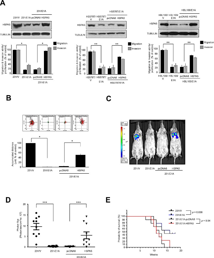
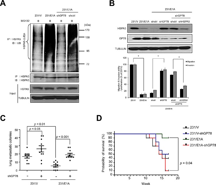
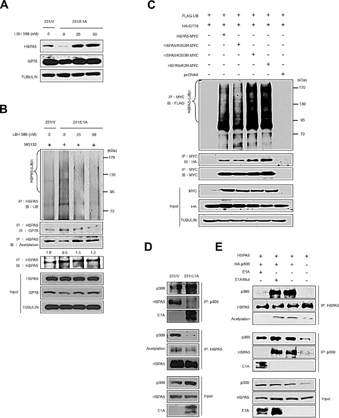
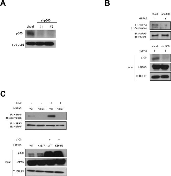
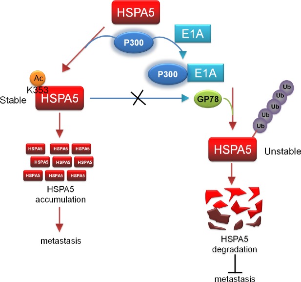
Elevated expression of heat shock protein 5 (HSPA5) promotes drug resistance and metastasis and is a marker of poor prognosis in breast cancer patients. Adenovirus type 5 E1A gene therapy has demonstrated antitumor efficacy but the mechanisms of metastasis-inhibition are unclear. Here, we report that E1A interacts with p300 histone acetyltransferase (HAT) and blocks p300-mediated HSPA5 acetylation at K353, which in turn promotes HSPA5 ubiquitination by GP78 (E3 ubiquitin ligase) and subsequent proteasome-mediated degradation. Our findings point out the Ying-Yang regulation of two different post-translational modifications (ubiquitination and acetylation) of HSPA5 in tumor metastasis.


Conflict of interest statement

Competing interests

The authors have declared that no conflict of interest exists.

Figures

Figure 1
Figure 1. HSPA5 repression is required for E1A-mediated metastasis suppression
(A) Three breast cancer cell lines were stably transfected with control vector (231/V, HS578T/V, HBL100/V) or E1A-expressing vector (231/E1A, HS578T/E1A, HBL100/E1A) with pcDNA6 or HSPA5 constructs. The expression of HSPA5 was analyzed by Western blot assay; cell migration and invasive ability were measured by transwell migration and matrigel invasion assay. Percentage of cell migration and invasion/proliferation is shown relative to control vector group. Data shown are mean ± s.e.m. of three independent experiments performed in triplicate. *p < 0.05; **p < 0.01 by t-test. (B) Measurement of migration ability by a time-lapse cell tracker migration assay. Top, representative images from the time-lapse cell tracker migration assay. Bottom, quantification of the time-lapse cell tracker migration assay results. Data shown are mean ± s.e.m. of three independent experiments performed in triplicate. *p < 0.05 by t-test. (C) 231/V, 231/E1A, 231/E1A/pcDNA6 and 231/E1A/HSPA5 cells stably expressing a luciferase reporter vector were injected into immunodeficient SCID mice through tail vein injection. Bioluminescence imaging of lung colonization measured 8 weeks after transplantation. (D) Values of photon flux from SCID mice were implanted 231/V, 231/E1A, 231/E1A/pcDNA6 and 231/E1A/HSPA5 cells stably expressing a luciferase reporter vector through tail vein injection; each data point represents mean ± s.e.m. (n = 10/group). ***p < 0.001 by t-test. (E) Kaplan-Meier plots of overall survival in SCID mice injected with 231/V, 231/E1A, 231/E1A/pcDNA6, or 231/E1A/HSPA5 cells (n = 10/group, log-rank test).
Figure 2
Figure 2. E1A decreases HSPA5 expression through GP78-mediated proteasomal degradation
(A) Determination of the protein stability of HSPA5 in 231/V and 231/E1A cells. 231/V and 231/E1A cells were treated with 100 μg/ml cycloheximide (CHX) for the indicated times, and then following by Western blot analysis. Quantification of HSPA5 expression was performed three independent experiments using the Image J system and was normalized to the vehicle control. (B) 231/V and 231/E1A cells were treated with or without the proteasome inhibitor MG132 (5 μM) for 12 h, and HSPA5 expression was analyzed by Western blot analysis. The fold change in the protein expression is shown below the lanes, with the expression levels normalized to lane 1. (C) 231/V and 231/E1A cells were treated with the proteasome inhibitor MG132 (5 μM) for 12 h. Total cell lysates were prepared for in vivo ubiquitination assay. IgG was used as a control for the immunoprecipitation (IP) analysis. (D) 231/E1A cells were transfected with the indicated shRNAs, and HSPA5 expression was measured by Western blot analysis (top) and real-time RT-PCR (bottom). Data shown are mean ± s.e.m. of three independent experiments performed in triplicate. **p < 0.01 by t-test. (E) 231/V and 231/E1A cells were treated with the proteasome inhibitor MG132 (5 μM) for 12 h, and total cell lysates were harvested for IP and Western blot analysis.
Figure 3
Figure 3. GP78 is an E3 ubiquitin ligase of HSPA5 and a tumor suppressor of breast cancer
(A) 231/E1A cells were transfected with shGP78 (231/E1A/shGP78) and shcontrol (231/E1A/shctrl) and submitted to an in vivo ubiquitination assay. (B) The expression of HSPA5 and GP78 in 231/E1A/shctrl cells, 231/E1A/shGP78 cells and 231/E1A/shGP78 cells transfected with shctrl or shHSPA5 were analyzed by Western blot analysis (top). Transwell migration assays and Matrigel invasion assays were also performed on these cells (bottom). Percentage of cell migration and invasion/proliferation is shown relative to control vector. Data shown are mean ± s.e.m. of three independent experiments performed in triplicate. *p < 0.05 by t-test. (C) 231/V, 231/V/shGP78, 231/E1A and 231/E1A/shGP78 cells stably expressing a luciferase reporter gene were i.v. injected into immunodeficient SCID mice. Lung metastatic colonies were counted with a stereoscopic microscope (n = 10/group, t-test). (D) Kaplan-Meier plots of overall survival in experimental SCID mice injected with 231/V, 231/V/shGP78, 231/E1A, or 231/E1A/shGP78 cells (n = 9/group, log-rank test).
Figure 4
Figure 4. E1A prevents p300-mediated HSPA5 acetylation and promotes ubiquitin-dependent proteolysis of HSPA5
(A) The expression levels of HSPA5 and GP78 in 231/V and 231/E1A cells exposed to the indicated dosages of an HDAC inhibitor (LBH589) were detected by Western blot analysis. (B) in vivo ubiquitination assay in 231/V and 231/E1A cells treated with LBH589. (C) HeLa cells were transfected with the indicated constructs, and an in vivo ubiquitination assay was performed. (D) 231/V and 231/E1A cells were treated with MG132 (5 μM), followed by IP and Western blot analysis. (E) HeLa cells were transfected with HSPA5 or HA-p300 and either E1A or E1A/Mut (loss of binding ability to p300), cell lysates were harvested for IP and Western blot analysis.
Figure 5
Figure 5. p300-dependent acetylation of HSPA5 at K353
(A) Hela cells were transfected with two p300 specific shRNAs (shp300#1, shp300#2) and control shRNA (shctrl) and analyzed the protein expression of p300 by Western blot analysis. TUBULIN was used as a loading control. (B) shctrl cells and shp300 cells were transfected with HSPA5 expression vector followed by IP and Western blot assays. (C) HeLa cells were transfected with HSPA5 wild type or HSPA5/K353R mutant construct in the presence or absence of p300 expressing vector, cell lysates were harvested for IP and Western blot assays.
Figure 6
Figure 6. p300/E1A complex-regulated HSPA5 deacetylation is critical for subsequent HSPA5 proteolysis and inhibition of metastasis
A proposed model describes the posttranslational modification of HSPA5. Acetylation of HSPA5 by p300 at K353 lysine residue is critical for HSPA5 accumulation and promotes breast cancer metastasis. p300/E1A complex deacetylates HSPA5 and induces HSPA5 ubiquitination then inhibit cancer metastasis.

References

    1. Siegel R, Naishadham D, Jemal A. Cancer statistics, 2013. CA: a cancer journal for clinicians. 2013;63(1):11–30. - PubMed
    1. Chalasani P, Downey L, Stopeck AT. Caring for the breast cancer survivor: a guide for primary care physicians. The American journal of medicine. 2010;123(6):489–495. - PubMed
    1. Ma Y, Hendershot LM. The role of the unfolded protein response in tumour development: friend or foe? Nature reviews Cancer. 2004;4(12):966–977. - PubMed
    1. Lee AS. The glucose-regulated proteins: stress induction and clinical applications. Trends Biochem Sci. 2001;26(8):504–510. - PubMed
    1. Ni M, Lee AS. ER chaperones in mammalian development and human diseases. FEBS Lett. 2007;581(19):3641–3651. - PMC - PubMed

Publication types

MeSH terms