. 2014 Nov 15;5(21):10584-95.
doi: 10.18632/oncotarget.2513.

Androgen receptor promotes gastric cancer cell migration and invasion via AKT-phosphorylation dependent upregulation of matrix metalloproteinase 9

Affiliations

Androgen receptor promotes gastric cancer cell migration and invasion via AKT-phosphorylation dependent upregulation of matrix metalloproteinase 9

Bao-gui Zhang et al. Oncotarget. .

Retraction in

Abstract

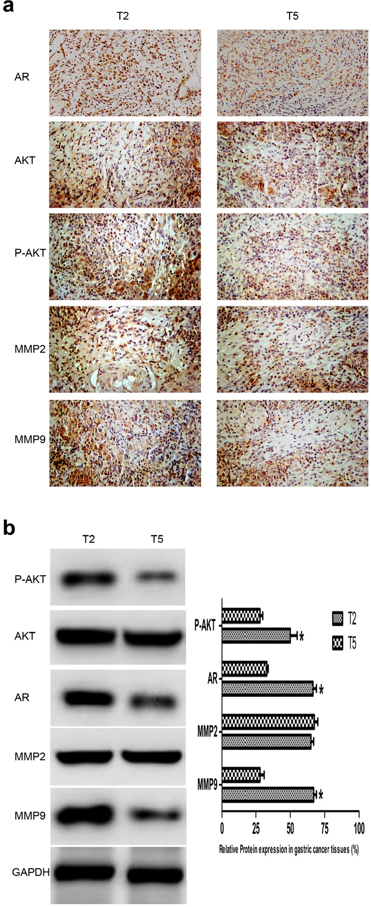
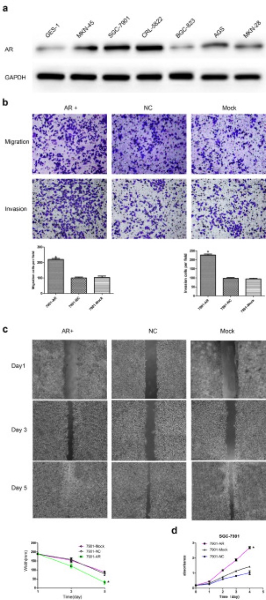
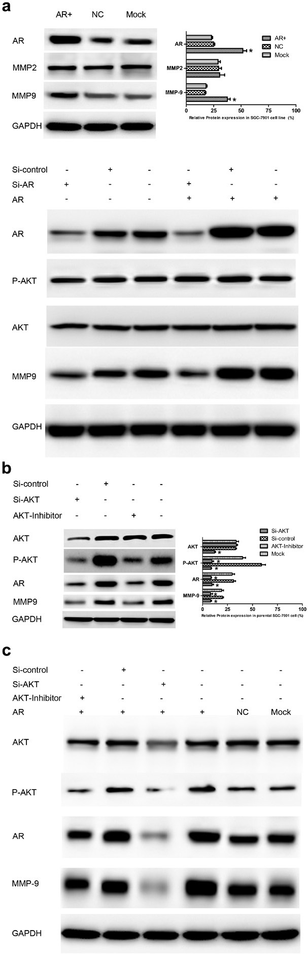
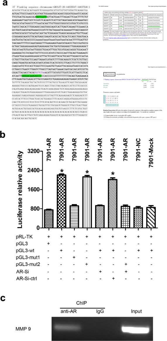
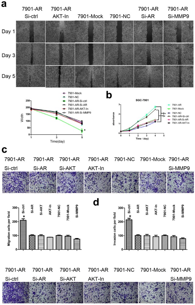
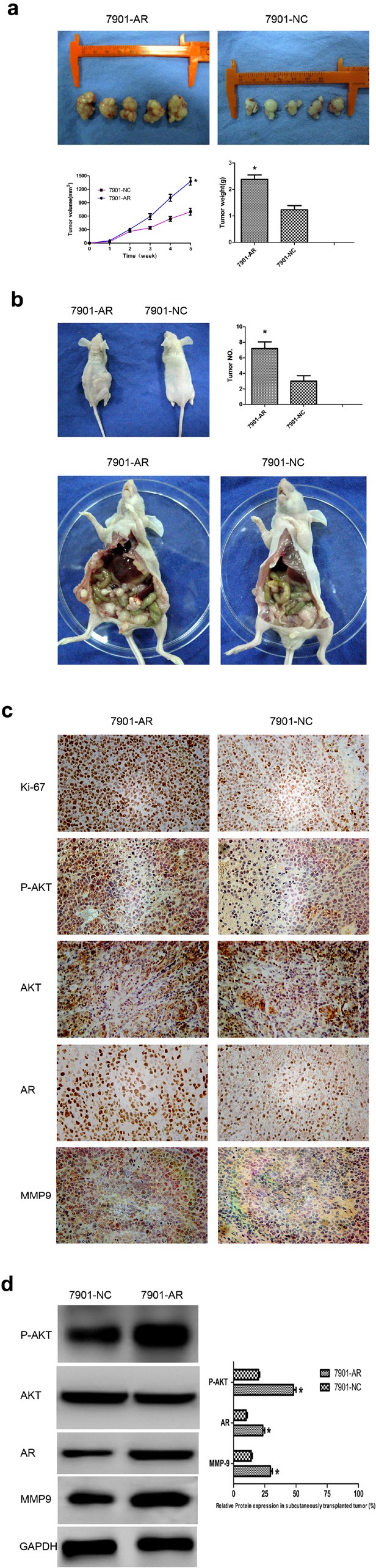
Androgen receptor (AR) plays an important role in many kinds of cancers. However, the molecular mechanisms of AR in gastric cancer (GC) are poorly characterized. Here, we investigated the role of AR in GC cell migration, invasion and metastatic potential. Our data showed that AR expression was positively correlated with lymph node metastasis and late TNM stages. These findings were accompanied by activation of AKT and upregulation of matrix metalloproteinase 9 (MMP9). AR overexpression induced increases in GC cell migration, invasion and proliferation in vitro and in vivo. These effects were attenuated by inhibition of AKT, AR and MMP9. AR overexpression upregulated MMP9 protein levels, whereas this effect was counteracted by AR siRNA. Inhibition of AKT by siRNA or an inhibitor (MK-2206 2HC) decreased AR protein expression in both stably transfected and parental SGC-7901 cells. Luciferase reporter and chromatin immunoprecipitation assays demonstrated that AR bound to the AR-binding sites of the MMP9 promoter. In summary, AR overexpression induced by AKT phosphorylation upregulated MMP9 by binding to its promoter region to promote gastric carcinogenesis. The AKT/AR/MMP9 pathway plays an important role in GC metastasis and may be a novel therapeutic target for GC treatment.


Conflict of interest statement

Disclosure of potential conflicts of interest

There no potential conflicts of interest to declare.

Figures

Fig.1
Fig.1. Expression of AR, P-AKT, MMP2, and MMP9 in GC tissues
(a) AR, P-AKT, MMP2 and MMP9 protein expression in GC tissues detected by IHC. T2 and T5 were cancer tissues deprived from individuals with or without lymph node metastasis (late or early TNM stage) as shown in Table 1, and they represented cancer tissues with positive and negative expression of AR, respectively. AR, mainly located in nucleus, AKT/P-AKT/MMPs mainly located in cytoplasm and nucleus. (b) Left: AR, P-AKT, MMP2 and MMP9 protein expression in GC tissues detected by WB. Right: densitometric analysis of the bands obtained for each signal. The P-AKT, AR and MMP9 protein levels were higher in T2 than that in T5. Results are expressed as relative expression to control samples. Data are the mean of three independent experiments ± SD (*P<0.05) (original magnification, ×200).
Fig.2
Fig.2. Effects of AR overexpression on the migration, invasion, and proliferation of SGC-7901 cells
(a) Endogenous expression of AR in seven adherent parental GC cell lines. AR expression level in SGC-7901 cells was lower than CRL-5822 but higher than other cell lines. (b) AR promoted the migration and invasion of SGC-7901 cells in vitro. Up: Representative photographs of transwell assays. Low: Histograms showed the cell numbers per field of migration (Left) and invasion (Right). (c) Up: Representative photographs of wound healing assays. Low: wound healing curve. AR promoted the wound healing rate of SGC-7901 cells in vitro. (d) Cell proliferation curve measured by a Cell Counting Kit-8 assay. Results are the mean of three independent experiments ± SD (*P<0.05) (original magnification, ×200).
Fig.3
Fig.3. AR upregulates MMP9, which depends on the phosphorylation of AKT in vitro
(a) Up: AR overexpression upregulated MMP9 (Left). Bar charts represented relative expression of different proteins (Right). Low: AR knockdown by siRNA or inhibitor decreased MMP9 expression. (b) Influence of AKT activity inhibition on endogenous expression of AR. The AR protein level was decreased significantly by siRNA or the inhibitor of AKT, which was accompanied by a decrease in MMP9 expression (Left). Bar charts represented relative expression of different proteins (Right). (c) Effect of AKT inhibition on AR protein expression in stably transfected SGC-7901 cells. AR protein expression was significantly downregulated by inhibition of AKT, which was accompanied by a decrease in MMP9 expression. Results are the mean of three independent experiments ± SD (*P<0.05).
Fig.4
Fig.4. AR enhances MMP9 expression by binding to its promoter region in SGC-7901 cells
(a) MMP9 promoter region and AR-binding sites predicated by two different on-line tools. Bold on the left indicated the amplified portion of the promoter region, and bold on the right (Up and Low) indicated the binding sites with high scores. (b) Luciferase report assays. A reporter plasmid for MMP9 (pGL3-MMP9) was generated by cloning the MMP9 promoter region (wt) or its mutants (mut1 and mut2) into the pGL3-basic vector. AR significantly increased the luciferase activity of MMP9 promoter region, which was significantly reduced by knockdown of AR. Mut1 may contained the AR-binding site. (c) MMP9 promoter region and AR interaction validated by ChIP assays. MMP9 cDNA was detectable in the immunoprecipitated chromatin samples of SGC7901-AR cells using an antibody against AR, suggesting that AR binds to the MMP9 promoter. Genomic DNA and IgG were used as controls. Results are the mean of three independent experiments ± SD (*P<0.05).
Fig.5
Fig.5. Effect of AR inhibition on the migration, invasion and proliferation of SGC-7901 cells
(a) Enhanced motility of AR-overexpressing cells in the wound healing assay was reversed by inhibition of AR, AKT or MMP9 (Up). Wound healing curve (Low). (b) CCK8 cell proliferation curve. Enhancement of AR-overexpressing cell proliferation was inhibited by suppression of AR or AKT. (c) Transwell assays. The migration-promoting effect induced by AR upregulation was prevented by downregulation of AR or MMP9 or inhibition of AKT in vitro (Up). Histograms showed the cell numbers per field of migration (Low). (d) Histograms showed the cell numbers per field of invasion (Up). The invasion-promoting effect induced by AR upregulation was prevented by downregulation of AR, MMP9 or inhibition of AKT in vitro (Low). Results are the mean of three independent experiments ± SD (*P<0.05) (original magnification, ×200).
Fig.6
Fig.6. Overexpression of AR promotes tumor growth and migration in vivo
(a) Photographs of tumors derived from SGC7901/AR and control SGC-7901 cells injected subcutaneously into nude mice. Growth kinetics curve of tumors in nude mice (Low left). Tumor diameters were measured every 7 days. Average tumor weights in nude mice (Low right). AR/Ki-67, mainly located in nucleus, AKT/P-AKT/MMPs mainly located in cytoplasm and nucleus. (b) Photographs of tumors derived from SGC7901/AR and control SGC-7901 cells injected peritonealy into nude mice. The general condition of mice (Up left). Bar charts showed numbers of tumors formed in the peritoneal cavity (Up right). Representative photographs of tumors formed in the peritoneal cavity (Low). (c) Representative IHC photographs of Ki-67 antigen, P-AKT, AR and MMP9 in tumors of nude mice. (d) Representative WB pictures of P-AKT, AR and MMP9 in tumors of nude mice (Left). Bar charts indicated relative expression levels of different proteins (Right). Results are the mean of three independent experiments ± SD (*P < 0.05) (original magnification, ×200).

Similar articles

Cited by

References

    1. Negi SS, Agarwal A, Chaudhary A. Flutamide in unresectable pancreatic adenocarcinoma: a randomized, double-blind, placebo-controlled trial. Invest New Drugs. 2006;24(3):189–194. - PubMed
    1. Chen PJ, Yeh SH, Liu WH, Lin CC, Huang HC, Chen CL, Chen DS, Chen PJ. Androgen pathway stimulates MicroRNA-216a transcription to suppress the tumor suppressor in lung cancer-1 gene in early hepatocarcinogenesis. Hepatology. 2012;56(2):632–643. - PubMed
    1. Catalano V, Labianca R, Beretta GD, Gatta G, de Braud F, Van Cutsem E. Gastric cancer. Crit Rev Oncol Hematol. 2009;71(2):127–164. - PubMed
    1. Ma WL, Hsu CL, Wu MH, Wu CT, Wu CC, Lai JJ, Jou YS, Chen CW, Yeh S, Chang C. Androgen receptor is a new potential therapeutic target for the treatment of hepatocellular carcinoma. Gastroenterology. 2008;135(3):947–955. 955.e941-945. - PMC - PubMed
    1. Feng H, Cheng ASL, Tsang DP, Li MS, Go MY, Cheung YS, Zhao GJ, Ng SS, Lin MC, Yu J, Lai PB, To KF, Sung JJY. Cell cycle-related kinase is a direct androgen receptor-regulated gene that drives beta-catenin/T cell factor-dependent hepatocarcinogenesis. J Clin Invest. 2011;121(8):3159–3175. - PMC - PubMed

Publication types

MeSH terms