. 2014 Nov 14;289(46):32044-32055.
doi: 10.1074/jbc.M114.585547. Epub 2014 Oct 9.

The endoplasmic reticulum-based acetyltransferases, ATase1 and ATase2, associate with the oligosaccharyltransferase to acetylate correctly folded polypeptides

Affiliations

The endoplasmic reticulum-based acetyltransferases, ATase1 and ATase2, associate with the oligosaccharyltransferase to acetylate correctly folded polypeptides

Yun Ding et al. J Biol Chem. .

Abstract

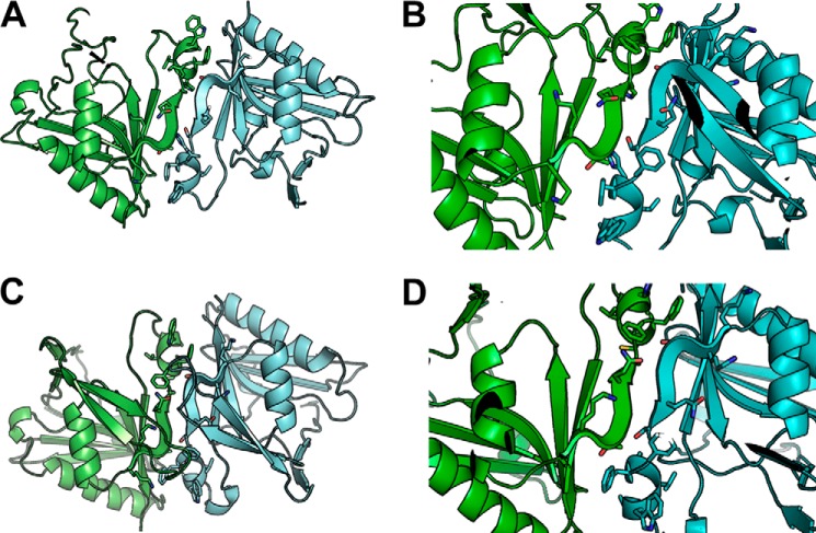
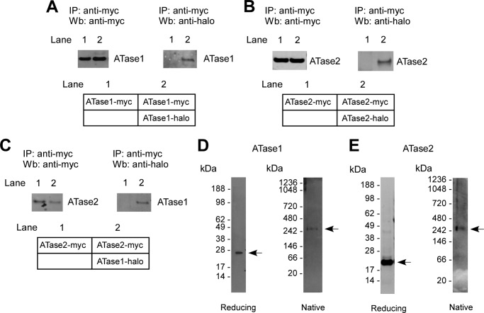
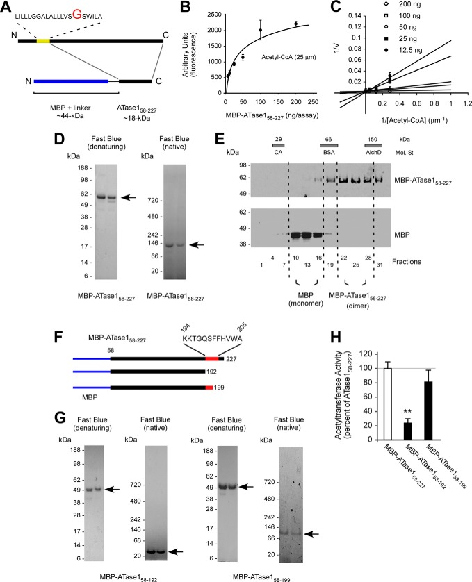
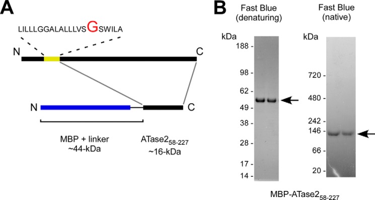
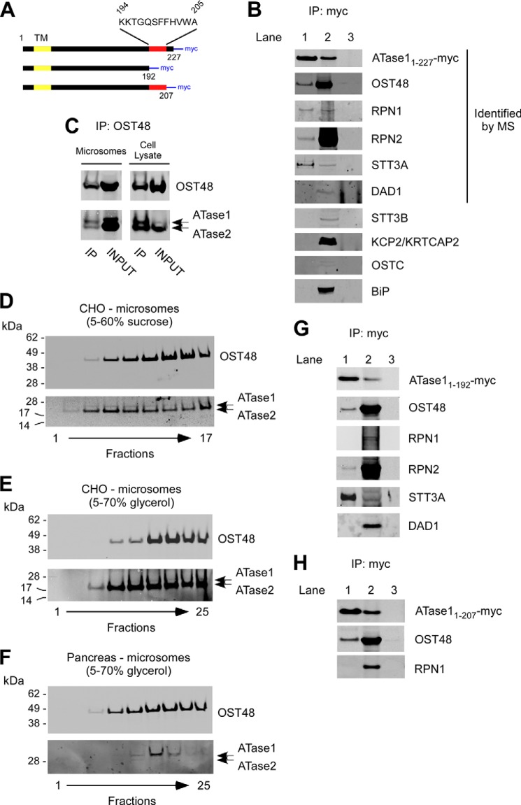
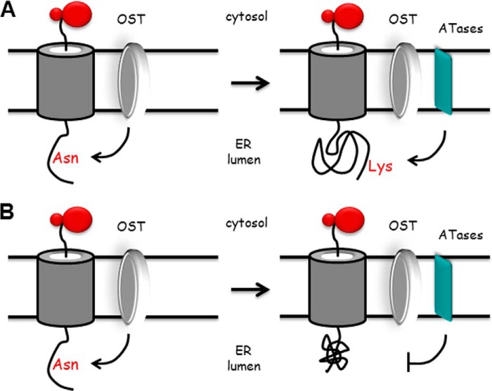
The endoplasmic reticulum (ER) has two membrane-bound acetyltransferases responsible for the endoluminal N(ϵ)-lysine acetylation of ER-transiting and -resident proteins. Mutations that impair the ER-based acetylation machinery are associated with developmental defects and a familial form of spastic paraplegia. Deficient ER acetylation in the mouse leads to defects of the immune and nervous system. Here, we report that both ATase1 and ATase2 form homo- and heterodimers and associate with members of the oligosaccharyltransferase (OST) complex. In contrast to the OST, the ATases only modify correctly folded polypetides. Collectively, our studies suggest that one of the functions of the ATases is to work in concert with the OST and "select" correctly folded from unfolded/misfolded transiting polypeptides.

Keywords: Acetyl Coenzyme A (acetyl-CoA); Acetyltransferase; Endoplasmic Reticulum (ER); Oligosaccharyltransferase; Post-translational Modification (PTM).


Figures

FIGURE 1.
FIGURE 1.
Structure modeling of ATase1 and ATase2. Homology modeling of the ATases suggests that they are dimers and that the dimerization involves the 194KKTGQSFFHVWA205 sequence at the C-terminal end of the protein. A and B, ATase1. C and D, ATase2. B and D show close-ups of the dimerization domain. Solvent-accessible surface area analysis is shown in Table 1.
FIGURE 2.
FIGURE 2.
ATase1 and ATase2 exist as homo- and heterodimers and are part of a high molecular mass complex. A–C, immunoblots showing co-IP of myc- and halo-tagged versions of ATase1 and ATase2 when co-expressed in the same cells. A, ATase1 can form homodimers. B, ATase2 can form homodimers. C, ATase1 and ATase2 can form heterodimers. A schematic view of each experiment is shown in the lower panels. D and E, affinity-purified ATase1 (D) and ATase2 (E) migrate with the molecular mass of 300–350 kDa under native conditions. IP, immunoprecipitation; Wb, Western blot.
FIGURE 3.
FIGURE 3.
The dimerization of the ATases occurs through the C terminus. A, schematic view of the MBP-ATase1 fusion protein (referred to as MBP-ATase158–227). The membrane domain of ATase1 is shown in yellow. B and C, MBP-ATase158–227 retains acetyl-CoA:lysine acetyltransferase activity. The transfer activity of the fusion protein at 25 μm concentration of acetyl-CoA ranged between 80 and 300 pmol/min/ng of enzyme. D, MBP-ATase158–227 migrates as a dimer under native conditions. E, MBP-ATase158–227 migrates as a dimer on sedimentation gradients. E, molecular standards (Mol. St.) were run in parallel; their sedimentation profile is shown as bars on top. CA, carbonic anhydrase (29 kDa); BSA, bovine serum albumin (66 kDa); AlchD, alcohol dehydrogenase (150 kDa). F, schematic view of the C-terminal deletions used to generate MBP-ATase158–192 and MBP-ATase158–199 fusion proteins. The domain predicted to be involved in the dimerization is shown in red. MBP is shown in blue. G, MBP-ATase158–192 migrates as a monomer under native conditions. H, acetyltransferase activity of the different MBP-ATase1 fusion proteins tested here. MBP-ATase158–192 displays reduced activity. The values are the averages (n = 5) ± S.D. **, p < 0.005.
FIGURE 4.
FIGURE 4.
The dimerization of ATase2 occurs through the C terminus. A, schematic view of the MBP-ATase258–227 fusion protein. The membrane domain of ATase2 is shown in yellow. B, MBP-ATase258–227 migrates as a dimer under native conditions.
FIGURE 5.
FIGURE 5.
ATase1 and ATase2 associate with OST members in a high-molecular-mass complex. A, schematic view of the C-terminal deletions used for the co-IP experiments described in B, G, and H. TM, transmembrane domain; myc, myc tag. B, immunoblots showing co-IP of OST48, RPN1, RPN2, and STT3A with transgenic ATase1 (referred to as ATase11–227-myc). Lanes 1 and 2 are from cells stable transfected with ATase1. Lane 1, immunoprecipitated product; lane 2, INPUT (prior to IP); lane 3, immunoprecipitation from mock-transfected cells (with empty plasmid). Proteins that were identified by MS analysis in the ATases high molecular mass complex are indicated. C, immunoblots showing co-IP of ATase1 and ATase2 with OST48. D and E, co-migration of OST48 and the ATases on two different sedimentation gradients. Microsomes from CHO cells overexpressing human ATase1 and ATase2 were used as starting material. F, co-migration of OST48 with endogenous ATases on a 5–70% glycerol sedimentation gradient. Microsomes from canine pancreas were used as starting material. G, immunoblots showing co-IP of OST48, RPN2, and STT3A with transgenic ATase11–192-myc. Lanes 1 and 2 are from cells stable transfected with ATase11–192. Lane 1, immunoprecipitated product; lane 2, INPUT (prior to IP); lane 3, immunoprecipitation from mock-transfected cells (with empty plasmid). H, immunoblots showing that the C-terminal tail (amino acids 208–227) of ATase1 is required for the ATase-RPN1 interaction. Co-IP of OST48 served as control. Lanes 1 and 2 are from cells stable transfected with ATase11–207. Lane 1, immunoprecipitated product; lane 2, INPUT (prior to IP); lane 3, immunoprecipitation from mock-transfected cells (with empty plasmid). IP, immunoprecipitation.
FIGURE 6.
FIGURE 6.
Nϵ-Lysine acetylation of BACE1 is post-translational and recognizes structural features of the folded protein. A, schematic view of the different deletion mutants of BACE1. The signal peptide (SP) and pro-domain (PD) at the N terminus, the single transmembrane domain (TM), and the myc tag at the C terminus are shown. The region of the protein containing the TM and the short cytosolic tail of BACE1 were left intact so as not to disrupt membrane insertion. All deletion mutants retained both SP and PD so as not to disturb correct translation and topology of the protein. *, Asn residues that are normally glycosylated; c, Cys residues required for disulfide bonds; bars, Lys residues that are normally acetylated; fl, full-length protein; mt1–mt3, deletion mutants 1–3. B, immunoblotting showing expression (left panel) and lysine acetylation (right panel) of full-length BACE1 and the deletion mutants 1–3. Mature (m) and immature (im) forms of full-length BACE1 are indicated. Ac.K., acetylated lysine. C, immunoblotting showing migration shift of all mutants following PNGase F digestion. Wb, Western blot.
FIGURE 7.
FIGURE 7.
Nϵ-Lysine acetylation of CD133 is post-translational and recognizes structural features of the folded protein. A, schematic view of full-length and deleted version of CD133. The signal peptide (SP) at the N terminus, the transmembrane domains (TM), and the myc tag at the C terminus are shown. Bars indicate the three Lys residues that are normally acetylated. fl, full-length protein; mt, deletion mutant. The deletion mutant retained the signal peptide so as not to disturb correct translation and topology of the protein. All predicted N-glycosylation sites (Asn220, Asn274, Asn395, Asn414, Asn548, Asn580, Asn729, and Asn730) were retained. B, schematic view of the topology of CD133. The truncation eliminates the first two transmembrane domains causing significant changes in the structural organization of the mutant protein. C, immunoblotting showing expression (left and middle panels) and lysine acetylation (right panel) of full-length CD133 and the deletion mutant. The expression of CD133 is shown in a total cell lysate (left panel) and after affinity purification (middle panel) with immobilized anti-myc antibodies. The slightly different migration profile of mutant CD133 is due to the deletion. Ac.K., acetylated lysine. D, immunoblotting showing migration shift of full-length and mutant CD133 following PNGase F digestion. Wb, Western blot.
FIGURE 8.
FIGURE 8.
Proposed model for our conclusions. Newly synthesized membrane proteins translocate across the translocon channel to enter the ER lumen. As soon as the NX(T/S) consensus motif for glycosylation is 30–40 Å (∼12–15 amino acids) away from the ER membrane, the OST will transfer the preformed GlcNAc2Man9Glc3 oligosaccharide to the asparagine residue of the consensus sequence. Following correct folding, the glycoprotein is recognized by the ATases, which proceed to attach one or more acetyl groups to the protein (A). This will “mark” the glycoprotein for correct trafficking along the secretory pathway. Incorrectly folded glycoproteins will not be recognized by the ATases and therefore will be “retained” for disposal (B). Therefore, Nϵ-lysine acetylation appears to act as a “selection marker” for correctly folded polypeptides. The model is based on results reported here and published elsewhere (2, 13). The model is not to scale and is only a “working interpretation” of the results.

References

    1. Pehar M., Puglielli L. (2013) Lysine acetylation in the lumen of the ER: a novel and essential function under the control of the UPR. Biochim. Biophys. Acta 1833, 686–697 - PMC - PubMed
    1. Costantini C., Ko M. H., Jonas M. C., Puglielli L. (2007) A reversible form of lysine acetylation in the ER and Golgi lumen controls the molecular stabilization of BACE1. Biochem. J. 407, 383–395 - PMC - PubMed
    1. Choudhary C., Kumar C., Gnad F., Nielsen M. L., Rehman M., Walther T. C., Olsen J. V., Mann M. (2009) Lysine acetylation targets protein complexes and co-regulates major cellular functions. Science 325, 834–840 - PubMed
    1. Pehar M., Lehnus M., Karst A., Puglielli L. (2012) Proteomic assessment shows that many endoplasmic reticulum (ER)-resident proteins are targeted by Nϵ-lysine acetylation in the lumen of the organelle and predicts broad biological impact. J. Biol. Chem. 287, 22436–22440 - PMC - PubMed
    1. Jonas M. C., Pehar M., Puglielli L. (2010) AT-1 is the ER membrane acetyl-CoA transporter and is essential for cell viability. J. Cell Sci. 123, 3378–3388 - PMC - PubMed

Publication types

MeSH terms

LinkOut - more resources