. 2013 Feb 26:8584:85840F.
doi: 10.1117/12.2008067.

Oxygen microenvironment affects the uptake of nanoparticles in head and neck tumor cells

Affiliations

Oxygen microenvironment affects the uptake of nanoparticles in head and neck tumor cells

Eunice Y Chen et al. Proc SPIE Int Soc Opt Eng. .

Abstract

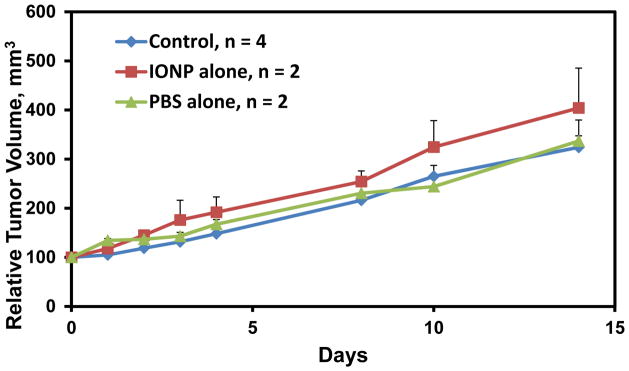
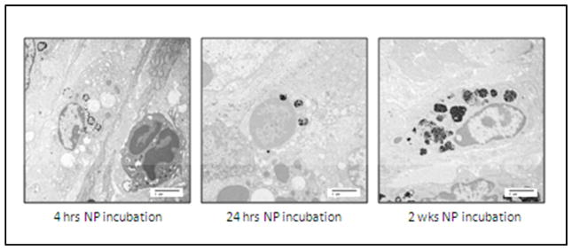
Survival of head and neck cancer patients has not improved in several decades despite advances in diagnostic and therapeutic techniques. Tumor hypoxia in head and neck cancers is a critical factor that leads to poor prognosis, resistance to radiation and chemotherapies, and increased metastatic potential. Magnetic nanoparticle hyperthermia (mNPHT) is a promising therapy for hypoxic tumors because nanoparticles (NP) can be directly injected into, or targeted to, hypoxic tumor cells and exposed to alternating magnetic fields (AMF) to induce hyperthermia. Magnetic NPHT can improve therapeutic effectiveness by two modes of action: 1) direct killing of hypoxic tumor cells; and 2) increase in tumor oxygenation, which has the potential to make the tumor more susceptible to adjuvant therapies such as radiation and chemotherapy. Prior studies in breast cancer cells demonstrated that a hypoxic microenvironment diminished NP uptake in vitro; however, mNPHT with intratumoral NP injection in hypoxic tumors increased tumor oxygenation and delayed tumor growth. In this study, head and neck squamous cell carcinoma (HNSCC) cell lines were incubated in normoxic, hypoxic, and hyperoxic conditions with iron oxide NP for 4-72 hours. After incubation, the cells were analyzed for iron uptake by mass spectrometry, Prussian blue staining, and electron microscopy. In contrast to breast cancer cells, uptake of NPs was increased in hypoxic microenvironments as compared to normoxic conditions in HNSCC cells. In future studies, we will confirm the effect of the oxygen microenvironment on NP uptake and efficacy of mNPHT both in vitro and in vivo.

Keywords: head and neck cancer; hypoxia; magnetic nanoparticle hyperthermia.


Figures

Figure 1
Figure 1
NP uptake in head and neck cancer cells as quantified by ICP-MS. A, Quantification of NP uptake in FaDu cells after 24 and 48 hrs of incubation in air and 1% oxygen conditions. B, Quantification of NP uptake in SCC-25 cells after 48 hrs of incubation in air, 1% oxygen, and 95% oxygen (carbogen) conditions. Error bars=1 STD.
Figure 2
Figure 2
Transmission electron microscopy of SCC-25 cells after 48 hrs of NP incubation under air (A, B) and 1% oxygen (C, D) conditions. Scale bars=2 micron.
Figure 3
Figure 3
Prussian blue staining in FaDu cells after incubation of NP at various time points under normoxic and hypoxic (1% O2) conditions. Scale bar=10 microns.
Figure 4
Figure 4
Quantification of Prussian blue staining in FaDu cells incubated at various time points under air and 1% oxygen conditions. Each condition was scored by a blinded pathologist three separate times and expressed as a percentage of stained to total cells. Error bars=1 STD.
Figure 5
Figure 5
Tumor oxygen measurements in ectopic FaDu tumors using EPR oximetry and OxyLite. During measurements, the FiO2 was switched from 30% oxygen to 95% oxygen and back to 30% oxygen for about 30 minutes each.
Figure 6
Figure 6
FaDu ectopic tumor growth over 14 days after the start of oxygen measurements. Control, no injection. IONP, iron oxide nanoparticle injection (5mg Fe/cm3 tumor). PBS, phoaphate buffered saline vehicle control. Error bars=1 STD
Figure 7
Figure 7
Transmission electron microscopy of FaDu tumors showing intracellular accumulation of NP. Scale bars=2 micron.
Figure 8
Figure 8
Histological analysis of NP and hypoxia localization in FaDu tumors. Serial sections stained with pimonidazole antibody to indicate hypoxic regions of tumor and Prussian blue to identify NPs. Magnification 10X

References

    1. Parkin D, Bray F, Ferlay J, Pisani P. CA Cancer J Clin. 2005;55:74–108. - PubMed
    1. Vaupel P. Oncologist. 2008;13:21–6. - PubMed
    1. Oleson J, Dewhirst M. Curr Probl Cancer. 1983;8:1–62. - PubMed
    1. Giustini A, Petryk A, Hoopes P. Nanomedicine. 2012;8:818–21. - PMC - PubMed
    1. Peng X, Qian X, Mao H, et al. Int J Nanomed. 2008;2:311–21. - PMC - PubMed

LinkOut - more resources